. 2017 Nov;40(5):1335-1346.
doi: 10.3892/ijmm.2017.3144. Epub 2017 Sep 20.

PKCζ as a promising therapeutic target for TNFα-induced inflammatory disorders in chronic cutaneous wounds

Affiliations

PKCζ as a promising therapeutic target for TNFα-induced inflammatory disorders in chronic cutaneous wounds

Jian Zhang et al. Int J Mol Med. 2017 Nov.

Abstract

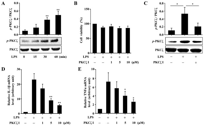
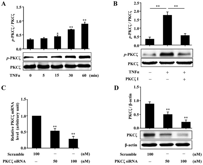
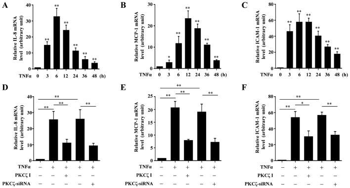
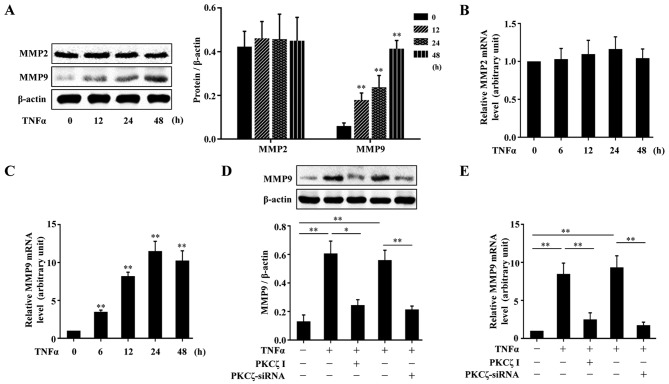
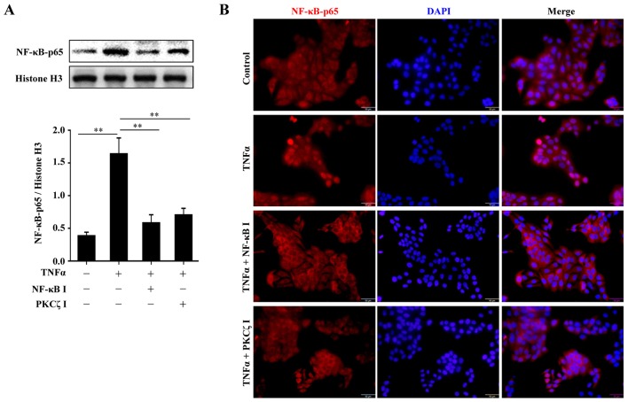
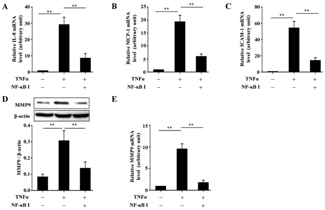
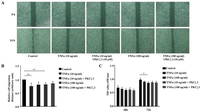
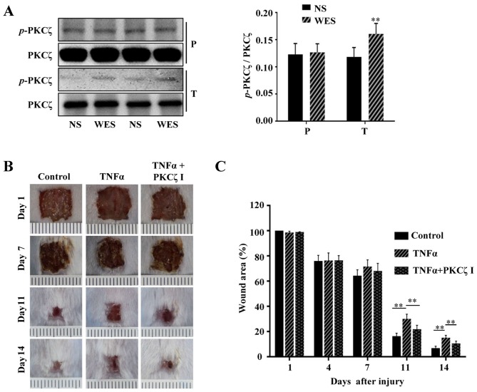
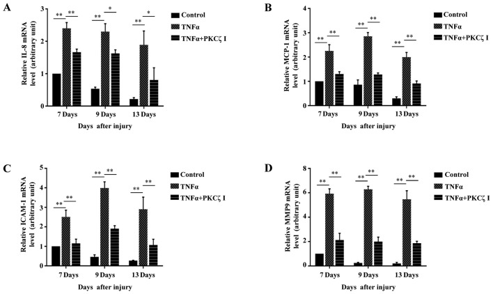
Protein kinase Cζ (PKCζ) is a member of the atypical protein kinase C family. Its roles in macrophages or skin-resident keratinocytes have not been fully evaluated. In this study, we provide evidence that PKCζ mediates lipopolysaccharide (LPS)-induced tumor necrosis factor α (TNFα) gene expression in the mouse macrophage cell line, RAW264.7. TNFα has been proven to be one of the main culprits of chronic wounds and impaired acute wounds, which are characterized by excessive inflammation, enhanced proteolysis and reduced matrix deposition. Among the multiple effects of TNFα on keratinocytes, the induction of chemokines which are indispensable factors involved in the massive infiltration of various inflammatory cells into skin lesions serves as a crucial mechanism. In the present study, we found that PKCζ inhibitor or its specific siRNA inhibited the TNFα-induced upregulation in the levels of the chemokines, interleukin (IL)-8, monocyte chemotactic protein-1 (MCP-1) and intercellular cell adhesion molecule-1 (ICAM-1) in HaCaT keratinocytes. Moreover, under a disrupted inflammatory environment, activated keratinocytes can synthesize large amounts of matrix metalloproteinases (MMP), which has a negative effect on tissue remodeling. We discovered that TNFα promoted the expression of MMP9 in a PKCζ-dependent manner. Further experiments revealed that nuclear factor-κB (NF-κB) was a key downstream molecule of PKCζ. In addition, as shown in vitro, PKCζ was not involved in the TNFα-induced decrease in HaCaT cell migration and proliferation. In vivo experiments demonstrated that TNFα-induced wound closure impairment and inflammatory disorders were significantly attenuated in the PKCζ inhibitor group. On the whole, our findings suggest that PKCζ is a crucial regulator in LPS- or TNFα-induced inflammatory responses in RAW264.7 cells and HaCaT keratinocytes, and that PKCζ/NF-κB signaling may be a potential target for interventional therapy for TNFα-induced skin inflammatory injury.


Figures

Figure 1
Figure 1
The roles of protein kinase Cζ (PKCζ) in lipopolysaccharide (LPS)-induced inflammatory responses in RAW264.7 macrophages. (A) Representative immunoblots assessing the changes in the levels of phosphorylated PKCζ in RAW264.7 cells following treatment with 100 ng/ml LPS at indicated time points (0, 15, 30 and 60 min). (B) CCK-8 assay assessing the effects of LPS (100 ng/ml) alone or with PKCζ specific pseudosubstrate inhibitor (PKCζ I) (1, 5 or 10 µM) on the viability of RAW264.7 cells. Data represent means ± SD of 3 independent experiments, the value in LPS-free group arbitrarily set as 100%. (C) Representative immunoblots showing the effects of PKCζ I (10 µM) on LPS-induced PKCζ phosphorylation. (D and E) RT-qPCR analysis showing the effects of PKCζ I on LPS-induced mRNA level changes of interleukin-1β (IL-1β) or tumor necrosis factor α (TNFα). Each bar represent the means ± SD of 3 independent experiments, and normalized to corresponding total PKCζ protein level (A and C) or GAPDH mRNA level with the value in the LPS-free group set as 1 arbitrarily (D and E); *p<0.05 and **p<0.01 vs. the value at 0 min (A) or the value in corresponding LPS alone group (C–E).
Figure 2
Figure 2
Effects of tumor necrosis factor α (TNFα) on the activation of protein kinase Cζ (PKCζ) in HaCaT cells. (A) Representative immunoblots showing the changes in the phosphorylation levels of PKCζ in HaCaT cells stimulated with 10 ng/ml TNFα at indicated time points (0, 5, 15, 30 and 60 min), respectively. (B) Representative immunoblots showing the effects of PKCζ I pre-treatment on TNFα-induced PKCζ phosphorylation. (C and D) RT-qPCR (C) or immunoblotting (D) assessing the effects of PKCζ specific interfering RNA (PKCζ-siRNA) on the knockdown of PKCζ at the mRNA and protein levels. Bars represent the means ± SD of 3 independent experiments, and normalized to corresponding total PKCζ protein level (A and B), glyceraldehyde 3-phosphate dehydrogenase (GAPDH) mRNA level with the value in the scramble group set as 1 arbitrarily (C) or β-actin protein level (D). *p<0.05 and **p<0.01 vs. the value at 0 min (A), in TNFα alone group (B), or in scramble group (C and D).
Figure 3
Figure 3
The roles of protein kinase Cζ (PKCζ) in tumor necrosis factor α (TNFα)-induced expression of interleukin-1β (IL-8), monocyte chemotactic protein-1 (MCP-1) and adhesion molecule intercellular cell adhesion molecule-1 (ICAM-1) in HaCaT cells. (A–C) The mRNA levels of (A) IL-8, (B) MCP-1 and (C) ICAM-1 in HaCaT cells exposed to TNFα (10 ng/ml) at indicated time points (0, 3, 6, 12, 24, 36 and 48 h) were assessed by RT-qPCR assays. (D–F) RT-qPCR assays showing the effects of PKCζ I (10 µM) or PKCζ-siRNA (100 nM) on the TNFα-induced upregulation of (D) IL-8, (E) MCP-1 and (F) ICAM-1. Bars represent the means ± SD of 3 independent experiments, and normalized to the GAPDH mRNA level with the value in the TNFα-free treatment group set as 1 arbitrarily. *p<0.05 and **p<0.01 vs. the value at 0 h (A–C).
Figure 4
Figure 4
Effect of tumor necrosis factor α (TNFα) on the expression of matrix metalloproteinase in HaCaT cells and the role of protein kinase Cζ (PKCζ). (A) Representative immunoblots showing the protein level changes of matrix metalloproteinase (MMP)2 and MMP9 in HaCaT cells exposed to TNFα (10 ng/ml). (B and C) RT-qPCR analysis showing the changes in the mRNA levels of MMP2 and MMP9 in HaCaT cells stimulated with TNFα. (D and E) Immunoblotting (D) or RT-qPCR (E) assessing the effect of PKCζ I or PKCζ-siRNA on the TNFα-induced upregulation of MMP9 at protein or mRNA level. Bars represent the means ± SD of 3 independent experiments, and normalized to the β-actin protein level (A and D) or GAPDH mRNA level with the value in TNFα-free group set as 1 arbitrarily (B, C and E). *p<0.05 and **p<0.01 vs. the value at 0 h (A–C).
Figure 5
Figure 5
Involvement of protein kinase Cζ (PKCζ) in tumor necrosis factor α (TNFα)-induced NF-κB-p65 nuclear translocation. (A) Representative immunoblots showing the effects of NF-κB inhibitor BAY11-7082 and PKCζ I on TNFα-induced NF-κB-p65 nuclear import. (B) The localization of NF-κB-p65 in HaCaT cells was analyzed by immunofluorescence. Bars represent the means ± SD of 3 independent experiments, and normalized to histone H3 protein level. **p<0.01. Scale bar, 50 µm.
Figure 6
Figure 6
The role of NF-κB in tumor necrosis factor α (TNFα)-induced expression of interleukin-1β (IL-8), monocyte chemotactic protein-1 (MCP-1), intercellular cell adhesion molecule-1 (ICAM-1) and MMP9 in HaCaT cells. (A–C) RT-qPCR analysis showing the effects of BAY11-7082 on the TNFα-induced changes in the mRNA levels of (A) IL-8, (B) MCP-1 and (C) ICAM-1 in HaCaT cells. (D and E) Immunoblotting (D) or RT-qPCR (E) assays showing the effect of BAY11-7082 on the TNFα-induced epression of MMP9 at protein or mRNA level in HaCaT cells. All the bars represent the means ± SD of 3 independent experiments, and normalized to the GAPDH mRNA level with the value in the TNFα-free group set as 1 arbitrarily (A–C and E) or to β-actin level (D). **p<0.01.
Figure 7
Figure 7
Effects of tumor necrosis factor α (TNFα) on the ability of migration and proliferation of HaCaT cells in vitro and the role of protein kinase Cζ (PKCζ) in these processes. (A) Representative images of the gap areas at 0 or 24 h after TNFα (10 or 100 ng/ml) treatment in the absence or presence of PKCζ I in scratch wound healing assay. Scale bar, 200 µm. (B) Histogram showing the relative cell migration distance. The migration distance was the difference value between the gap width at 0 and 24 h and the value at control group was set as 1 arbitrarily. (C) CCK-8 assay showing the proliferation of HaCaT cells after TNFα (10 or 100 ng/ml) treatment with or without PKCζ I. The absorbance at 450 nm is in direct proportion to the cell proliferation ability. Bars represent the means ± SD of 3 independent experiments. *p<0.05 and **p<0.01.
Figure 8
Figure 8
The activity of protein kinase Cζ (PKCζ) in tumor necrosis factor α (TNFα)-treated wounds and effects of PKCζ inhibitor on the attenuation of prolonged TNFα environment-induced skin wound closure delay. (A) Representative immunoblots showing the phosphorylation levels of PKCζ in paired tissue samples of the normal skin (NS) and PBS or TNFα-treated wound edge skin (WES). P, PBS-treated mouse; T, TNFα-treated mouse. (B and C) Representative images of the wound areas in PBS group (control), TNFα group and TNFα+PKCζ inhibitor (PKCζ I) group on days 1, 7, 11 and 14 post-incision. The wound closure rate was the ratio of the remanent wound area on the indicated days and the original wound area on day 1 post-incision. Bars represent the means ± SD of 3 independent experiments, n=8. **p<0.01.
Figure 9
Figure 9
Roles of protein kinase Cζ (PKCζ) in prolonged tumor necrosis factor α (TNFα) environment-induced upregulation of interleukin-1β (IL-8), monocyte chemotactic protein-1 (MCP-1), intercellular cell adhesion molecule-1 (ICAM-1) and MMP9. (A–D) RT-qPCR analysis showing the effects of PKCζ inhibitor (PKCζ I) on the TNFα-induced increased mRNA levels of (A) IL-8, (B) MCP-1 and (C) ICAM-1 and (D) MMP9 in wounds edges at different days of the late phase of wound healing (days 7, 9 and 13 after wounding). Bars represent the means ± SD of 3 independent experiments, and normalized to the GAPDH mRNA level. The value on day 7 in control group was set as 1 arbitrarily. *p<0.05 and **p<0.01.

Similar articles

Cited by

References

    1. Martin P, Nunan R. Cellular and molecular mechanisms of repair in acute and chronic wound healing. Br J Dermatol. 2015;173:370–378. doi: 10.1111/bjd.13954. - DOI - PMC - PubMed
    1. Streit M, Beleznay Z, Braathen LR. Topical application of the tumour necrosis factor-alpha antibody infliximab improves healing of chronic wounds. Int Wound J. 2006;3:171–179. doi: 10.1111/j.1742-481X.2006.00233.x. - DOI - PMC - PubMed
    1. Xu F, Zhang C, Graves DT. Abnormal cell responses and role of TNF-α in impaired diabetic wound healing. BioMed Res Int. 2013;2013:754802. doi: 10.1155/2013/754802. - DOI - PMC - PubMed
    1. Barker JN, Mitra RS, Griffiths CE, Dixit VM, Nickoloff BJ. Keratinocytes as initiators of inflammation. Lancet. 1991;337:211–214. doi: 10.1016/0140-6736(91)92168-2. - DOI - PubMed
    1. Wetzler C, Kämpfer H, Stallmeyer B, Pfeilschifter J, Frank S. Large and sustained induction of chemokines during impaired wound healing in the genetically diabetic mouse: Prolonged persistence of neutrophils and macrophages during the late phase of repair. J Invest Dermatol. 2000;115:245–253. doi: 10.1046/j.1523-1747.2000.00029.x. - DOI - PubMed

MeSH terms