Age-dependent electrocardiographic changes in Pgc-1β deficient murine hearts
- PMID: 28949414
- PMCID: PMC5814877
- DOI: 10.1111/1440-1681.12863
Age-dependent electrocardiographic changes in Pgc-1β deficient murine hearts
Abstract
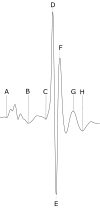
Increasing evidence implicates chronic energetic dysfunction in human cardiac arrhythmias. Mitochondrial impairment through Pgc-1β knockout is known to produce a murine arrhythmic phenotype. However, the cumulative effect of this with advancing age and its electrocardiographic basis have not been previously studied. Young (12-16 weeks) and aged (>52 weeks), wild type (WT) (n = 5 and 8) and Pgc-1β-/- (n = 9 and 6), mice were anaesthetised and used for electrocardiographic (ECG) recordings. Time intervals separating successive ECG deflections were analysed for differences between groups before and after β1-adrenergic (intraperitoneal dobutamine 3 mg/kg) challenge. Heart rates before dobutamine challenge were indistinguishable between groups. The Pgc-1β-/- genotype however displayed compromised nodal function in response to adrenergic challenge. This manifested as an impaired heart rate response suggesting a functional defect at the level of the sino-atrial node, and a negative dromotropic response suggesting an atrioventricular conduction defect. Incidences of the latter were most pronounced in the aged Pgc-1β-/- mice. Moreover, Pgc-1β-/- mice displayed electrocardiographic features consistent with the existence of a pro-arrhythmic substrate. Firstly, ventricular activation was prolonged in these mice consistent with slowed action potential conduction and is reported here for the first time. Additionally, Pgc-1β-/- mice had shorter repolarisation intervals. These were likely attributable to altered K+ conductance properties, ultimately resulting in a shortened QTc interval, which is also known to be associated with increased arrhythmic risk. ECG analysis thus yielded electrophysiological findings bearing on potential arrhythmogenicity in intact Pgc-1β-/- systems in widespread cardiac regions.
Keywords: cardiac arrhythmias; cardiac conduction; electrocardiogram, ECG; peroxisome proliferator activated receptor-γ-coactivator-1 (PGC-1).
© 2017 The Authors. Clinical and Experimental Pharmacology and Physiology Published by John Wiley & Sons Australia, Ltd.
Figures


References
-
- Lin PH, Lee SH, Su CP, Wei YH. Oxidative damage to mitochondrial DNA in atrial muscle of patients with atrial fibrillation. Free Radic Biol Med. 2003;35:1310‐1318. - PubMed
-
- Bukowska A, Schild L, Keilhoff G, et al. Mitochondrial dysfunction and redox signaling in atrial tachyarrhythmia. Exp Biol Med. 2008;233:558‐574. - PubMed
Publication types
MeSH terms
Substances
Grants and funding
LinkOut - more resources
Full Text Sources
Other Literature Sources
Medical

