

Developmental and adult characterization of secretagogin expressing amacrine cells in zebrafish retina
- PMID: 28949993
- PMCID: PMC5614429
- DOI: 10.1371/journal.pone.0185107
Developmental and adult characterization of secretagogin expressing amacrine cells in zebrafish retina
Abstract
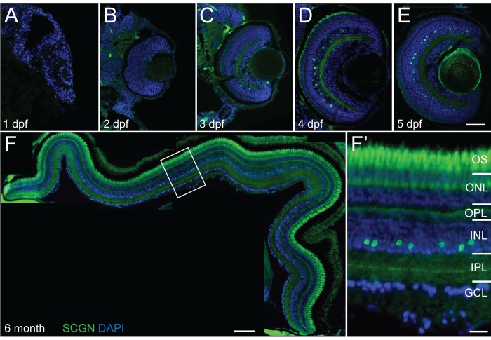
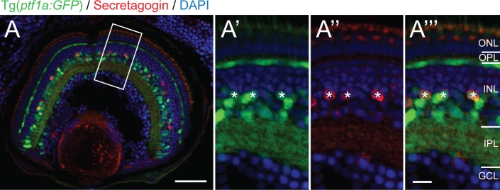
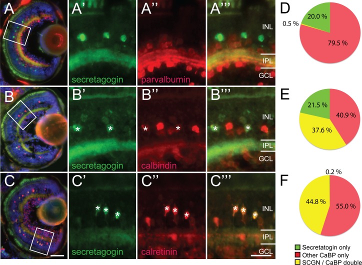
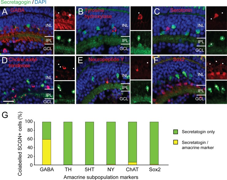
Calcium binding proteins show stereotypical expression patterns within diverse neuron types across the central nervous system. Here, we provide a characterization of developmental and adult secretagogin-immunolabelled neurons in the zebrafish retina with an emphasis on co-expression of multiple calcium binding proteins. Secretagogin is a recently identified and cloned member of the F-hand family of calcium binding proteins, which labels distinct neuron populations in the retinas of mammalian vertebrates. Both the adult distribution of secretagogin labeled retinal neurons as well as the developmental expression indicative of the stage of neurogenesis during which this calcium binding protein is expressed was quantified. Secretagogin expression was confined to an amacrine interneuron population in the inner nuclear layer, with monostratified neurites in the center of the inner plexiform layer and a relatively regular soma distribution (regularity index > 2.5 across central-peripheral areas). However, only a subpopulation (~60%) co-labeled with gamma-aminobutyric acid as their neurotransmitter, suggesting that possibly two amacrine subtypes are secretagogin immunoreactive. Quantitative co-labeling analysis with other known amacrine subtype markers including the three main calcium binding proteins parvalbumin, calbindin and calretinin identifies secretagogin immunoreactive neurons as a distinct neuron population. The highest density of secretagogin cells of ~1800 cells / mm2 remained relatively evenly along the horizontal meridian, whilst the density dropped of to 125 cells / mm2 towards the dorsal and ventral periphery. Thus, secretagogin represents a new amacrine label within the zebrafish retina. The developmental expression suggests a possible role in late stage differentiation. This characterization forms the basis of functional studies assessing how the expression of distinct calcium binding proteins might be regulated to compensate for the loss of one of the others.
Conflict of interest statement
Figures

Similar articles
-
Characterization of secretagogin-immunoreactive amacrine cells in marmoset retina.J Comp Neurol. 2014 Feb 1;522(2):435-55. doi: 10.1002/cne.23420. J Comp Neurol. 2014. PMID: 23852983
-
Parvalbumin-immunoreactive neurons in the inner nuclear layer of zebrafish retina.Exp Eye Res. 2009 Mar;88(3):553-60. doi: 10.1016/j.exer.2008.11.014. Epub 2008 Dec 3. Exp Eye Res. 2009. PMID: 19084520
-
In vivo expression of Nurr1/Nr4a2a in developing retinal amacrine subtypes in zebrafish Tg(nr4a2a:eGFP) transgenics.J Comp Neurol. 2017 Jun 1;525(8):1962-1979. doi: 10.1002/cne.24185. Epub 2017 Mar 15. J Comp Neurol. 2017. PMID: 28177524
-
Revival of calcium-binding proteins for neuromorphology: secretagogin typifies distinct cell populations in the avian brain.Brain Behav Evol. 2014;83(2):82-92. doi: 10.1159/000357834. Epub 2014 Apr 24. Brain Behav Evol. 2014. PMID: 24776989
-
The renaissance of Ca2+-binding proteins in the nervous system: secretagogin takes center stage.Cell Signal. 2012 Feb;24(2):378-387. doi: 10.1016/j.cellsig.2011.09.028. Epub 2011 Oct 1. Cell Signal. 2012. PMID: 21982882 Free PMC article. Review.
Cited by
-
Immunohistochemical distribution of secretagogin in the mouse brain.Front Neuroanat. 2023 Aug 30;17:1224342. doi: 10.3389/fnana.2023.1224342. eCollection 2023. Front Neuroanat. 2023. PMID: 37711587 Free PMC article.
-
20 Years of Secretagogin: Exocytosis and Beyond.Front Mol Neurosci. 2019 Feb 12;12:29. doi: 10.3389/fnmol.2019.00029. eCollection 2019. Front Mol Neurosci. 2019. PMID: 30853888 Free PMC article.
-
Onecut Regulates Core Components of the Molecular Machinery for Neurotransmission in Photoreceptor Differentiation.Front Cell Dev Biol. 2021 Mar 18;9:602450. doi: 10.3389/fcell.2021.602450. eCollection 2021. Front Cell Dev Biol. 2021. PMID: 33816460 Free PMC article.
References
-
- Greer PL, Greenberg ME. From synapse to nucleus: calcium-dependent gene transcription in the control of synapse development and function. Neuron. 2008;59(6):846–60. doi: 10.1016/j.neuron.2008.09.002 . - DOI - PubMed
-
- Martin ZS, Neugebauer V, Dineley KT, Kayed R, Zhang W, Reese LC, et al. alpha-Synuclein oligomers oppose long-term potentiation and impair memory through a calcineurin-dependent mechanism: relevance to human synucleopathic diseases. J Neurochem. 2012;120(3):440–52. doi: 10.1111/j.1471-4159.2011.07576.x ; PubMed Central PMCID: PMCPMC3253169. - DOI - PMC - PubMed
-
- Bai Y, Sun Y, Peng J, Liao H, Gao H, Guo Y, et al. Overexpression of secretagogin inhibits cell apoptosis and induces chemoresistance in small cell lung cancer under the regulation of miR-494. Oncotarget. 2014;5(17):7760–75. doi: 10.18632/oncotarget.2305 ; PubMed Central PMCID: PMCPMC4202159. - DOI - PMC - PubMed
-
- Andressen C, Blumcke I, Celio MR. Calcium-binding proteins: selective markers of nerve cells. Cell Tissue Res. 1993;271(2):181–208. . - PubMed
-
- Raju CS, Spatazza J, Stanco A, Larimer P, Sorrells SF, Kelley KW, et al. Secretagogin is Expressed by Developing Neocortical GABAergic Neurons in Humans but not Mice and Increases Neurite Arbor Size and Complexity. Cereb Cortex. 2017:1–13. doi: 10.1093/cercor/bhx101 . - DOI - PMC - PubMed
MeSH terms
Substances
LinkOut - more resources
Full Text Sources
Other Literature Sources
Molecular Biology Databases
