

Ethanol extract of propolis and its constituent caffeic acid phenethyl ester inhibit breast cancer cells proliferation in inflammatory microenvironment by inhibiting TLR4 signal pathway and inducing apoptosis and autophagy
- PMID: 28950845
- PMCID: PMC5615448
- DOI: 10.1186/s12906-017-1984-9
Ethanol extract of propolis and its constituent caffeic acid phenethyl ester inhibit breast cancer cells proliferation in inflammatory microenvironment by inhibiting TLR4 signal pathway and inducing apoptosis and autophagy
Abstract
Background: Propolis and its major constituent - caffeic acid phenethyl ester (CAPE) have good abilities on antitumor and anti-inflammation. However, little is known about the actions of propolis and CAPE on tumor in inflammatory microenvironment, and inflammatory responses play decisive roles at different stages of tumor development. To understand the effects and mechanisms of ethanol-extracted Chinese propolis (EECP) and its major constituent - CAPE in inflammation-stimulated tumor, we investigated their effects on Toll-like receptor 4 (TLR4) signaling pathway which plays a crucial role in breast cancer MDA-MB-231 cell line.
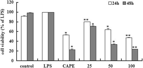
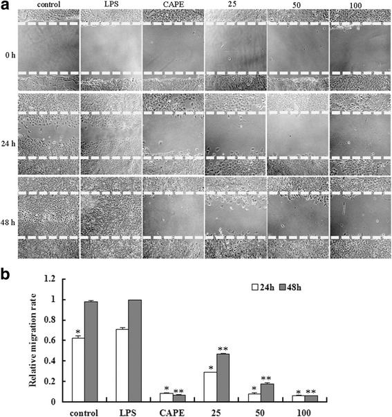
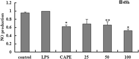
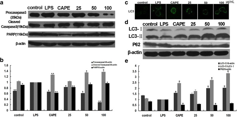
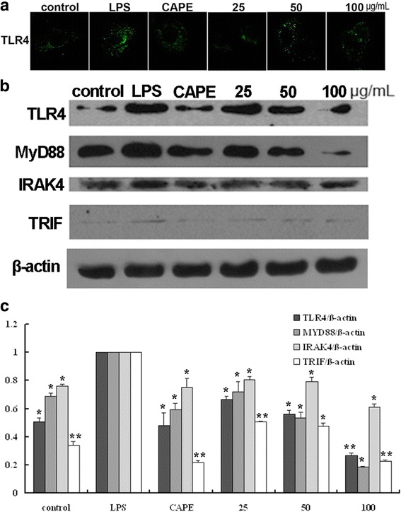
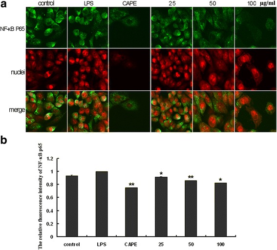
Methods: 80% confluent breast cancer MDA-MB-231 cells were stimulated with 1 μg/mL lipopolysaccaride (LPS). Then the cells were divided for treatment by CAPE (25 μg/mL) and EECP (25, 50 and 100 μg/mL), respectively. Cell viability, nitric oxide (NO) production and cell migration were measured by sulforhodamine B assay, chemical method and scratch assay. The levels of TLR4, MyD88, IRAK4, TRIF, caspase 3, PARP, LC3B and p62 were investigated through western blotting. The expression of TLR4, LC3B and nuclear factor-κB p65 (NF-κB p65) were tested by immunofluorescence microscopy assay.
Results: Treatment of different concentrations of EECP (25, 50 and 100 μg/mL) and CAPE (25 μg/mL) significantly inhibited LPS-stimulated MDA-MB-231 cell line proliferation, migration and NO production. Furthermore, EECP and CAPE activated caspase3 and PARP to induce cell apoptosis, and also upregulated LC3-II and decreased p62 level to induce autophagy during the process. TLR4 signaling pathway molecules such as TLR4, MyD88, IRAK4, TRIF and NF-κB p65 were all down-regulated after EECP and CAPE treatment in LPS-stimulated MDA-MB-231 cells.
Conclusions: These findings indicated that EECP and its major constituent - CAPE inhibited breast cancer MDA-MB-231 cells proliferation in inflammatory microenvironment through activating apoptosis, autophagy and inhibiting TLR4 signaling pathway. EECP and CAPE may hold promising prospects in treating inflammation-induced tumor.
Keywords: Anti-inflammatory; Antitumor; Caffeic acid phenethyl ester; Propolis; Toll like receptor 4.
Conflict of interest statement
Ethics approval and consent to participate
N/A
Consent for publication
Not applicable
Competing interests
The authors declare that they have no competing interests.
Publisher’s Note
Springer Nature remains neutral with regard to jurisdictional claims in published maps and institutional affiliations.
Figures
Similar articles
-
Caffeic acid phenethyl ester attenuates lipopolysaccharide-stimulated proinflammatory responses in human gingival fibroblasts via NF-κB and PI3K/Akt signaling pathway.Eur J Pharmacol. 2017 Jan 5;794:61-68. doi: 10.1016/j.ejphar.2016.11.003. Epub 2016 Nov 7. Eur J Pharmacol. 2017. PMID: 27832944
-
Caffeic Acid phenethyl ester and ethanol extract of propolis induce the complementary cytotoxic effect on triple-negative breast cancer cell lines.Molecules. 2015 May 20;20(5):9242-62. doi: 10.3390/molecules20059242. Molecules. 2015. PMID: 26007182 Free PMC article.
-
Caffeic acid phenethyl ester (CAPE), derived from a honeybee product propolis, exhibits a diversity of anti-tumor effects in pre-clinical models of human breast cancer.Cancer Lett. 2011 Sep 1;308(1):43-53. doi: 10.1016/j.canlet.2011.04.012. Epub 2011 May 13. Cancer Lett. 2011. PMID: 21570765 Free PMC article.
-
In vivo and in vitro antıneoplastic actions of caffeic acid phenethyl ester (CAPE): therapeutic perspectives.Nutr Cancer. 2013;65(4):515-26. doi: 10.1080/01635581.2013.776693. Nutr Cancer. 2013. PMID: 23659443 Review.
-
Caffeic acid phenethyl ester and therapeutic potentials.Biomed Res Int. 2014;2014:145342. doi: 10.1155/2014/145342. Epub 2014 May 29. Biomed Res Int. 2014. PMID: 24971312 Free PMC article. Review.
Cited by
-
[Effects of mineral trioxide aggregate and ethanolic extracts of Shandong propolis on the biological properties of human dental pulp fibroblasts].Beijing Da Xue Xue Bao Yi Xue Ban. 2019 Dec 18;51(6):1108-1114. doi: 10.19723/j.issn.1671-167X.2019.06.023. Beijing Da Xue Xue Bao Yi Xue Ban. 2019. PMID: 31848513 Free PMC article. Chinese.
-
Propolis and Their Active Constituents for Chronic Diseases.Biomedicines. 2023 Jan 18;11(2):259. doi: 10.3390/biomedicines11020259. Biomedicines. 2023. PMID: 36830794 Free PMC article. Review.
-
The Effect of Natural Substances Contained in Bee Products on Prostate Cancer in In Vitro Studies.Molecules. 2023 Jul 28;28(15):5719. doi: 10.3390/molecules28155719. Molecules. 2023. PMID: 37570691 Free PMC article.
-
Anticancer Activity of Propolis and Its Compounds.Nutrients. 2021 Jul 28;13(8):2594. doi: 10.3390/nu13082594. Nutrients. 2021. PMID: 34444754 Free PMC article. Review.
-
Food as medicine: targeting the uraemic phenotype in chronic kidney disease.Nat Rev Nephrol. 2021 Mar;17(3):153-171. doi: 10.1038/s41581-020-00345-8. Epub 2020 Sep 22. Nat Rev Nephrol. 2021. PMID: 32963366 Review.
References
-
- Rzepecka-Stojko A, Kabała-Dzik A, Moździerz A, Kubina R, Wojtyczka RD, Stojko R, Dziedzic A, Jastrzębska-Stojko Ż, Jurzak M, Buszman E. Caffeic Acid phenethyl ester and ethanol extract of propolis induce the complementary cytotoxic effect on triple-negative breast cancer cell lines. Molecules. 2015;20(5):9242–9262. doi: 10.3390/molecules20059242. - DOI - PMC - PubMed
MeSH terms
Substances
LinkOut - more resources
Full Text Sources
Other Literature Sources
Medical
Research Materials
Miscellaneous
