. 2017 Sep 26;10(1):445.
doi: 10.1186/s13071-017-2385-0.

Stimulation of IL-1β and IL-6 through NF-κB and sonic hedgehog-dependent pathways in mouse astrocytes by excretory/secretory products of fifth-stage larval Angiostrongylus cantonensis

Affiliations

Stimulation of IL-1β and IL-6 through NF-κB and sonic hedgehog-dependent pathways in mouse astrocytes by excretory/secretory products of fifth-stage larval Angiostrongylus cantonensis

Kuang-Yao Chen et al. Parasit Vectors. .

Abstract

Background: Angiostrongylus cantonensis is an important causative agent of eosinophilic meningitis and eosinophilic meningoencephalitis in humans. Previous studies have shown that the Sonic hedgehog (Shh) signaling pathway may reduce cell apoptosis by inhibiting oxidative stress in A. cantonensis infection. In this study, we investigated the relationship between cytokine secretion and Shh pathway activation after treatment with excretory/secretory products (ESP) of fifth-stage larval A. cantonensis (L5).

Results: The results showed that IL-1β and IL-6 levels in mouse astrocytes were increased. Moreover, ESP stimulated the protein expression of Shh pathway molecules, including Shh, Ptch, Smo and Gli-1, and induced IL-1β and IL-6 secretion. The transcription factor nuclear factor-κB (NF-κB) plays an important role in inflammation, and it regulates the expression of proinflammatory genes, including cytokines and chemokines, such as IL-1β and TNF-α. After ESP treatment, NF-κB induced IL-1β and IL-6 secretion in astrocytes by activating the Shh signaling pathway.

Conclusions: Overall, the data presented in this study showed that ESP of fifth-stage larval A. cantonensis stimulates astrocyte activation and cytokine generation through NF-κB and the Shh signaling pathway.

Keywords: Angiostrongylus cantonensis; Astrocytes; Cytokine; Excretory/secretory products; NF-κB; Sonic hedgehog signaling pathway.


Conflict of interest statement

Ethics approval

All animal protocols in this study were approved by the Chang Gung University Institutional Animal Care and Use Committee (CGU15–193). Mice were housed in plastic cages and provided with food and water ad libitum. The experimental animals were sacrificed by anesthesia with chloral hydrate.

Consent for publication

Not applicable.

Competing interests

The authors declare that they have no competing interests.

Publisher’s Note

Springer Nature remains neutral with regard to jurisdictional claims in published maps and institutional affiliations.

Figures
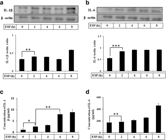
Fig. 1
Fig. 1
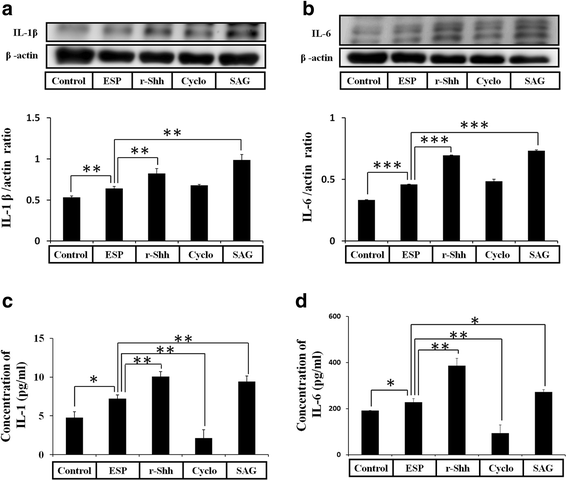
ESP induce IL-1β and IL-6. Astrocytes were treated with 500 μg/ml ESP at different time points. The protein levels of IL-1β (a) and IL-6 (b) were detected by Western blot analysis. The concentrations of IL-1β (c) and IL-6 (d) were determined in astrocyte culture medium by ELISA. Statistical significance was determined by Student’s t-test: *P < 0.05, **P < 0.01, ***P < 0.001 (n = 3)
Fig. 2
Fig. 2
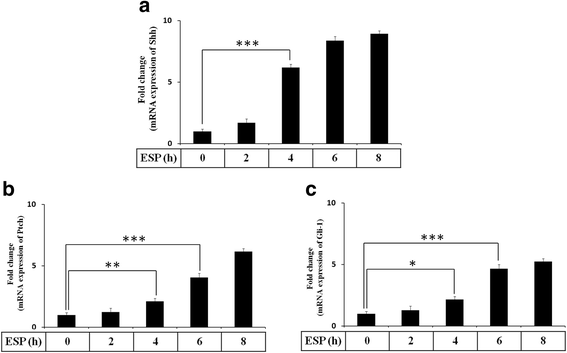
ESP stimulate the gene expression of Shh signaling pathway. Astrocytes were treated with 500 μg/ml ESP for the indicated time points. The mRNA levels of Shh, Ptch, and Gli-1 were determined in astrocytes by Quantitative real-time PCR. Statistical significance was determined by Student’s t-test: *P < 0.05, **P < 0.01, ***P < 0.001 (n = 3)
Fig. 3
Fig. 3
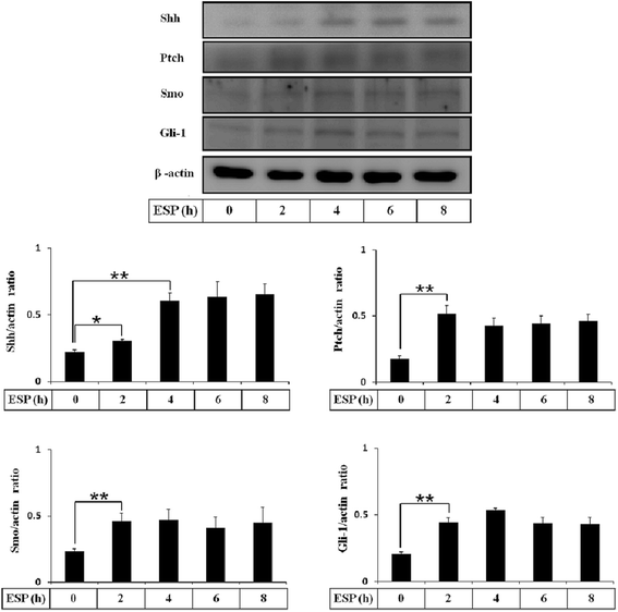
ESP induce Shh signaling pathway activation. Astrocytes were treated with 500 μg/ml ESP for the indicated time points. The protein levels of Shh, Ptch, Smo, and Gli-1 were determined in astrocytes by Western blots. Statistical significance was determined by Student’s t-test: *P < 0.05, **P < 0.01 (n = 3)
Fig. 4
Fig. 4
ESP induce the expressions of IL-1β and IL-6 via the Shh pathway. Western blot analysis of IL-1β (a) and IL-6 (b) in astrocytes co-cultured with ESP alone or pretreated with a recombinant Sonic hedgehog peptide from mouse (Shh) (3 μg), cyclopamine (20 μM) or SAG for 2 h and then with 500 μg/ml ESP for 4 h. β-actin is shown as a control. Data are expressed as the mean ± SD from three independent experiments (**P < 0.01). (c, d) Changes in the concentrations of IL-1β and IL-6 protein in the culture medium of astrocytes detected by the ELISA. Data are expressed as the mean ± SD from three independent experiments (*P < 0.05, **P < 0.01, ***P < 0.001)
Fig. 5
Fig. 5
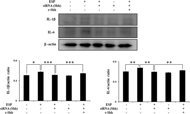
The expressions of IL-1β and IL-6 were increased through the Shh in ESP treatment. Western blot analysis of IL-1β and IL-6 in astrocytes co-cultured with ESP alone or pretreated with Shh siRNA for 48 h and then with a recombinant Sonic hedgehog peptide from mouse (Shh) (3 μg) for 2 h and 500 μg/ml ESP for 4 h. Data are expressed as the mean ± SD from three independent experiments (*P < 0.05, **P < 0.01, ***P < 0.001)
Fig. 6
Fig. 6
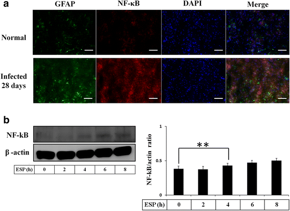
ESP induce NF-κB expression. a Fluorescence microscopy demonstrated the expression of NF-κB in astrocytes from the hippocampus of BALB/c mice infected with 25 A. cantonensis third-stage larvae on day 28 post-infection (GFAP: green; NF-κB: red; colocalization of NF-κB and Shh: yellow). b Western blot analysis of NF-κB in astrocytes treated with ESP for 0–8 h. Data are expressed as the mean ± SD from three independent experiments (**P < 0.01). Scale-bars: 100 μm
Fig. 7
Fig. 7
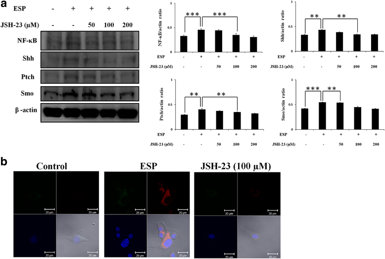
NF-κB induces Shh signaling pathway activation. a Western blot analysis of NF-κB, Shh, Ptch, and Smo in astrocytes co-cultured with ESP alone or JSH-23 for 2 h and then with 500 μg/ml ESP for 4 h; β-actin is shown as a control. Data are expressed as the mean ± SD from three independent experiments (**P < 0.01, ***P < 0.001). b The expression levels of Shh and GFAP in astrocytes. The Shh and GFAP protein expressions were determined by immunofluorescence staining of astrocytes with ESP alone or JSH-23 for 2 h and then with 500 μg/ml ESP for 4 h. (GFAP: green; Shh: red; DAPI: blue)
Fig. 8
Fig. 8
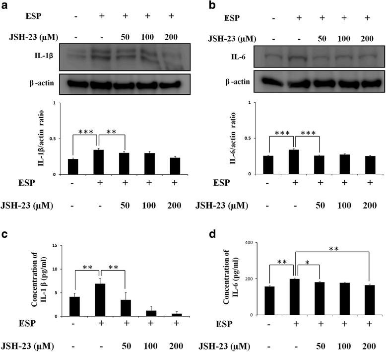
NF-κB induces the expressions of IL-1β and IL-6. Western blot analysis of IL-1β (a) and IL-6 (b) in astrocytes co-cultured with ESP alone or JSH-23 for 2 h and then with 500 μg/ml ESP for 4 h; β-actin is shown as a control. Data are expressed as the mean ± SD from three independent experiments (**P < 0.01, ***P < 0.001). c, d Changes in the concentrations of IL-1β and IL-6 protein in the culture medium of astrocytes detected by the ELISA. Data are expressed as the mean ± SD from three independent experiments (*P < 0.05, ** P < 0.01)

Similar articles

Cited by

References

    1. Chauhan A. Unperturbed post-transcriptional regulatory rev protein function and HIV-1 replication in astrocytes. PLoS One. 2014;9:e106910. doi: 10.1371/journal.pone.0106910. - DOI - PMC - PubMed
    1. Xia YP, Dai RL, Li YN, Mao L, Xue YM, He QW, et al. The protective effect of sonic hedgehog is mediated by the propidium iodide 3-kinase/AKT/Bcl-2 pathway in cultured rat astrocytes under oxidative stress. Neuroscience. 2012;209:1–11. doi: 10.1016/j.neuroscience.2012.02.019. - DOI - PubMed
    1. Lau LT, Yu AC. Astrocytes produce and release interleukin-1, interleukin-6, tumor necrosis factor alpha and interferon-gamma following traumatic and metabolic injury. J Neurotrauma. 2001;18:351–359. doi: 10.1089/08977150151071035. - DOI - PubMed
    1. Norris JG, Benveniste EN. Interleukin-6 production by astrocytes: induction by the neurotransmitter norepinephrine. J Neuroimmunol. 1993;45:137–145. doi: 10.1016/0165-5728(93)90174-W. - DOI - PubMed
    1. Van Wagoner NJ, Oh JW, Repovic P, Benveniste EN. Interleukin-6 (IL-6) production by astrocytes: autocrine regulation by IL-6 and the soluble IL-6 receptor. J Neurosci. 1999;19:5236–5244. - PMC - PubMed

MeSH terms

LinkOut - more resources