

Tyrosines-740/751 of PDGFRβ contribute to the activation of Akt/Hif1α/TGFβ nexus to drive high glucose-induced glomerular mesangial cell hypertrophy
- PMID: 28951244
- PMCID: PMC5889140
- DOI: 10.1016/j.cellsig.2017.09.017
Tyrosines-740/751 of PDGFRβ contribute to the activation of Akt/Hif1α/TGFβ nexus to drive high glucose-induced glomerular mesangial cell hypertrophy
Abstract
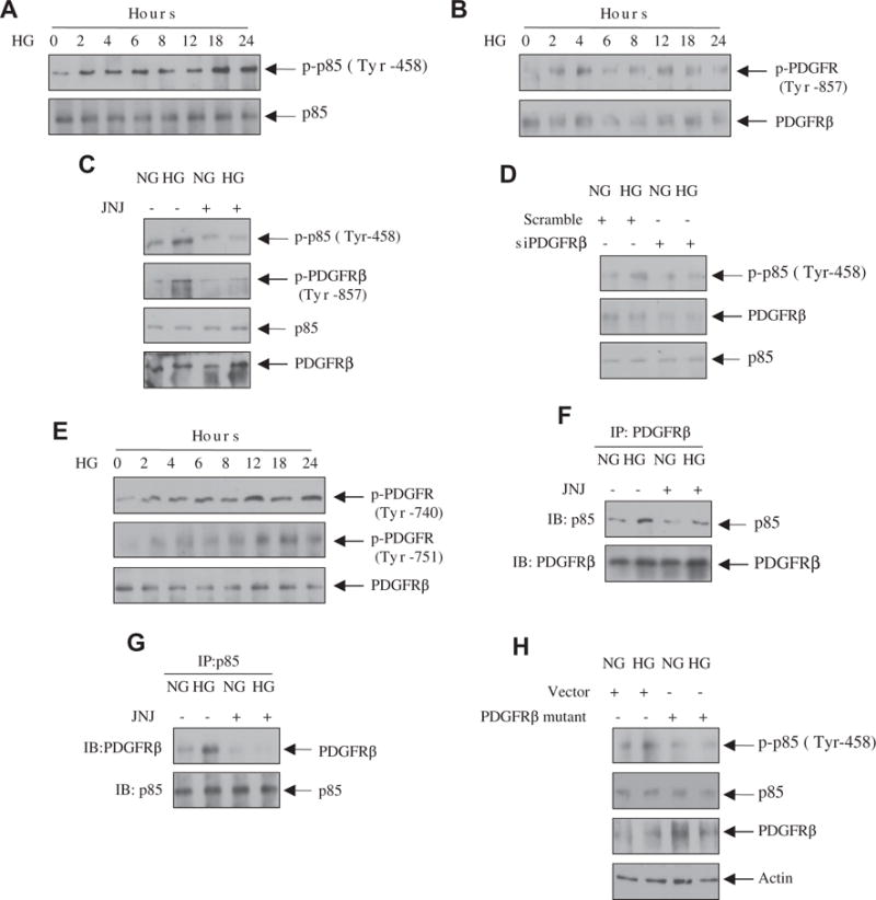
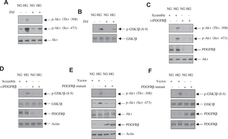
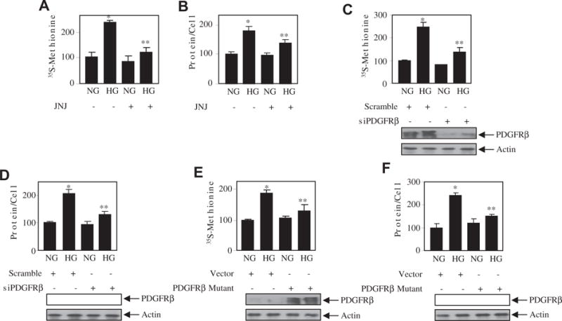
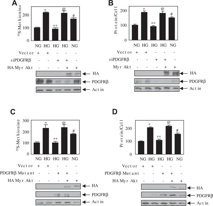
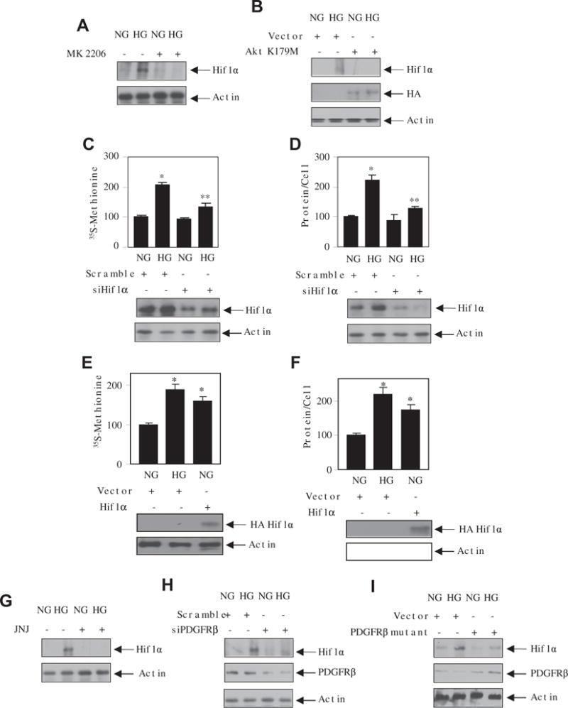
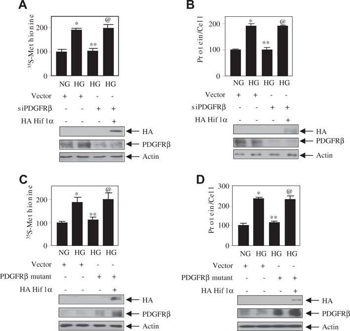
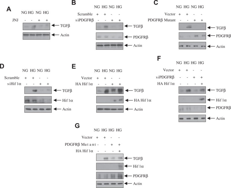
Glomerular mesangial cell hypertrophy contributes to the complications of diabetic nephropathy. The mechanism by which high glucose induces mesangial cell hypertrophy is poorly understood. Here we explored the role of the platelet-derived growth factor receptor-β (PDGFRβ) tyrosine kinase in driving the high glucose-induced mesangial cell hypertrophy. We show that high glucose stimulates the association of the PDGFRβ with PI 3 kinase leading to tyrosine phosphorylation of the latter. High glucose-induced Akt kinase activation was also dependent upon PDGFRβ and its tyrosine phosphorylation at 740/751 residues. Inhibition of PDGFRβ activity, its downregulation and expression of its phospho-deficient (Y740/751F) mutant inhibited mesangial cell hypertrophy by high glucose. Interestingly, expression of constitutively active Akt reversed this inhibition, indicating a role of Akt kinase downstream of PDGFRβ phosphorylation in this process. The transcription factor Hif1α is a target of Akt kinase. siRNAs against Hif1α inhibited the high glucose-induced mesangial cell hypertrophy. In contrast, increased expression of Hif1α induced hypertrophy similar to high glucose. We found that inhibition of PDGFRβ and expression of PDGFRβ Y740/751F mutant significantly inhibited the high glucose-induced expression of Hif1α. Importantly, expression of Hif1α countered the inhibition of mesangial cell hypertrophy induced by siPDGFRβ or PDGFRβ Y740/751F mutant. Finally, we show that high glucose-stimulated PDGFRβ tyrosine phosphorylation at 740/751 residues and the tyrosine kinase activity of the receptor regulate the transforming growth factor-β (TGFβ) expression by Hif1α. Thus we define the cell surface PDGFRβ as a major link between high glucose and its effectors Hif1α and TGFβ for induction of diabetic mesangial cell hypertrophy.
Keywords: Diabetic nephropathy; Receptor tyrosine kinase; Renal hypertrophy.
Copyright © 2017. Published by Elsevier Inc.
Figures
Similar articles
-
TGFβ acts through PDGFRβ to activate mTORC1 via the Akt/PRAS40 axis and causes glomerular mesangial cell hypertrophy and matrix protein expression.J Biol Chem. 2020 Oct 16;295(42):14262-14278. doi: 10.1074/jbc.RA120.014994. Epub 2020 Jul 30. J Biol Chem. 2020. PMID: 32732288 Free PMC article.
-
PDGF receptor-β uses Akt/mTORC1 signaling node to promote high glucose-induced renal proximal tubular cell collagen I (α2) expression.Am J Physiol Renal Physiol. 2017 Aug 1;313(2):F291-F307. doi: 10.1152/ajprenal.00666.2016. Epub 2017 Apr 19. Am J Physiol Renal Physiol. 2017. PMID: 28424212 Free PMC article.
-
Hydrophobic motif site-phosphorylated protein kinase CβII between mTORC2 and Akt regulates high glucose-induced mesangial cell hypertrophy.Am J Physiol Cell Physiol. 2016 Apr 1;310(7):C583-96. doi: 10.1152/ajpcell.00266.2015. Epub 2016 Jan 6. Am J Physiol Cell Physiol. 2016. PMID: 26739493 Free PMC article.
-
TGFβ-stimulated microRNA-21 utilizes PTEN to orchestrate AKT/mTORC1 signaling for mesangial cell hypertrophy and matrix expansion.PLoS One. 2012;7(8):e42316. doi: 10.1371/journal.pone.0042316. Epub 2012 Aug 3. PLoS One. 2012. PMID: 22879939 Free PMC article.
-
Role of growth arrest-specific gene 6 in diabetic nephropathy.Vitam Horm. 2008;78:375-92. doi: 10.1016/S0083-6729(07)00015-5. Vitam Horm. 2008. PMID: 18374201 Review.
Cited by
-
Akt2 causes TGFβ-induced deptor downregulation facilitating mTOR to drive podocyte hypertrophy and matrix protein expression.PLoS One. 2018 Nov 16;13(11):e0207285. doi: 10.1371/journal.pone.0207285. eCollection 2018. PLoS One. 2018. PMID: 30444896 Free PMC article.
-
Deacetylation of S6 kinase promotes high glucose-induced glomerular mesangial cell hypertrophy and matrix protein accumulation.J Biol Chem. 2019 Jun 14;294(24):9440-9460. doi: 10.1074/jbc.RA118.007023. Epub 2019 Apr 26. J Biol Chem. 2019. PMID: 31028173 Free PMC article.
-
High glucose couples DJ-1 with PTEN to activate PDGFRβ for renal proximal tubular cell injury.PLoS One. 2025 Jan 6;20(1):e0311828. doi: 10.1371/journal.pone.0311828. eCollection 2025. PLoS One. 2025. PMID: 39761275 Free PMC article.
References
-
- Kanwar YS, Wada J, Sun L, Xie P, Wallner EI, Chen S, Chugh S, Danesh FR. Diabetic nephropathy: mechanisms of renal disease progression. Exp Biol Med (Maywood) 2008;233:4–11. - PubMed
-
- Satriano J. Kidney growth, hypertrophy and the unifying mechanism of diabetic complications. Amino Acids. 2007;33:331–339. - PubMed
-
- Mahimainathan L, Das F, Venkatesan B, Choudhury GG. Mesangial cell hypertrophy by high glucose is mediated by downregulation of the tumor suppressor PTEN. Diabetes. 2006;55:2115–2125. - PubMed
MeSH terms
Substances
Grants and funding
LinkOut - more resources
Full Text Sources
Other Literature Sources
Research Materials
Miscellaneous
