. 2017 Nov 15;77(22):6119-6130.
doi: 10.1158/0008-5472.CAN-17-1605. Epub 2017 Sep 26.

Genomic Landscape of Atypical Adenomatous Hyperplasia Reveals Divergent Modes to Lung Adenocarcinoma

Affiliations

Genomic Landscape of Atypical Adenomatous Hyperplasia Reveals Divergent Modes to Lung Adenocarcinoma

Smruthy Sivakumar et al. Cancer Res. .

Abstract

There is a dearth of knowledge about the pathogenesis of premalignant lung lesions, especially for atypical adenomatous hyperplasia (AAH), the only known precursor for the major lung cancer subtype adenocarcinoma (LUAD). In this study, we performed deep DNA and RNA sequencing analyses of a set of AAH, LUAD, and normal tissues. Somatic BRAF variants were found in AAHs from 5 of 22 (23%) patients, 4 of 5 of whom had matched LUAD with driver EGFR mutations. KRAS mutations were present in AAHs from 4 of 22 (18%) of patients. KRAS mutations in AAH were only found in ever-smokers and were exclusive to BRAF-mutant cases. Integrative analysis revealed profiles expressed in KRAS-mutant cases (UBE2C, REL) and BRAF-mutant cases (MAX) of AAH, or common to both sets of cases (suppressed AXL). Gene sets associated with suppressed antitumor (Th1; IL12A, GZMB) and elevated protumor (CCR2, CTLA-4) immune signaling were enriched in AAH development and progression. Our results reveal potentially divergent BRAF or KRAS pathways in AAH as well as immune dysregulation in the pathogenesis of this premalignant lung lesion. Cancer Res; 77(22); 6119-30. ©2017 AACR.


Conflict of interest statement

Conflict of Interest: The authors declare no potential conflicts of interest within the scope of this work.

Figures

Figure 1
Figure 1. Study design to understand the development and progression of adenomatous atypical hyperplasia
Diagnosis and histopathological determination of specimens following H&E staining was performed to determine and classify normal lung tissues (NL), atypical adenomatous hyperplasia (AAH) and lung adenocarcinoma (LUAD). A two pronged approach was used to study the pathogenesis of AAH. Deep targeted sequencing of 409 cancer-associated genes was performed to identify somatic point mutations and transcriptome sequencing was carried out to study expression profiles.
Figure 2
Figure 2. Somatic mutation profiles in atypical adenomatous hyperplasia
Deep targeted sequencing of a cancer gene panel (n = 409) and identification of somatic nonsynonymous mutations in AAHs and LUADs was performed as described in the Methods section. (A) We examined, in greater detail, mutations in previously established lung cancer drivers from the TCGA [12] as well as other known cancer-associated genes [11]. AAH specimens (n = 17) that exhibited a mutation in either driver gene set were plotted. The paired LUADs were also plotted depicting mutations in genes previously established by the TCGA to be significantly mutated in LUAD [12]. Shown within the red panel is the enrichment of EGFR mutations in LUAD (80%) paired to BRAF-mutant AAH. (B) A tissue level analysis of mutations in AAH and LUAD specimens was performed to identify mutated genes, from the same set of driver genes surveyed in panel A, that were common or disparate between AAH and LUAD. (C) Lollipop plot for mutations (p.K601E; n = 4 and p.N581S; n = 1) in the BRAF gene and their prevalence in AAH specimens.
Figure 3
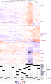
Figure 3. Expression profiles differentially modulated in development of atypical adenomatous hyperplasia and lung adenocarcinoma
Transcriptome sequencing was performed as described in the Methods section. Genes (n = 1008) differentially expressed between the three tissues (AAH vs NL, LUAD vs NL or LUAD vs AAH) were determined using ANOVA (P < 0.001, 2-fold change) and analyzed by hierarchical clustering (red, up-regulated relative to median sample; blue, down-regulated relative to median sample). Genes were grouped into eight different patterns based on two one-sided t-tests for NL to AAH and AAH to LUAD comparisons. Patterns of differential expression in each gene cluster are schematically depicted on the right. Pathways and gene set enrichment analysis was performed using Ingenuity Pathways Analysis. Pathways deregulated in each cluster of genes are depicted in red (activation) and blue (inhibition) alongside the heatmap. Mutations status of EGFR, KRAS and BRAF for AAH and LUAD specimens is depicted below.
Figure 4
Figure 4. Differential gene expression based on driver mutation status in atypical adenomatous hyperplasia
AAHs were subgrouped based on BRAF and KRAS mutation status: BRAF-mutant, KRAS-mutant and BRAF/KRAS wild type. Genes (n = 327) differentially expressed between the three AAH subgroups were identified using ANOVA (P < 0.01, 1.5 fold-change) and analyzed by hierarchical clustering (red, up-regulated relative to median sample; blue, down-regulated relative to median sample).
Figure 5
Figure 5. Deregulation of immune signaling in the molecular pathogenesis of atypical adenomatous hyperplasia
Expression profiles for an a priori list (n = 730) of immune markers from the nCounter PanCancer Immune Profiling Panel (nanoString technologies) was compiled (see Materials and Methods section) and studied to identify differentially expressed immune genes (n = 131; ANOVA; P < 0.001 and 1.5 fold-change). The genes were divided into different clusters based on patterns of differential expression between NL, AAH and LUAD derived from two one-sided t-tests (AAH vs NL and LUAD vs AAH). Patterns of differential expression in each gene cluster and select immune markers are schematically depicted on the right. present in major clusters are also depicted on the right.
Figure 6
Figure 6. Proposed models for the pathogenesis of atypical adenomatous hyperplasia
Two potential divergent modes in the pathogenesis of these preneoplastic lesions are proposed based on the mutual exclusivity of mutations and disparate expression profiles. A subgroup of AAHs, occurring in both non-smokers and ever-smokers, are initiated by BRAF and tend to be associated with development of LUADs with driver mutations in the EGFR oncogene (not excluding the possibility that the EGFR-mutant LUADs may have arised from different AAHs). Mechanisms involved in the potential progression of BRAF-mutant AAH to LUAD (e.g. EGFR-mutant tumors) warrant further studies. Another subset of AAHs are driven by KRAS, occur predominantly in ever-smokers and lead to LUADs with mutations in other driver genes besides KRAS (e.g., TP53). Transcriptome sequencing analysis pointed to aberrant immune signaling (e.g., up-regulated CTLA-4) in the pathogenesis of AAH. Further analysis (e.g. of a larger cohort of AAH) may help underscore profiles, immune markers and pathways unique to each molecular group of AAHs thus paving the way for new strategies (e.g., immune-based) for (chemo)prevention and early intervention.

References

    1. Herbst RS, Heymach JV, Lippman SM. Lung cancer. N Engl J Med. 2008;359:1367–1380. - PMC - PubMed
    1. Siegel RL, Miller KD, Jemal A. Cancer statistics, 2017. CA Cancer J Clin. 2017;67:7–30. - PubMed
    1. Kadara H, Scheet P, Wistuba II, Spira AE. Early Events in the Molecular Pathogenesis of Lung Cancer. Cancer Prev Res. 2016;9:518–527. - PubMed
    1. Mori M, Rao SK, Popper HH, Cagle PT, Fraire AE. Atypical Adenomatous Hyperplasia of the Lung: A Probable Forerunner in the Development of Adenocarcinoma of the Lung. Mod Pathol. 2001;14:72–84. - PubMed
    1. Sakamoto H, Shimizu J, Horio Y, Ueda R, Takahashi T, Mitsudomi T, et al. Disproportionate representation of KRAS gene mutation in atypical adenomatous hyperplasia, but even distribution of EGFR gene mutation from preinvasive to invasive adenocarcinomas. J Pathol. 2007;212:287–294. - PubMed

Publication types

Substances