. 2017 Sep 26;8(5):e00859-17.
doi: 10.1128/mBio.00859-17.

Wholly Rickettsia! Reconstructed Metabolic Profile of the Quintessential Bacterial Parasite of Eukaryotic Cells

Affiliations

Wholly Rickettsia! Reconstructed Metabolic Profile of the Quintessential Bacterial Parasite of Eukaryotic Cells

Timothy P Driscoll et al. mBio. .

Abstract

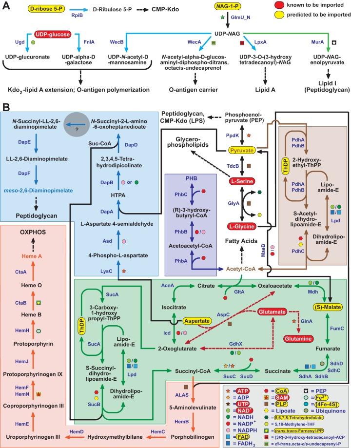
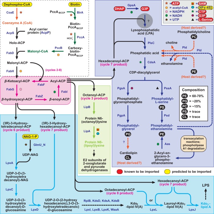
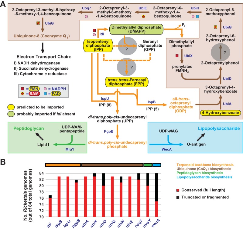
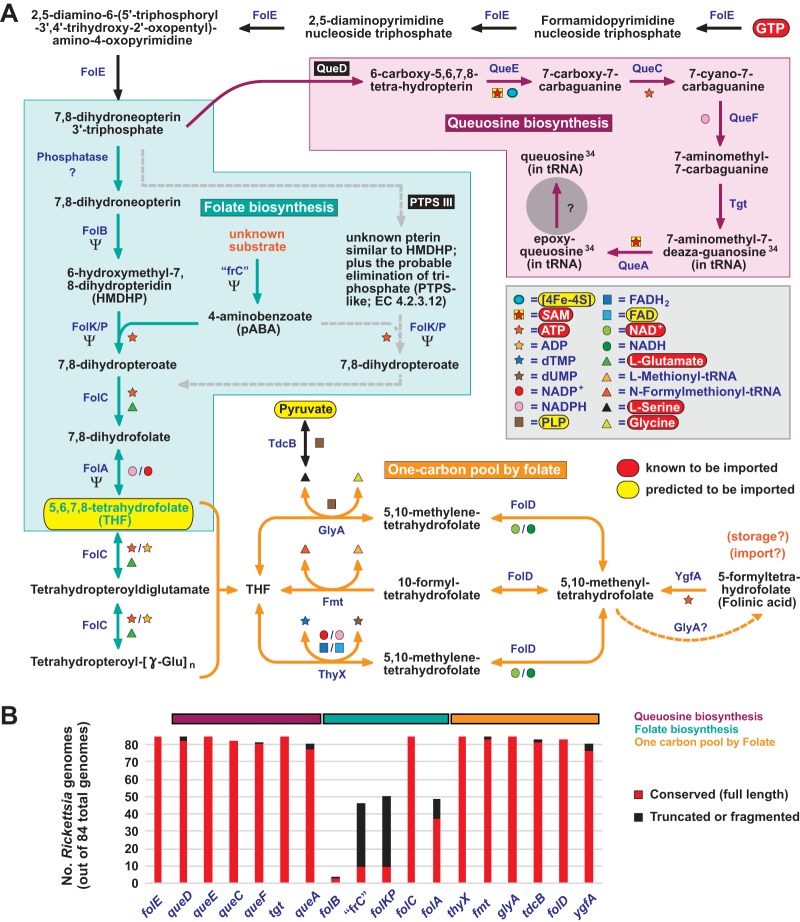
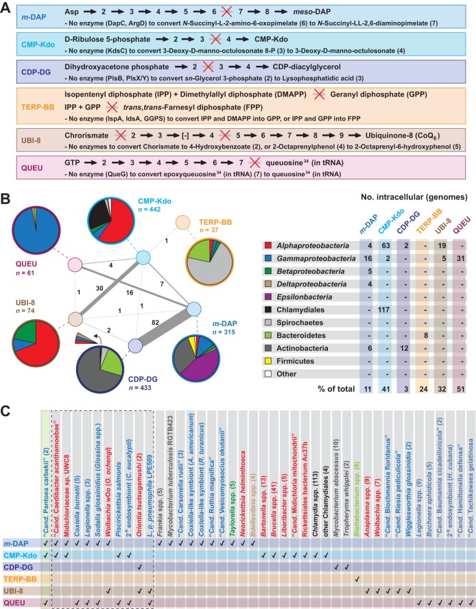
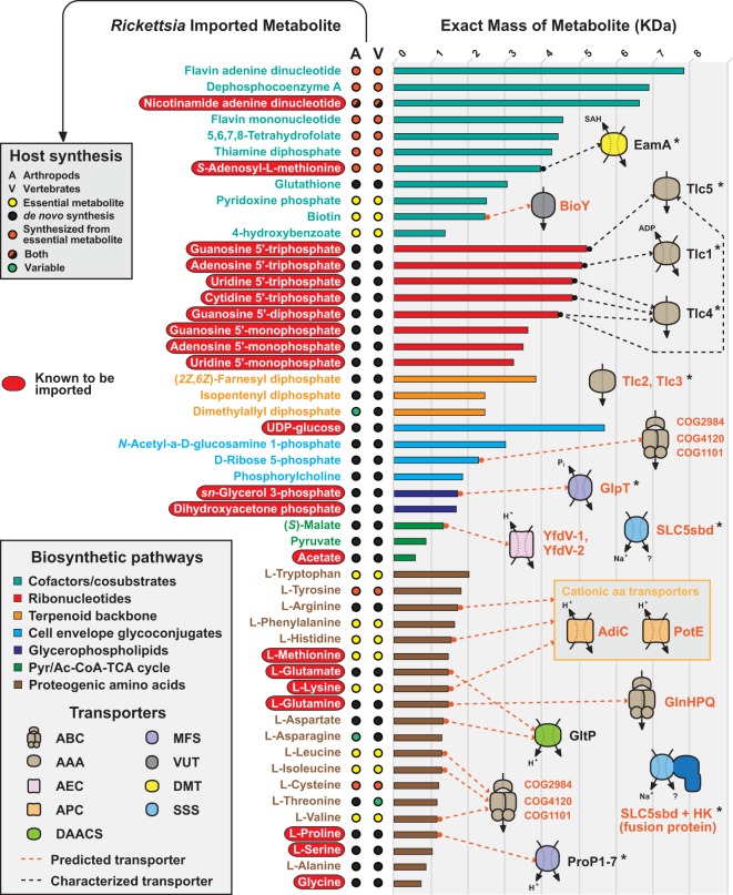
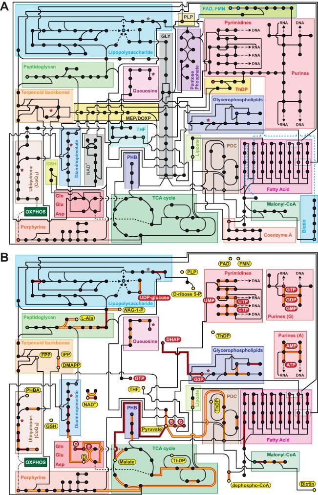
Reductive genome evolution has purged many metabolic pathways from obligate intracellular Rickettsia (Alphaproteobacteria; Rickettsiaceae). While some aspects of host-dependent rickettsial metabolism have been characterized, the array of host-acquired metabolites and their cognate transporters remains unknown. This dearth of information has thwarted efforts to obtain an axenic Rickettsia culture, a major impediment to conventional genetic approaches. Using phylogenomics and computational pathway analysis, we reconstructed the Rickettsia metabolic and transport network, identifying 51 host-acquired metabolites (only 21 previously characterized) needed to compensate for degraded biosynthesis pathways. In the absence of glycolysis and the pentose phosphate pathway, cell envelope glycoconjugates are synthesized from three imported host sugars, with a range of additional host-acquired metabolites fueling the tricarboxylic acid cycle. Fatty acid and glycerophospholipid pathways also initiate from host precursors, and import of both isoprenes and terpenoids is required for the synthesis of ubiquinone and the lipid carrier of lipid I and O-antigen. Unlike metabolite-provisioning bacterial symbionts of arthropods, rickettsiae cannot synthesize B vitamins or most other cofactors, accentuating their parasitic nature. Six biosynthesis pathways contain holes (missing enzymes); similar patterns in taxonomically diverse bacteria suggest alternative enzymes that await discovery. A paucity of characterized and predicted transporters emphasizes the knowledge gap concerning how rickettsiae import host metabolites, some of which are large and not known to be transported by bacteria. Collectively, our reconstructed metabolic network offers clues to how rickettsiae hijack host metabolic pathways. This blueprint for growth determinants is an important step toward the design of axenic media to rescue rickettsiae from the eukaryotic cell.IMPORTANCE A hallmark of obligate intracellular bacteria is the tradeoff of metabolic genes for the ability to acquire host metabolites. For species of Rickettsia, arthropod-borne parasites with the potential to cause serious human disease, the range of pilfered host metabolites is unknown. This information is critical for dissociating rickettsiae from eukaryotic cells to facilitate rickettsial genetic manipulation. In this study, we reconstructed the Rickettsia metabolic network and identified 51 host metabolites required to compensate patchwork Rickettsia biosynthesis pathways. Remarkably, some metabolites are not known to be transported by any bacteria, and overall, few cognate transporters were identified. Several pathways contain missing enzymes, yet similar pathways in unrelated bacteria indicate convergence and possible novel enzymes awaiting characterization. Our work illuminates the parasitic nature by which rickettsiae hijack host metabolism to counterbalance numerous disintegrated biosynthesis pathways that have arisen through evolution within the eukaryotic cell. This metabolic blueprint reveals what a Rickettsia axenic medium might entail.

Keywords: Rickettsia; evolution; host-parasite relationship; host-pathogen interactions; intracellular parasites; metabolic modeling; phylogenetic analysis; phylogenomics.


Figures

FIG 1
FIG 1
Rickettsia species synthesize cell envelope glycoconjugates from imported host sugars and fuel the TCA cycle with a range of host-acquired metabolites. (A) Previously shown to be imported, UDP-glucose is predicted to yield UDP-glucuronate and UDP-α-d-galactose, sugars likely to be used in LPS synthesis (light blue pathway lines). Synthesis of UDP-N-acetyl-d-mannosamine, another sugar likely incorporated into LPS, as well as pathways for O-antigen, lipid A, and lipid I of PGN (green pathway line), initiates with UDP-NAG. Without glycolysis enzymes, rickettsiae are predicted to import host NAG-1-P and convert this charged sugar to UDP-NAG via the uridyltransferase GlmU. d-Ribose 5-P, which is required to initiate CMP-Kdo synthesis, is also predicted to be imported from the host, provided that Rickettsia species lack enzymes of the pentose phosphate pathway. (B) Rickettsia species must acquire pyruvate for generation of PEP and acetyl-CoA. Pyruvate interconversions with Ser, Gly (via Ser), and malate are likely mediated by additional import of these molecules, ensuring that enough pyruvate enters the PDC to yield acetyl-CoA (brown). Aside from entering the TCA cycle (green), acetyl-CoA is also used in fatty acid biosynthesis and production of PHB (dark blue), a storage molecule that is metabolized when host energy sources are unavailable. Imported malate, glutamine (Gln), and glutamate (Glu) likely regulate the flow of acetyl-CoA into the TCA cycle, with Gln/Glu interconversions with 2-oxaloglutarate and oxaloacetate providing additional energy (dashed burgundy pathway lines). Generated aspartate is essential for initiation of the synthesis of DAP, which is used in PGN biosynthesis (light blue), a pathway nearly conserved except for a central hole (gray circle). DAP synthesis also requires generated succinyl-CoA, which is also used to synthesize porphyrins (orange). HTPA, (2S,4S)-4-hydroxy-2,3,4,5-tetrahydrodipicolinate.
FIG 2
FIG 2
Rickettsia species synthesize fatty acids and glycerophospholipids from host precursors. (Dashed box) Predicted imported substrates dephospho-CoA and biotin are required for holo-ACP synthesis and loading of the biotin carboxyl carrier protein, respectively, which collectively lead to the formation of malonyl-ACP. As Rickettsia species lack ACC, a conserved PCC complex is predicted to generate malonyl-CoA (green). Type II fatty acid synthesis (pink) is utilized by Rickettsia species to generate octanoyl-ACP for lipoate synthesis (light green), β-hydroxyacyl-ACPs (14C and 16C) and acyl-ACPs (16C and 18C) for Kdo2-lipid A synthesis (light blue), and hexadecanoyl-ACP for glycerophospholipid synthesis (dark blue). Acyl chain incorporation into lipid A follows the structure deduced for R. typhi (171). While both DHAP and G3P are known to be imported from the host (51, 52), Rickettsia species lack enzymes to generate LPA via the first incorporation of hexadecanoyl-ACP (the gray circle represents this pathway hole). All enzymes subsequent to this step are highly conserved (see Fig. S10), generating the predominant glycerophospholipids characterized in Rickettsia membranes (inset at upper right) (81). Dashed lines illustrate possible Pld-mediated salvage pathways for bacterial PE, as well as host PE and PC (orange). If PC (82) and cardiolipin (81) are incorporated into Rickettsia membranes, both must be acquired from the host (orange).
FIG 3
FIG 3
Rickettsia species must import host isoprenes and terpenoids for the synthesis of ubiquinone and the lipid carrier of lipid I and O-antigen. (A) In the absence of a mevalonate or MEP/DOXP pathway for terpenoid synthesis, Rickettsia species must import IPP from the host. If Idi is present, as it is in some species, DMAPP can be synthesized to provide dimethylallyl phosphate for the ubiquinone (CoQ8) pathway. Otherwise, DMAPP must also be imported from the host. Dashed orange lines indicate that no enzymes are present to use IPP and DMAPP for GPP generation, and thus, FPP must also be acquired from the host. Gray circles depict holes in the pathways for the generation of both GPP and FPP. Host-acquired IPP and FPP can then be used by undecaprenyl diphosphate synthase (IspU) and octaprenyl-diphosphate synthase (IspB) to generate terpenoid backbones UPP and ODP, respectively. Via phosphatidylglycerophosphatase B (PgpB), UPP is then converted to di-trans,poly-cis-undecaprenyl phosphate, the lipid carrier for lipid I (green) and O-antigen (light blue). OPP and PHBA are used by PHBA polyprenyltransferase (UbiA) to initiate CoQ8 synthesis (brown). The lack of enzymes to either synthesize chorismate or convert it to PHBA indicates that rickettsiae must import host PHBA, which is an essential host metabolite provided by diet and/or the microbiome. The gray circle indicates a hole (UbiC) in the CoQ8 synthesis pathway. Note that all rickettsial genomes encode UbiB, a putative kinase with an unknown role in CoQ8 biosynthesis (172). (B) Genes involved in terpenoid backbone and CoQ8 biosynthesis are largely conserved. The complete distributions of these genes in 84 Rickettsia genomes (see Fig. S4G) indicate that the most basal lineage of spotted fever group rickettsiae (R. tamurae, R. monacensis, REIP, and R. buchneri strains) lacks idi and thus must also acquire DMAPP from the host.
FIG 4
FIG 4
Rickettsia species lack the capability for de novo folate biosynthesis. (A) Rickettsiae use GTP cyclohydrolase I (FolE) to convert host-acquired GTP (red) to DHN-P3, a precursor of both queuosine (purple) and folate (aquamarine) biosynthesis. The classical THF synthesis pathway (aquamarine arrows), wherein DHN-P3 is dephosphorylated and subsequently converted to HMDHP by dihydropteridin aldolase (FolB), is disintegrating from Rickettsia genomes (Ψ denotes pseudogenization in over 50% of genomes). In the FolB bypass proposed by Hunter et al. (89) (gray dashed arrows), DHN-P3 is directly converted to HMDHP or a structurally similar molecule via PTPS-III (black box). Our analysis instead suggests that this enzyme is QueD (black box), which performs the first committed step in queuosine biosynthesis (see the text for further details). The one-carbon pool by folate (orange arrows) illustrates the role of host-acquired THF and several intermediates in the essential one-carbon transfer reactions that yield pyrimidine deoxynucleoside triphosphates, N-formylmethionyl-tRNA, and Ser/Gly. “frC,” fol_rel_CADD domain-containing protein (TIGR04305). The gray circle represents a hole in the pathway for queuosine synthesis (see Fig. S6). (B) Conservation of 18 genes involved in queuosine and THF biosynthesis and reactions within the one-carbon pool by folate. The complete distributions of these genes in 84 Rickettsia genomes reveals that no single Rickettsia species is capable of de novo folate biosynthesis, while the queuosine biosynthesis and one-carbon pool by folate pathways are highly conserved (see Fig. S5D).
FIG 5
FIG 5
Comparative analysis of six biosynthetic pathways containing holes. The reconstructed Rickettsia metabolic network revealed holes in six biosynthetic pathways, DAP (m-DAP), CMP-Kdo, CDP-diacylglycerol (CDP-DG), terpenoid backbones (TERP-BB), ubiquinone-8 (UBI-8), and queuosine (QUEU). The distribution of these pathways across other prokaryotic genomes was determined via comparative metabolic pathway analyses. (A) Illustration and description of each hole-containing biosynthetic pathway. A red X indicates the missing enzyme(s) within each pathway, compared to well-characterized biosynthetic pathways for other prokaryotes. (B) Distribution of Rickettsia-like biosynthetic pathways across other prokaryotic genomes. The pie charts at the left indicate the taxonomic breakdown of genomes per biosynthetic pathway, with connections between pathways illustrating the number of genomes containing multiple Rickettsia-like pathways. Note that Rickettsia genomes were excluded from these analyses. At the right, the taxonomic color scheme is shown, with the number of genomes from intracellular species provided. (C) Intracellular species containing one or more Rickettsia-like biosynthetic pathways. The green box depicts the only genome found to contain three Rickettsia-like pathways, that of “Ca. Pantoea carbekii”) (117), which is an extracellular primary symbiont of the brown marmorated stink bug, where it is found in the gastric cecal lumina (see the text for further details). The dashed box indicates genomes containing two Rickettsia-like pathways. Taxa are colored in accordance with the color scheme in panel B.
FIG 6
FIG 6
Synopsis of known and predicted metabolites imported from the eukaryotic cytoplasm by rickettsiae. On the left, metabolites are grouped into biosynthetic pathways (colors are described in the inset at the bottom left), with red ellipses depicting 21 metabolites previously shown to be imported. The remaining 30 metabolites are predicted to be imported on the basis of the metabolic network reconstruction presented in this report. In the center are the biosynthesis capabilities of the metabolites in arthropod and vertebrate genomes (further described in the inset at the top left). Information was obtained from KEGG pathways for arthropods and vertebrates. On the right, within each group, metabolites are ranked by exact mass. Dashed lines connect metabolites with their known (black) or predicted (orange) transport systems (transporter families are listed in the inset at the bottom left). One ABC transporter (COG1101/COG4120/COG2984) is shown twice, as annotations indicate uptake of branched-chain amino acids, as well as monosaccharides (including ribose, galactose, and arabinose). SLC5sbd and SLC5sbd+HK (fusion protein with His kinase domain) transporters are not linked with specific metabolites because of their known broad range of substrates (e.g., sugars, amino acids, organo-cations such as choline, nucleosides, inositols, vitamins, urea, or anions). Asterisks indicate transporters previously shown to be associated with mobile genetic elements and/or predicted to be spread by lateral gene transfer across diverse intracellular bacteria (60, 88, 126, 127). Transporter names and family identifications (123) are as follows: ABC, ATP-binding cassette (3.A.1); AAA, ATP:ADP antiporter (2.A.12); DMT, drug/metabolite transporter (2.A.7); VUT, vitamin uptake transporter (2.A.88); AEC (2.A.69); MFS, major facilitator superfamily (2.A.1); APC, amino acid polyamine organo-cation (2.A.3); DAACS, dicarboxylate/amino acid:cation (Na+ or H+) symporter (2.A.23); SSS, solute:sodium symporter (2.A.21). Phylogenomics analysis indicates that these transporters are highly conserved in rickettsial genomes (see Fig. S10).
FIG 7
FIG 7
Rickettsia metabolic network reconstruction highlights reductive genome evolution and addiction to host cell metabolites. The network focuses on the biosynthesis pathways discussed in the text. For brevity, pathways for most amino acids are not shown. Red stars indicate six pathway holes (see Fig. 5). GLY, glycolysis; MEP/DOXP, nonmevalonate terpenoid biosynthesis; THF, 5,6,7,8-THF; GSH, glutathione. (A) Theoretical Rickettsia metabolic network in the absence of imported metabolites. Rickettsia metabolic pathways are supplemented with typical Gram-negative biosynthetic pathways to create a complete metabolic network. (B) Reconstructed Rickettsia metabolic network, including imported metabolites. Pathways removed from panel A have been purged from Rickettsia genomes throughout evolution, a consequence of pilfering of metabolites from the eukaryotic host. Red ellipses, metabolites known to be imported by Rickettsia species; yellow ellipses, metabolites predicted to be imported on the basis of metabolic network reconstruction. The import of S-adenosyl-l-methionine, phosphorylcholine, and the majority of amino acids is not included in the reconstruction. Pathway lines are highlighted in red to indicate cofactors that are synthesized directly from imported metabolites. Additionally, if the cofactor is directly imported from the host, the pathway line is yellow. The network is based on a phylogenomics analyses of 84 Rickettsia genomes (see Fig. S10).

References

    1. Gillespie JJ, Nordberg EK, Azad AA, Sobral BW. 2012. Phylogeny and comparative genomics: the shifting landscape in the genomics era, p 84–141. In Azad AF, Palmer GH (ed), Intracellular pathogens II: rickettsiales. ASM Press, Washington, DC.
    1. Driscoll T, Gillespie JJ, Nordberg EK, Azad AF, Sobral BW. 2013. Bacterial DNA sifted from the Trichoplax adhaerens (Animalia: Placozoa) genome project reveals a putative rickettsial endosymbiont. Genome Biol Evol 5:621–645. doi:10.1093/gbe/evt036. - DOI - PMC - PubMed
    1. Szokoli F, Castelli M, Sabaneyeva E, Schrallhammer M, Krenek S, Doak TG, Berendonk TU, Petroni G. 2016. Disentangling the taxonomy of Rickettsiales and description of two novel symbionts (“Candidatus Bealeia paramacronuclearis” and “Candidatus Fokinia cryptica”) sharing the cytoplasm of the ciliate protist Paramecium biaurelia. Appl Environ Microbiol 82:7236–7247. doi:10.1128/AEM.02284-16. - DOI - PMC - PubMed
    1. Beier-Sexton M, Driscoll TP, Azad AF, Gillespie JJ. 2015. The family Rickettsiaceae, p 547–566. In Goldman E, Green LH (ed), Practical handbook of microbiology, 3rd edition. CRC Press, Boca Raton, FL.
    1. Darby AC, Cho NH, Fuxelius HH, Westberg J, Andersson SGE. 2007. Intracellular pathogens go extreme: genome evolution in the Rickettsiales. Trends Genet 23:511–520. doi:10.1016/j.tig.2007.08.002. - DOI - PubMed

Publication types

LinkOut - more resources