Review
. 2017 Jul 8;6(9):943-957.
doi: 10.1016/j.molmet.2017.06.019. eCollection 2017 Sep.

Human beta cell mass and function in diabetes: Recent advances in knowledge and technologies to understand disease pathogenesis

Affiliations
Review

Human beta cell mass and function in diabetes: Recent advances in knowledge and technologies to understand disease pathogenesis

Chunguang Chen et al. Mol Metab. .

Abstract

Background: Plasma insulin levels are predominantly the product of the morphological mass of insulin producing beta cells in the pancreatic islets of Langerhans and the functional status of each of these beta cells. Thus, deficiency in either beta cell mass or function, or both, can lead to insufficient levels of insulin, resulting in hyperglycemia and diabetes. Nonetheless, the precise contribution of beta cell mass and function to the pathogenesis of diabetes as well as the underlying mechanisms are still unclear. In the past, this was largely due to the restricted number of technologies suitable for studying the scarcely accessible human beta cells. However, in recent years, a number of new platforms have been established to expand the available techniques and to facilitate deeper insight into the role of human beta cell mass and function as cause for diabetes and as potential treatment targets.

Scope of review: This review discusses the current knowledge about contribution of human beta cell mass and function to different stages of type 1 and type 2 diabetes pathogenesis. Furthermore, it highlights standard and newly developed technological platforms for the study of human beta cell biology, which can be used to increase our understanding of beta cell mass and function in human glucose homeostasis.

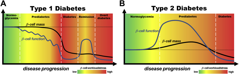
Major conclusions: In contrast to early disease models, recent studies suggest that in type 1 and type 2 diabetes impairment of beta cell function is an early feature of disease pathogenesis while a substantial decrease in beta cell mass occurs more closely to clinical manifestation. This suggests that, in addition to beta cell mass replacement for late stage therapies, the development of novel strategies for protection and recovery of beta cell function could be most promising for successful diabetes treatment and prevention. The use of today's developing and wide range of technologies and platforms for the study of human beta cells will allow for a more detailed investigation of the underlying mechanisms and will facilitate development of treatment approaches to specifically target human beta cell mass and function.

Keywords: Beta cell function; Beta cell mass; Diabetes; Human; In situ; In vitro; In vivo; Islet of Langerhans; Pathogenesis.


Figures

Figure 1
Figure 1
Models of the contribution of beta cell mass and function to pathogenesis of type 1 diabetes (A) and type 2 diabetes (B). (A): Beta cell mass and function in the development of type 1 diabetes. Initiation of islet autoimmunity by genetic and environmental factors leads to a relapsing-remitting decline of beta cell function, continuously increasing beta cell workload, and stress in the asymptomatic prediabetes phase. Shortly before clinical manifestation of diabetes the prolonged intensified beta cell workload and autoimmunity results in total cellular exhaustion and enhanced cell death leading to a massive decrease in beta cell mass and the onset of hyperglycemia. In some patients, initial insulin treatment induces temporary remission called the “honeymoon phase,” which is attributed to a moderate reduction in beta cell workload and antigenicity, resulting in functional recovery of residual beta cells. However, ongoing autoimmunity and elevated workload lead to recurrence of cellular exhaustion, cell death, and the development of overt diabetes. Black line: beta cell mass; Blue line: beta cell function. The color-coded background indicates the intensity of beta cell workload and stress caused by immune infiltration, metabolic demand and hyperglycemia. (B): Beta cell mass and function in the development of type 2 diabetes. In many individuals, genetic predisposition and unhealthy lifestyle lead to an increased insulin resistance, which is typically met by massive functional and moderate morphological compensation to maintain normoglycemia, thus increasing the workload of each beta cell. In some of these individuals, functional compensation halts, despite prolonged insulin resistance and results in a further escalation of beta cell workload and glucose intolerance. In this prediabetic phase, chronic glucose intolerance and elevated blood glucose levels continuously exacerbate beta cell workload and stress, culminating in cellular exhaustion, cell death, and clinical manifestation of hyperglycemia. Thereafter, uncontrolled hyperglycemia, often in concert with other cytotoxic factors, leads to accelerated beta cell mass loss and functional deterioration in overt diabetic patients. Black line: beta cell mass; Blue line: beta cell function. The color-coded background indicates the intensity of beta cell workload and stress caused by insulin resistance, metabolic demand, hyperglycemia and additional cytotoxic factors.
Figure 2
Figure 2
Human pancreas tissue slices for the in situ study of human beta cell morphology and function. (A) Maximum intensity projection of a human islet in a pancreas tissue slice from a non-diabetic human patient that underwent partial pancreatectomy. Slices were stained for insulin (green), glucagon (blue), somatostatin (yellow), and fluorescently labeled lectin (red) following recently published protocols . (B) Kinetic insulin release from 4 human pancreas tissue slices obtained from the same pancreatic tissue as used in (A). Insulin release is expressed as simulation index (SI) over mean basal secretion (0–10 min). Slices were perfused in a closed chamber with Krebs–Ringer bicarbonate HEPES buffer and indicated glucose concentrations at a flow rate of 200 μl/min using a perifusion system (Biorep). Insulin concentrations of the perfusate were assessed by ELISA. Human tissue was kindly provided by Michele Solimena and Jürgen Weitz (University Hospital Carl Gustav Carus, TU Dresden, Germany).

References

    1. Eisenbarth G.S. Type I diabetes mellitus. A chronic autoimmune disease. The New England Journal of Medicine. 1986;314(21):1360–1368. - PubMed
    1. Chatenoud L., Bluestone J.A. CD3-specific antibodies: a portal to the treatment of autoimmunity. Nature Reviews Immunology. 2007;7(8):622–632. - PubMed
    1. von Herrath M., Sanda S., Herold K. Type 1 diabetes as a relapsing-remitting disease? Nature Reviews Immunology. 2007;7(12):988–994. - PubMed
    1. van Belle T.L., Coppieters K.T., von Herrath M.G. Type 1 diabetes: etiology, immunology, and therapeutic strategies. Physiological Reviews. 2011;91(1):79–118. - PubMed
    1. Ziegler A.G., Rewers M., Simell O., Simell T., Lempainen J., Steck A. Seroconversion to multiple islet autoantibodies and risk of progression to diabetes in children. JAMA. 2013;309(23):2473–2479. - PMC - PubMed

Publication types

MeSH terms