. 2018 Mar;13(1):100-112.
doi: 10.1007/s11481-017-9766-1. Epub 2017 Sep 26.

Cross-Talk between Glia, Neurons and Mast Cells in Neuroinflammation Associated with Parkinson's Disease

Affiliations

Cross-Talk between Glia, Neurons and Mast Cells in Neuroinflammation Associated with Parkinson's Disease

Duraisamy Kempuraj et al. J Neuroimmune Pharmacol. 2018 Mar.

Abstract

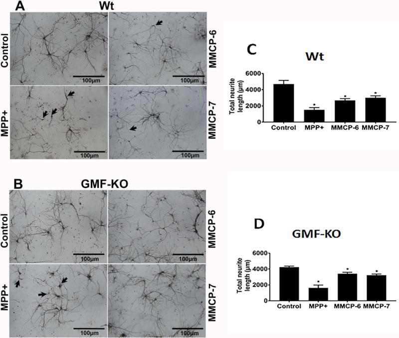
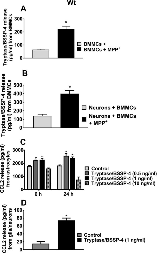
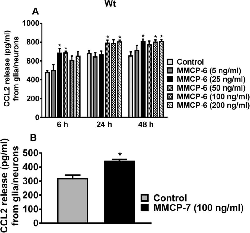
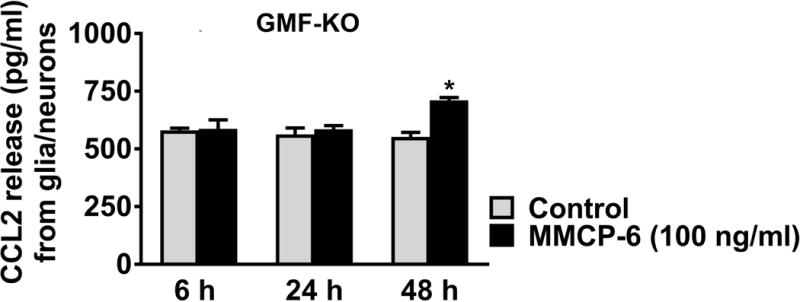
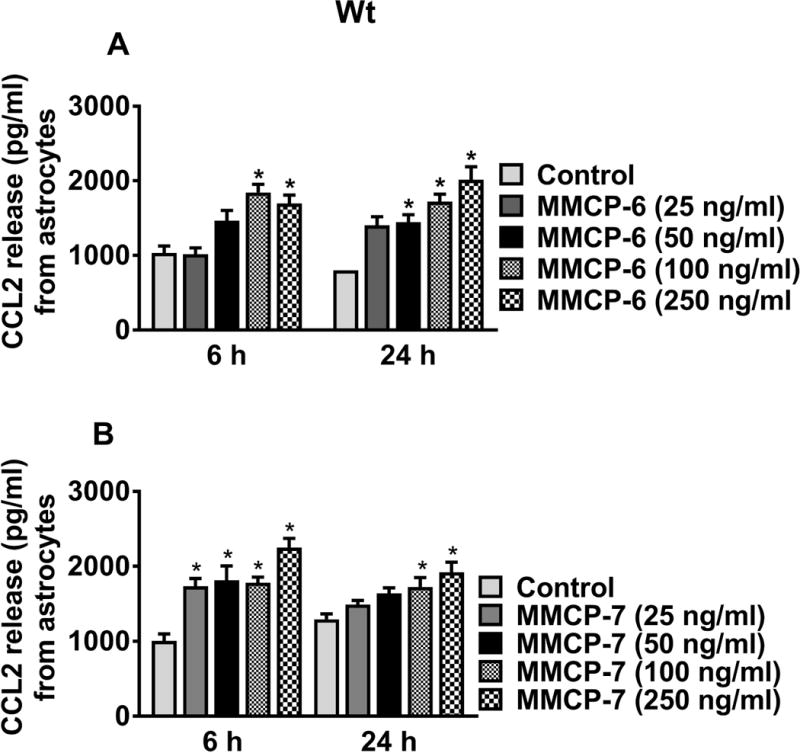
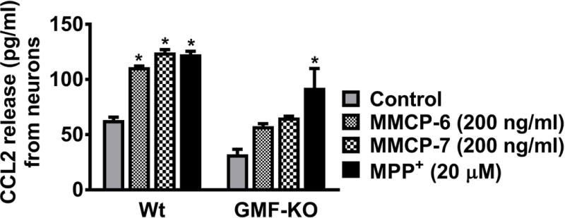
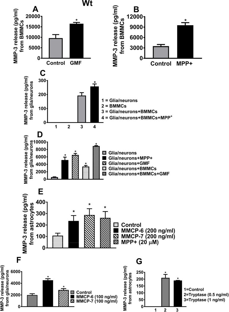
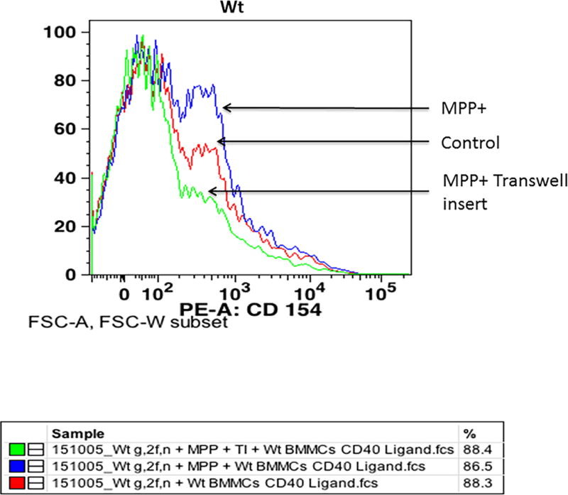
Parkinson's disease (PD) is a progressive movement disorder characterized by neuroinflammation and dopaminergic neurodegeneration in the brain. 1-methyl-4-phenylpyridinium (MPP+), a metabolite of the parkinsonian neurotoxin 1-methyl-4-phenyl-1,2,3,6-tetrahydropyridine (MPTP) induces the release of inflammatory mediators from glial cells and neurons. Glia maturation factor (GMF), a brain proinflammatory protein, MPP+, and mast cell-derived inflammatory mediators induce neurodegeneration which eventually leads to PD. However, the precise mechanisms underlying interaction between glial cells, neurons and mast cells in PD still remain elusive. In the present study, mouse bone marrow-derived mast cells (BMMCs) and mouse fetal brain-derived mixed glia/neurons, astrocytes and neurons were incubated with MPP+, GMF and mast cell-derived inflammatory mediators mouse mast cell protease-6 (MMCP-6), MMCP-7 or tryptase/brain-specific serine protease-4 (tryptase/BSSP-4). Inflammatory mediators released from these cells in the culture medium were quantitated by enzyme-linked immunosorbent assay. Neurodegeneration was quantified by measuring total neurite outgrowth following microtubule-associated protein-2 immunocytochemistry. MPP+-induced significant neurodegeneration with reduced total neurite outgrowth. MPP+induced the release of tryptase/BSSP-4 from the mouse mast cells, and tryptase/BSSP-4 induced chemokine (C-C motif) ligand 2 (CCL2) release from astrocytes and glia/neurons. Overall our results suggest that MPP+, GMF, MMCP-6 or MMCP-7 stimulate glia/neurons, astrocytes or neurons to release CCL2 and matrix metalloproteinase-3. Additionally, CD40L expression is increased in BMMCs after incubation with MPP+ in a co-culture system consisting of BMMCs and glia/neurons. We propose that mast cell interaction with glial cells and neurons during neuroinflammation can be explored as a new therapeutic target for PD.

Keywords: 1-methyl-4-phenylpyridinium; CCL2; Cytokines; Glia maturation factor; Mast cells; Microtubule-associated protein-2; Neuroinflammation; Parkinson’s disease.


Conflict of interest statement

Conflict of interest disclosure

Authors declare that they have no competing interests.

Figures

Fig. 1
Fig. 1
MPP+ and mast cell proteases-induce neuronal degeneration. Primary mouse neurons from Wt mice and GMF-KO mice fetal brains were cultured for 2 weeks. These cells were incubated with MPP+ (10 μM) or mast cell proteases (MMCP-6 or MMCP-7 at 100 ng/ml) for 24 h. MAP-2 ICC was performed to analyze the neuronal morphology. (A) MPP+, MMCP-6 and MMCP-7-induced significant neuronal degeneration (arrows) in the neurons obtained from Wt mice fetal brains as compared to non-treated control neuronal cells. (B) GMF-KO neurons showed less neuronal degeneration when compared to the extent of neurodegeneration observed in the neurons grown from Wt mice fetal brains. Control neuronal cells did not show any degeneration. Scale bar = 100 μm. (C, D) The total neuronal outgrowth length was measured in the whole photomicrographs using MetaMorph software to determine the neuronal degeneration. MPP+, MMCP-6 and MMCP-7 significantly reduced the total neurite length as compared with control cells as shown in the bar graphs (One-way ANOVA and Tukey-Kramer post hoc, n=3, *p<0.05). Neurodegeneration is relatively higher in Wt neurons than in GMF-KO neurons.
Fig. 2
Fig. 2
MPP+-induces tryptase/BSSP-4 release from Wt BMMCs, and tryptase release CCL2 from astrocytes and glia/neurons. (A) Wt BMMCs were incubated with MPP+ (10 μM) and the culture media were used for tryptase assay by ELISA (n=4). MPP+ induced significant tryptase/BSSP-4 release from BMMCs as compared to untreated control cells. (B) Wt mouse neurons and Wt BMMCs co-cultured cells were treated with MPP+ (10 μM) for 24 h (n=4) and the tryptase released in the medium was assayed. MPP+ released significantly more tryptase in this co-culture system as compared to co-cultured control cells (*p<0.05; t test, control vs treated). (C, D) Wt mouse astrocytes (n=6) or glia/neurons (n=4) were incubated with tryptase/BSSP-4 (0.5–10 ng/ml) and the release of CCL2 was assayed in the culture media by ELISA. Tryptase/BSSP-4 induced CCL2 release from the astrocytes and glia/neurons. Results were shown as mean ± SEM (*p<0.05; control vs treated, C= One-way ANOVA and Tukey-Kramer post hoc, D = t test).
Fig. 3
Fig. 3
MMCP-6 -induces CCL2 release from Wt mouse glia/neurons. (A) Wt mouse glia/neurons were incubated with MMCP-6 for dose-response (5 to 200 ng/ml) and time-course studies (6 to 48 h), and CCL2 levels in the culture media were determined by ELISA (n=3). MMCP-6 induced significant CCL2 release from 6 h (at 25 ng/ml concentration) onwards from glia/neurons. Results were presented as mean ± SEM (*p<0.05 control vs MMCP-6 treated cells, One-way ANOVA and Tukey-Kramer post hoc). (B) Wt glia/neurons were incubated with MMCP-7 (100 ng/ml) for 24 h and CCL2 release was measured by ELISA (n=5). MMCP-7 induced significant CCL2 release from glia/neurons as compared to untreated control cells (*p<0.05 control vs cells incubated with MMCP-7, t-test).
Fig. 4
Fig. 4. Effect of
MMCP-6 on CCL2 release from GMF-KO mouse glia/neurons. Glia/neurons obtained from GMF-KO mice were incubated with MMCP-6 (100 ng/ml) for 6, 24 and 48 h. CCL2 release was measured in the culture media by ELISA (n=4). MMCP-6 significantly released CCL2 only at 48 h when compared to untreated control cells. Results were presented as mean ± SEM (*p<0.05 control vs MMCP-6 treated cells, t-test).
Fig. 5
Fig. 5
MMCP-6 and MMCP-7-induce CCL2 release from Wt mouse astrocytes. Wt mouse astrocytes were grown and stimulated with MMCP-6 and MMCP-7 for 6 h and 24 h, and CCL2 release was measured in the culture media by ELISA (n=3–8). Both (A) MMCP-6 and (B) MMCP-7 induced CCL2 release in a dose-dependent manner from mouse astrocytes when compared with non-treated control cells. Results were presented as mean ± SEM (*p<0.05 control vs MMCP-6 or MMCP-7 treated cells, One-way ANOVA and Tukey-Kramer post hoc).
Fig. 6
Fig. 6
MMCP-6, MMCP-7, and MPP+ -induce CCL2 release from mouse neurons. We examined whether MMCP-6, MMCP-7 and MPP+ activate mouse neurons to release CCL2. Mouse neuronal cells grown from Wt mice as well as from GMF-KO mice were incubated with MMCP-6 (200 ng/ml), MMCP-7 (200 ng/ml) or MPP+ (20 μM) for 24 h and the CCL2 release was assayed in the culture media by ELISA (n=3). Neurons obtained from Wt mice significantly released CCL2 after incubation with MMCP-6, MMCP-7, and MPP+ when compared to control non-treated cells. However, only MPP+ significantly released CCL2 from neuronal cultures obtained from GMF-KO mice. Results were presented as mean ± SEM (*p<0.05 control vs treated cells, One-way ANOVA and Tukey-Kramer post hoc).
Fig. 7
Fig. 7
GMF, MPP+, MMCP-6, MMCP-7 and tryptase release MMP-3 from Wt brain cells or mast cells. (A, B) BMMCs grown from Wt mice were incubated with GMF (100 ng/ml) or MPP+ (20 μM) for 24 h and MMP-3 release in the cell culture media was measured by ELISA. (A) GMF (n=3) and (B) MPP+ (n=4) significantly increased the release of MMP-3 when compared with non-treated control BMMCs (*p<0.05; t test, control vs treated). (C) Wt mouse BMMCs plus glia/neurons incubated with MPP+ significantly induced MMP-3 release as compared to non-treated control cells (n=3, *p<0.05; t test, control vs treated). (D) Wt mouse glia/neurons were then incubated with MPP+ (15 μM) or GMF (100 ng/ml) for 24 h and the release of MMP-3 was measured in the culture media (n=3). MPP+ and GMF significantly induced the release of MMP-3 from glia/neurons. Wt glia/neurons and BMMCs were co-cultured and then stimulated with GMF. GMF augments the release of MMP-3 in this co-culture system as compared to single glia/neurons culture condition (*p<0.05; t test, control vs treated). (E, F, G) Wt mouse astrocytes or glia/neurons were incubated with MMCP-6, MMCP-7, MPP+ or tryptase/BSSP-4 and the release of MMCP-3 in the culture media was measured by ELISA. MMCP-7 (n=6), MMP-6 (n=6), MPP+ (n=6) and tryptase/BSSP-4 (n=3)-induced significantly increased MMP-3 release as compared to control cells. Results were presented as mean ± SEM (*p<0.05 control vs treated cells, One-way ANOVA and Tukey-Kramer post hoc).
Fig. 8
Fig. 8
Expression of CD40L in Wt BMMCs co-cultured with glia/neurons as analyzed by flow cytometry. BMMCs and glia/neurons were co-cultured in 24 well culture plates for 72 h and subsequently incubated with MPP+ (15 μM) for 72 h at 37°C. In additional wells, transwell inserts were placed to avoid direct contact between glia/neurons and BMMCs. BMMCs were added on top of transwell inserts and incubated with MPP+. The expression of CD40L on BMMCs was analyzed by flow cytometry using anti-mCD40L/TNFSF5 antibody (n=3). MPP+ increased the expression of CD40L on BMMCs as compared to control untreated cells where there was no direct cell-to-cell contact inhibition. However, CD40L expression was reduced in the wells where transwell inserts were used to avoid direct contact of glia/neurons and BMMCs.

Similar articles

Cited by

References

    1. Bennett JL, Blanchet MR, Zhao L, Zbytnuik L, Antignano F, Gold M, Kubes P, McNagny KM. Bone marrow-derived mast cells accumulate in the central nervous system during inflammation but are dispensable for experimental autoimmune encephalomyelitis pathogenesis. J Immunol. 2009;182:5507–5514. - PubMed
    1. Brahmachari S, Jana A, Pahan K. Sodium benzoate, a metabolite of cinnamon and a food additive, reduces microglial and astroglial inflammatory responses. J Immunol. 2009;183:5917–5927. - PMC - PubMed
    1. Caughey GH. Mast cell tryptases and chymases in inflammation and host defense. Immunol Rev. 2007;217:141–154. - PMC - PubMed
    1. Chikahisa S, Kodama T, Soya A, Sagawa Y, Ishimaru Y, Sei H, Nishino S. Histamine from brain resident MAST cells promotes wakefulness and modulates behavioral states. PLoS One. 2013;8:e78434. - PMC - PubMed
    1. Cottrell GS, Amadesi S, Schmidlin F, Bunnett N. Protease-activated receptor 2: activation, signalling and function. Biochem Soc Trans. 2003;31:1191–1197. - PubMed

Publication types

LinkOut - more resources