Review
. 2017 Jan 24;4(1):8.
doi: 10.3390/bioengineering4010008.

Vasculature-On-A-Chip for In Vitro Disease Models

Affiliations
Review

Vasculature-On-A-Chip for In Vitro Disease Models

Seunggyu Kim et al. Bioengineering (Basel). .

Abstract

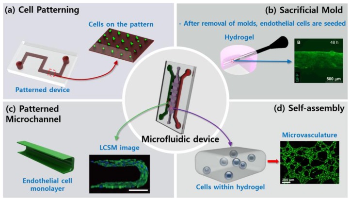
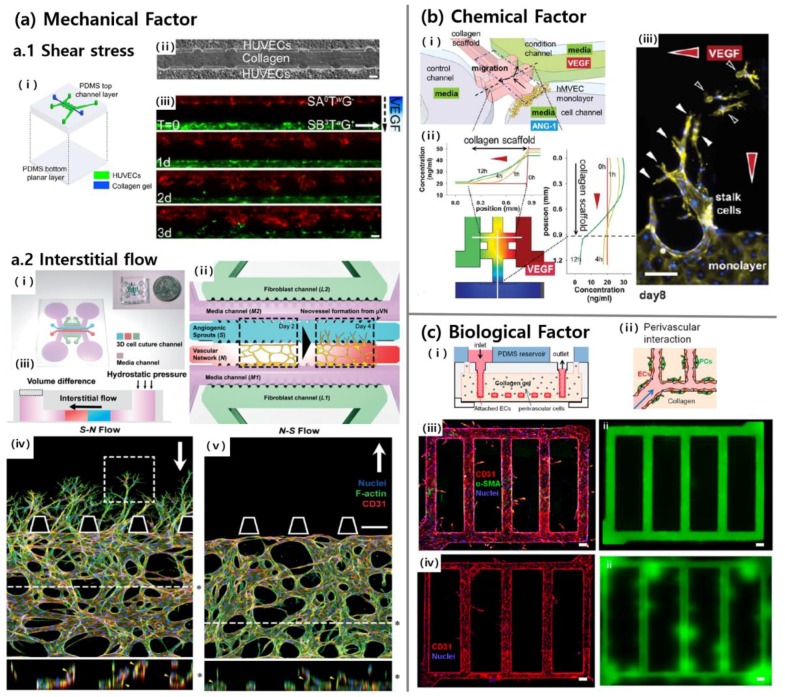
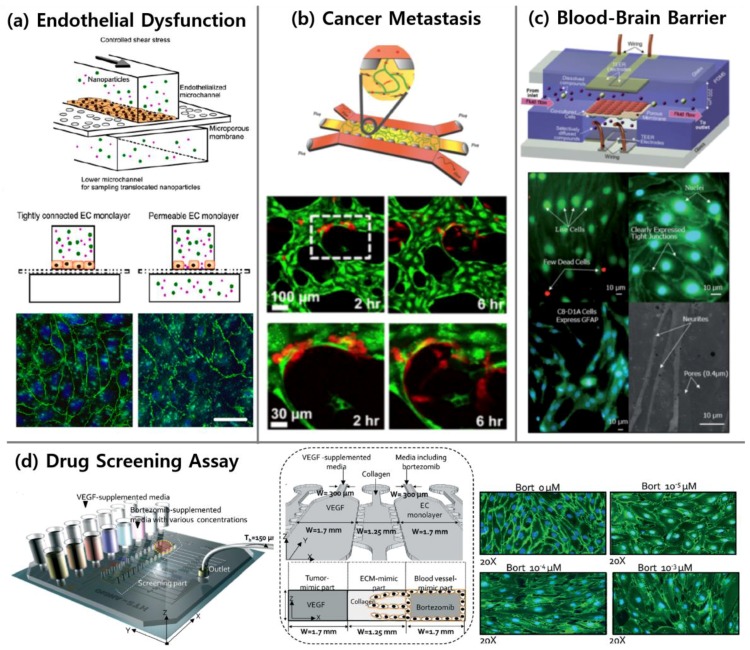
Vascularization, the formation of new blood vessels, is an essential biological process. As the vasculature is involved in various fundamental physiological phenomena and closely related to several human diseases, it is imperative that substantial research is conducted on characterizing the vasculature and its related diseases. A significant evolution has been made to describe the vascularization process so that in vitro recapitulation of vascularization is possible. The current microfluidic systems allow elaborative research on the effects of various cues for vascularization, and furthermore, in vitro technologies have a great potential for being applied to the vascular disease models for studying pathological events and developing drug screening platforms. Here, we review methods of fabrication for microfluidic assays and inducing factors for vascularization. We also discuss applications using engineered vasculature such as in vitro vascular disease models, vasculature in organ-on-chips and drug screening platforms.

Keywords: angiogenesis; in vitro disease models; microfluidics; vasculature-on-a-chip.


Conflict of interest statement

The authors declare no conflict of interest.

Figures

Figure 1
Figure 1
Schematic representation of fabrication methods. (a) Endothelial cells (ECs) (green) are cultured on the designed pattern or the specified membrane. (b) After removal of sacrificial molds, ECs (green) are seeded [30]. (c) ECs form monolayer alongside the hydrogel. Immunostaining of vascular endothelial cadherin (VE-cadherin, green) and nuclei (blue) confirm the functionality of the vessel [16]. Scale bar 100 µm. (d) ECs (green) are seeded within the hydrogel, and microvasculature is then formed through vasculogenesis [23]. Reproduced with permission.
Figure 2
Figure 2
Three major factors (mechanical, chemical, and biological factors) in vessel formation on a chip. (a.1) Fluid flow was given by syringe pumps that were connected to reservoirs of the chip (i), which had one channel for collagen and two channels for human umbilical vein endothelial cells (HUVECs) (ii). Shear stress inhibited sprouting in the presence of interstitial flow and vascular endothelial growth factor (VEGF) gradient (iii) [12]. Scale bars 100 μm. (a.2) Angiogenic sprouting from the vascular network (i–ii) was observed in the presence of interstitial flow (iii). Sprouting was more active when the interstitial flow acts on sprouting in reverse direction (iv), than forward direction (v) [45]. Scale bar 200 μm. (b) The direction of sprouting could be guided along spatial VEGF gradient (iii) by forming two orthogonal gradient profiles on the gel region (i–ii) [21]. Scale bars 100 μm. (c) HUVECs were seeded on the hollow microchannels in the pericyte-embedded collagen gel (i) to generate stable vascular structures through heterotypic cell–cell interactions (ii). Pericytes (α-SMA, green) enclosed endothelial wall (CD31, red) and contributed to structural stability (iii) confirmed by perfusing of fluorescent microbeads (green). A number of leakages could be observed when only HUVECs were seeded (iv) [56]. Scale bars 100μm. Reproduced with permission.
Figure 3
Figure 3
Representative figures of applications using vasculature-on-a-chip. (a) Increased permeability of inflamed endothelium was demonstrated with translocation of nanoparticles over an EC monolayer and immunostaining of VE-cadherin (green) and nuclei (blue) [98]. Scale bars 20 µm. (b) The bone microenvironment was replicated to investigate extravasation of breast cancer cells (red) from microvasculature (green) [23]. (c) Microfluidic model of the blood-brain barrier (µBBB) was characterized, and its validity was demonstrated [121]. (d) HUVECs morphology was investigated quantitatively depending on anti-angiogenic drug concentrations to find optimal effective concentration which minimizes both the number of migrated HUVECs and their morphological changes [24]. Reproduced with permission.

References

    1. Jain R.K. Molecular regulation of vessel maturation. Nat. Med. 2003;9:685–693. doi: 10.1038/nm0603-685. - DOI - PubMed
    1. Carmeliet P., Jain R.K. Molecular mechanisms and clinical applications of angiogenesis. Nature. 2011;473:298–307. doi: 10.1038/nature10144. - DOI - PMC - PubMed
    1. Munzel T., Sinning C., Post F., Warnholtz A., Schulz E. Pathophysiology, diagnosis and prognostic implications of endothelial dysfunction. Ann. Med. 2008;40:180–196. doi: 10.1080/07853890701854702. - DOI - PubMed
    1. Roudsari L.C., West J.L. Studying the influence of angiogenesis in in vitro cancer model systems. Adv. Drug Deliv. Rev. 2016;97:250–259. doi: 10.1016/j.addr.2015.11.004. - DOI - PubMed
    1. Carmeliet P., Jain R.K. Angiogenesis in cancer and other diseases. Nature. 2000;407:249–257. doi: 10.1038/35025220. - DOI - PubMed