Review
. 2017 Jan 25;4(1):10.
doi: 10.3390/bioengineering4010010.

3D Printing of Organs-On-Chips

Affiliations
Review

3D Printing of Organs-On-Chips

Hee-Gyeong Yi et al. Bioengineering (Basel). .

Abstract

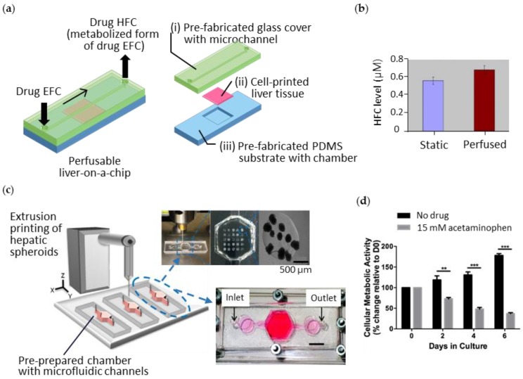
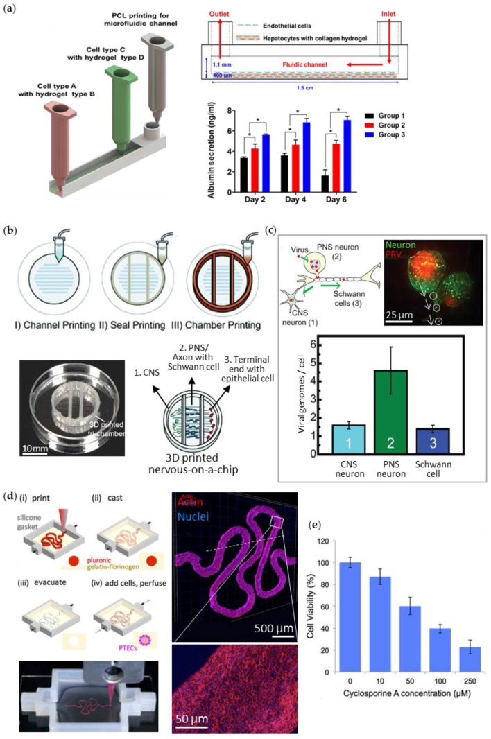
Organ-on-a-chip engineering aims to create artificial living organs that mimic the complex and physiological responses of real organs, in order to test drugs by precisely manipulating the cells and their microenvironments. To achieve this, the artificial organs should to be microfabricated with an extracellular matrix (ECM) and various types of cells, and should recapitulate morphogenesis, cell differentiation, and functions according to the native organ. A promising strategy is 3D printing, which precisely controls the spatial distribution and layer-by-layer assembly of cells, ECMs, and other biomaterials. Owing to this unique advantage, integration of 3D printing into organ-on-a-chip engineering can facilitate the creation of micro-organs with heterogeneity, a desired 3D cellular arrangement, tissue-specific functions, or even cyclic movement within a microfluidic device. Moreover, fully 3D-printed organs-on-chips more easily incorporate other mechanical and electrical components with the chips, and can be commercialized via automated massive production. Herein, we discuss the recent advances and the potential of 3D cell-printing technology in engineering organs-on-chips, and provides the future perspectives of this technology to establish the highly reliable and useful drug-screening platforms.

Keywords: 3D printing; bioprinting; cell-printing; in vitro disease model; in vitro tissue model; organ-on-a-chip.


Conflict of interest statement

The authors declare no conflict of interest.

Figures

Figure 1
Figure 1
3D printing of biological constructs with heterogeneous and complex structures. Photographs of the 3D-printed (a) a mandible bone construct, (b) an ear cartilage with ear lobule, (c) a kidney with renal pelvis, (d) a liver, (e) a heart cross-section, and (f) an arterial tree. Reproduced with permissions from [18,19,21,22,24].
Figure 2
Figure 2
Schematics of 3D-cell printing methods with different working principles: (a) micro-extrusion, (b) inkjet, (c) laser-assisted printing, and (d) stereolithographic printing. Reproduced with permission from [76].
Figure 3
Figure 3
3D cell-printed livers-on-chips. (a) Schematic diagram of inkjet printing into multiple micro-wells to create liver tissues with three layering compositions: 1L (HepG2), 2L (HUVEC/HepG2), and 3L (HUVEC/HepG2, HUVEC); (b) Hepatotoxic responses with increase in CYP450 3A4 (CYP3A4) secretions under troglitazone treatment; (c) Illustration of the sequential SLA process for building the liver lobule structure and (d) rifampicin-induced changes in expression of the CYP450 series in HPCs grown in the 2D monolayer, the 3D HPC-only model, and the 3D-printed model (3D Tri-culture). Reproduced with permissions from [79,80]
Figure 4
Figure 4
3D cell-printed cancers-on-chips (a) Schematic diagram and microscopic observations (lower left: H&E staining, lower right: immunofluorescent staining) of the extruded breast stromal cells and cancer cells into a multi-well plate; (b) Increased chemosensitivity to tamoxifen from breast cancer cells cultured in 3D-printed chip compared to those in a 2D monolayer; (c) Process of the micro-extrusion of cervical cancer cells (Hela) with gelatin-alginate-fibrinogen bioink; (d) Photograph of the printed cervical cancer-on-a-chip showing the lattice pattern and its fluorescent microscopic image showing the cell morphology; (e) Increased chemosensitivity to paclitaxel from Hela cells cultured in 3D-printed chip compared to those in a 2D monolayer. Reproduced with permission from [82,83].
Figure 5
Figure 5
3D cell-printed liver constructs integrated with a pre-prepared microfluidic device. (a) Illustration of the 3D-printed perfusable liver-on-a-chip and its exploded view; (b) The higher level of metabolized drug 7-hydroxy-4-trifluoromethyl coumarin (HFC) under perfusion condition compared to that under static condition; (c) Schematics of extrusion printing of liver cells onto pre-prepared microfluidic device and its photographs, showing the array of hepatic spheroids in the chamber; (d) Hepatotoxic effect of acute acetaminophen on the liver tissue. Reproduced with permissions from [84,85].
Figure 6
Figure 6
3D-printed organs-on-chips in a one-step fabrication process. (a) Schematic diagrams for extrusion printing of liver-on-a-chip resembling sinusoid (Group 1 = 3D cell-printed hepatocytes alone in static culture, Group 2 = 3D cell-printed hepatocytes–endothelial cells in static culture, and Group 3 = 3D cell-printed liver-on-a-chip); (b) Extrusion-printed nervous system-on-a-chip with compartmentalized chambers: central neurons (CNS) in chamber 1, peripheral neurons (PNS), and the axons with Schwann cells in chamber 2, and terminal cell junctions with epithelial cells in chamber 3; (c) Study of viral infection in the neuron system. Microscopic observation of the pseudorabies virus (PRV) transmission and the level of the transported virus in each chamber; (d) Illustration (upper left) and photograph (lower left) of the extrusion-printing process of a kidney proximal tubule-on-a-chip and the immunofluorescent stained images of the tubule (upper and lower right); (e) Nephrotoxicity effect of cyclosporine A on kidney epithelial cells in the chip. Reproduced with permissions from [50,87,88].

Similar articles

Cited by

References

    1. Pedron S., Becka E., Harley B.A. Spatially gradated hydrogel platform as a 3D engineered tumor microenvironment. Adv. Mater. 2015;27:1567–1572. doi: 10.1002/adma.201404896. - DOI - PubMed
    1. Jeon J.S., Bersini S., Gilardi M., Dubini G., Charest J.L., Moretti M., Kamm R.D. Human 3D vascularized organotypic microfluidic assays to study breast cancer cell extravasation. Proc. Natl. Acad. Sci. USA. 2015;112:214–219. doi: 10.1073/pnas.1417115112. - DOI - PMC - PubMed
    1. Shin Y., Han S., Jeon J.S., Yamamoto K., Zervantonakis I.K., Sudo R., Kamm R.D., Chung S. Microfluidic assay for simultaneous culture of multiple cell types on surfaces or within hydrogels. Nat. Protoc. 2012;7:1247–1259. doi: 10.1038/nprot.2012.051. - DOI - PMC - PubMed
    1. Wang L., Liu W., Wang Y., Wang J.-C., Tu Q., Liu R., Wang J. Construction of oxygen and chemical concentration gradients in a single microfluidic device for studying tumor cell–drug interactions in a dynamic hypoxia microenvironment. Lab Chip. 2013;13:695–705. doi: 10.1039/C2LC40661F. - DOI - PubMed
    1. Sackmann E.K., Fulton A.L., Beebe D.J. The present and future role of microfluidics in biomedical research. Nature. 2014;507:181–189. doi: 10.1038/nature13118. - DOI - PubMed

LinkOut - more resources