. 2017 May 25;4(2):51.
doi: 10.3390/bioengineering4020051.

Development and Characterization of a Parallelizable Perfusion Bioreactor for 3D Cell Culture

Affiliations

Development and Characterization of a Parallelizable Perfusion Bioreactor for 3D Cell Culture

Dominik Egger et al. Bioengineering (Basel). .

Abstract

The three dimensional (3D) cultivation of stem cells in dynamic bioreactor systems is essential in the context of regenerative medicine. Still, there is a lack of bioreactor systems that allow the cultivation of multiple independent samples under different conditions while ensuring comprehensive control over the mechanical environment. Therefore, we developed a miniaturized, parallelizable perfusion bioreactor system with two different bioreactor chambers. Pressure sensors were also implemented to determine the permeability of biomaterials which allows us to approximate the shear stress conditions. To characterize the flow velocity and shear stress profile of a porous scaffold in both bioreactor chambers, a computational fluid dynamics analysis was performed. Furthermore, the mixing behavior was characterized by acquisition of the residence time distributions. Finally, the effects of the different flow and shear stress profiles of the bioreactor chambers on osteogenic differentiation of human mesenchymal stem cells were evaluated in a proof of concept study. In conclusion, the data from computational fluid dynamics and shear stress calculations were found to be predictable for relative comparison of the bioreactor geometries, but not for final determination of the optimal flow rate. However, we suggest that the system is beneficial for parallel dynamic cultivation of multiple samples for 3D cell culture processes.

Keywords: 3D cell culture; computational fluid dynamics; dynamic cultivation; fluid shear stress; perfusion bioreactor system.


Conflict of interest statement

The authors declare no conflict of interest.

Figures

Figure 1
Figure 1
(A) Explosion view, (B) cross section with inserted biomaterial and (C) pictures of the bioreactor chamber prototypes. The first prototype (BR1) was made of stainless steel whereas the second prototype (BR2) made of polyoxymethylene (POM) features two sieve-like medium distribution units (MDU). The flow channel of BR2 opens up to the full diameter of the chamber (detailed view) and the chamber is adjustable in height to allow scaffolds of different thickness to be inserted. The placement of the scaffold in both bioreactors is represented with a yellow box in panel B.
Figure 2
Figure 2
(A) Tailor made incubator system developed by Fraunhofer IGB, Stuttgart. A heating plate controls the temperature of the incubator chamber. Two 4 channel pumps can operate several tubing systems at once. A pinch valve can be used to apply hydrostatic pressure (HP). All functions are controlled via a touch screen control panel and all data can be recorded via USB port; (B) Single reactor setup in the incubator system: pressure sensors (P) measure the pressure differential inside the bioreactor system non-invasively; (C) Multi reactor setup: up to eight independent bioreactors can be operated in parallel (modified from [23], with permission from S. Karger AG, Medical and Scientific Publishers).
Figure 3
Figure 3
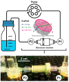
Scheme and picture of the bioreactor setup used during fluid shear stress prediction measurements. Water at 37 °C was pumped through the bioreactor chamber containing the porous scaffold Sponceram. Pressure sensors measured the pressure upstream (P1) and downstream (P2) of the bioreactor chamber non-invasively. Sponceram is depicted as volume rendering of a microCT scan and scaffold dimensions are given as h = height, d = diameter, and A = area.
Figure 4
Figure 4
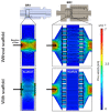
Computational model of the flow profile with streamlines of the bioreactor chambers BR1 and BR2 without and with a scaffold inserted (porosity: 66.7%, permeability 1.74 × 10⁻¹⁰ m2) at a flow rate of 1.5 mL/min (3.6 mm/s). Streamlines and the flow profile indicate a more homogeneous flow distribution in BR2 due to the medium distribution units.
Figure 5
Figure 5
(A) Hydrodynamic residence time and (B) residence time distribution of BR1 and BR2 without inserted scaffold. (C) Hydrodynamic residence time and (D) residence time distribution of BR1 and BR2 with inserted scaffold.
Figure 6
Figure 6
(A) Sensor characterization at different flow rates. ΔP depicts the differential of both sensors; (B) Determination of the permeability of the porous scaffold Sponceram. ΔP and the resulting permeability k at different flow rates; (C) Shear stress prediction based on the determination of the permeability of Sponceram and further calculation with the Vossenberg model or CFD data. Depending on the flow rate, the analytical model predicts shear forces in the physiological in vivo range of 0.3–3 Pa (shaded area) whereas the computational modeling predicts shear forces that are approximately three orders of magnitude lower; (D) Computational simulation of the shear stress conditions. BR1 and BR2 with porous scaffold (porosity: 66.7%, specific permeability 1.74 × 10⁻¹⁰ m2) at a flow rate of 15 mL/min. The model indicates a more homogeneous distribution of shear stress in BR2.
Figure 7
Figure 7
(A) Cell numbers, (B) course of alkaline phosphatase (ALP) activity and (C) ALP activity per cell of adipose derived mesenchymal stem cells (ASCs) cultivated on Sponceram in a perfusion bioreactor or under 3D and 2D static conditions. Data are represented as mean ± SD (n = 3); * significant difference of the indicated conditions in panel (A) or to 3D static in panel (C) with a confidence interval of 99% and p < 0.001.
Figure 8
Figure 8
(A) 4′,6-diamidin-2-phenylindol (DAPI) stain and (B) DAPI-calcein double stain of Sponceram after 21 days of cultivation with ASCs in a perfusion bioreactor or under static conditions.
Figure 9
Figure 9
Course of (A) glucose consumption and (C) lactate production, (B) glucose consumption and (D) lactate production per cell between day 19 and 21 of ASCs cultivated on Sponceram in a perfusion bioreactor or under 3D and 2D static conditions. Data are represented as mean ± SD (n = 3); * indicates significant difference to 3D static with a confidence interval of 99% and p < 0.001.
Figure 10
Figure 10
Von Kossa stain of Sponceram after 21 days of cultivation with ASCs in a perfusion bioreactor or under static conditions.

References

    1. Volkmer E., Drosse I., Otto S., Stangelmayer A., Stengele M., Kallukalam B.C., Mutschler W., Schieker M. Hypoxia in static and dynamic 3D culture systems for tissue engineering of bone. Tissue Eng. Part A. 2008;14:1331–1340. doi: 10.1089/ten.tea.2007.0231. - DOI - PubMed
    1. Goldstein A.S., Juarez T.M., Helmke C.D., Gustin M.C., Mikos A.G. Effect of convection on osteoblastic cell growth and function in biodegradable polymer foam scaffolds. Biomaterials. 2001;22:1279–1288. doi: 10.1016/S0142-9612(00)00280-5. - DOI - PubMed
    1. Stiehler M., Bünger C., Baatrup A., Lind M., Kassem M., Mygind T. Effect of dynamic 3-D culture on proliferation, distribution, and osteogenic differentiation of human mesenchymal stem cells. J. Biomed. Mater. Res. Part A. 2009;89:96–107. doi: 10.1002/jbm.a.31967. - DOI - PubMed
    1. Wang T., Wu H., Wang H., Lin F., Sun J. Regulation of adult human mesenchymal stem cells into osteogenic and chondrogenic lineages by different bioreactor systems. J. Biomed. Mater. Res. Part A. 2009;88:935–946. doi: 10.1002/jbm.a.31914. - DOI - PubMed
    1. Gooch K.J., Kwon J.H., Blunk T., Langer R., Freed L.E., Vunjak-Novakovic G. Effects of mixing intensity on tissue-engineered cartilage. Biotechnol. Bioeng. 2001;72:402–407. doi: 10.1002/1097-0290(20000220)72:4<402::AID-BIT1002>3.0.CO;2-Q. - DOI - PubMed