Review
. 2017 Nov 15;9(11):1501-1512.
doi: 10.1039/c7mt00221a.

The mitochondrion: a central architect of copper homeostasis

Affiliations
Review

The mitochondrion: a central architect of copper homeostasis

Zakery N Baker et al. Metallomics. .

Abstract

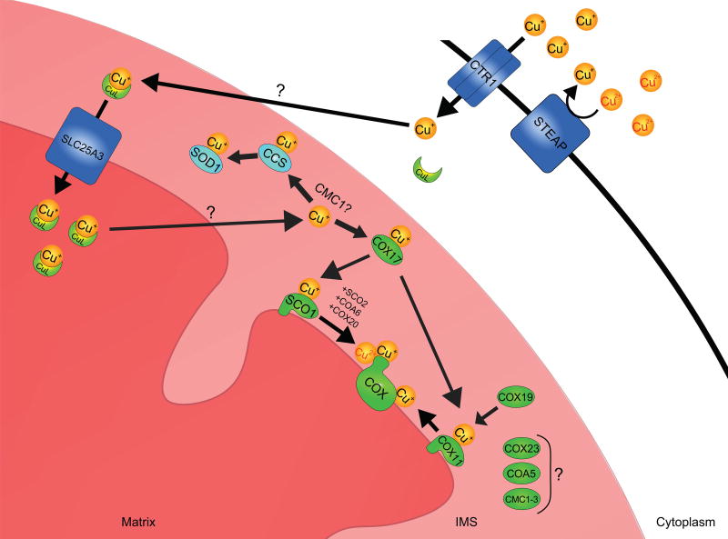
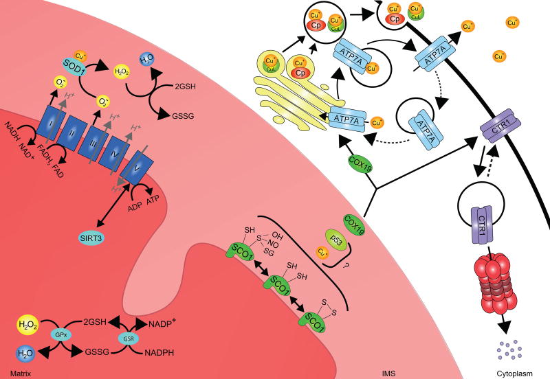
All known eukaryotes require copper for their development and survival. The essentiality of copper reflects its widespread use as a co-factor in conserved enzymes that catalyze biochemical reactions critical to energy production, free radical detoxification, collagen deposition, neurotransmitter biosynthesis and iron homeostasis. However, the prioritized use of copper poses an organism with a considerable challenge because, in its unbound form, copper can potentiate free radical production and displace iron-sulphur clusters to disrupt protein function. Protective mechanisms therefore evolved to mitigate this challenge and tightly regulate the acquisition, trafficking and storage of copper such that the metal ion is rarely found in its free form in the cell. Findings by a number of groups over the last ten years emphasize that this regulatory framework forms the foundation of a system that is capable of monitoring copper status and reprioritizing copper usage at both the cellular and systemic levels of organization. While the identification of relevant molecular mechanisms and signaling pathways has proven to be difficult and remains a barrier to our full understanding of the regulation of copper homeostasis, mounting evidence points to the mitochondrion as a pivotal hub in this regard in both healthy and diseased states. Here, we review our current understanding of copper handling pathways contained within the organelle and consider plausible mechanisms that may serve to functionally couple their activity to that of other cellular copper handling machinery to maintain copper homeostasis.


Figures

Figure 1
Figure 1. Mitochondrial copper acquisition and trafficking pathways in mammals
Extracellular copper in the cupric (Cu2+) ion form is reduced by a member of the STEAP family of oxidoreductases. The cuprous (Cu+) ion is then imported into the cell by the high affinity copper transporter CTR1. How intracellular copper is trafficked to the mitochondrion is not clear; however, it is thought that copper binding to an unknown ligand triggers copper translocation from the cytosol to the mitochondrial intermembrane space (IMS). There, the copper-ligand complex is transported across the inner membrane for its storage in the matrix by SLC25A3, a member of the mitochondrial carrier family and the mammalian homolog of yeast PIC2. Matrix copper is ultimately mobilized and translocated across the inner membrane to the IMS by an unknown transporter, to metallate the cupro-enzymes SOD1 and COX during their maturation. Copper loading onto apo-SOD1 requires CCS. Copper destined for the metallation of the CuA and CuB sites of COX is initially transferred to COX17, which delivers it to the copper chaperones SCO1 and COX11. SCO1 function during the maturation of the CuA site is facilitated by at least 3 other COX assembly factors; SCO2, COA6 and COX20. Several additional COX assembly factors (COX23, COA5, CMC1–3) with unknown or poorly understood functions may also facilitate copper delivery to COX and/or regulate copper trafficking within the IMS.
Figure 2
Figure 2. Potential inputs that contribute to SCO1-dependent mitochondrial signaling and the regulation of copper homeostasis
SCO1 function is integral to a mitochondrial signaling pathway that impinges upon copper homeostasis. The redox state of its cysteinyl sulphurs, which are located within the Cx3C motif, is significantly perturbed in patients with combined COX and total copper deficiencies. Thus, signaling may be directly modulated by the proportion of the SCO1 pool with copper-loaded versus apo-Cx3C motifs, or by the balance of reversible post-translational modifications to one or both of its cysteinyl sulphurs (S-S, SH, SOH, SNO or SSG). How the functional status of SCO1 is sensed and transduced outside of the mitochondrion to effect changes in the abundance or localization of the copper import or efflux machinery remains poorly understood. COX19 partitions between the IMS and the cytosol in a copper-dependent manner, and acts alone or in concert with unknown factors to stimulate ATP7A–mediated copper export from the cell by regulating its trafficking to the plasma membrane and/or increasing the rate of secretion of copper bound to cupro-proteins (e.g Cp, ceruloplasmin) or small molecules (e.g. CuL, copper ligand). Mechanisms that couple SCO1-dependent mitochondrial signaling to the regulation of CTR1 localization or degradation are currently unknown. However, redox and/or metabolic switch mechanisms may contribute to the modulation of SCO1-dependent mitochondrial signaling. Complexes (labeled I-IV) of the electron transport chain (ETC) oxidize NADH and FADH2 while establishing a proton gradient that is harnessed by Complex V to generate ATP. Perturbations in this pathway lead to changes in the NAD+:NADH and AMP:ATP ratios and the activation of the AMPK/p53 pathway and SIRT3, both of which trigger downstream changes in metabolism that may affect cellular copper levels. Complexes I and III of the ETC also generate superoxide anions and ROS are known to promote signaling by post-translationally modifying proteins and/or altering redox balance. Changes in the mitochondrial redox state are sensed primarily by the GSH:GSSG ratio, which is buffered within the matrix by the activity of the enzymes glutathione peroxidase (GPx) and glutathione reductase (GSR).

References

    1. Institute of Medicine (US) Panel on Micronutrients. Dietary Reference Intakes for Vitamin A, Vitamin K, Arsenic, Boron, Chromium, Copper, Iodine, Iron, Manganese, Molybdenum, Nickel, Silicon, Vanadium, and Zinc. National Academies Press (US); Washington (DC): 2001. pp. 224–257. - PubMed
    1. Prohaska JR. Impact of copper deficiency in humans. Ann. N.Y. Acad. Sci. 2014;1314:1–5. - PubMed
    1. Nevitt T, Ohrvik H, Thiele DJ. Charting the travels of copper in eukaryotes from yeast to mammals. Biochim. Biophys. Acta. 2012;1823:1580–1593. - PMC - PubMed
    1. Kim BE, Nevitt T, Thiele DJ. Mechanisms for copper acquisition, distribution and regulation. Nat. Chem. Biol. 2008;4:176–185. - PubMed
    1. Tisato F, Marzano C, Porchia M, Pellei M, Santini C. Copper in diseases treatments, and copper-based anticancer strategies. Med. Res. Rev. 2010;30:708–749. - PubMed