

Comparing 3-Dimensional Ultrasound to 3-Dimensional Magnetic Resonance Imaging in the Detection of Levator Ani Defects
- PMID: 28953075
- PMCID: PMC5869069
- DOI: 10.1097/SPV.0000000000000485
Comparing 3-Dimensional Ultrasound to 3-Dimensional Magnetic Resonance Imaging in the Detection of Levator Ani Defects
Abstract
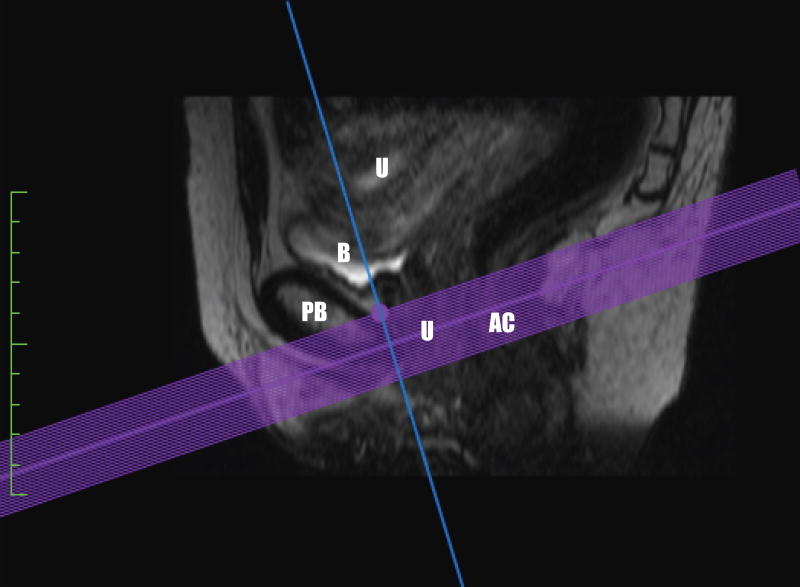
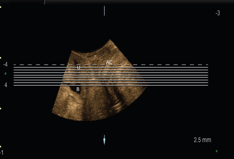
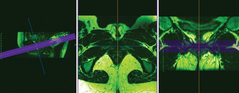
Objective: The aim of this study was to compare the detection of levator ani defects (LAD) between 3-dimensional (3D) ultrasound (US) and 3D magnetic resonance imaging (MRI).
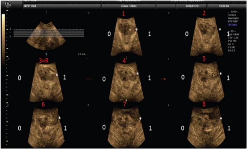
Methods: This is a secondary analysis of the Pelvic Floor Nerve Injury Following Childbirth Study. Nulliparous women underwent a standardized protocol of pelvic floor evaluations between January 2008 and December 2013, prior to pregnancy (V1) and at 2 points postpartum: 6 weeks (V2) and 6 months (V3). Those women who underwent a high-resolution 3D MRI pelvic floor sequence were selected. Comparisons were made to concomitantly acquired 3D perineal US. Eight tomographic slices were examined in the axial plane, each side independently scored with 0 (no defect) or 1 (defect). A similar tomographic approach was applied to the MRI. For both MRI and US, the right and left sides were each scored. A total score of 0 to 8 was given to each side. A dichotomous variable "complete LAD" was defined. Cohen κ was used as a measurement of agreement of complete LAD between MRI and US. Kendall τ b was used to correlate total scores.
Results: On the right side, 80 (90%) of 89 pairs were in agreement (concordant in the diagnosis or not of a "defect"). On the left side, 72 (81%) of 89 pairs were in agreement. Correlations (Cohen κ) of complete LAD were 0.65 (P < 0.001) on the right and 0.37 (P < 0.001) on the left. Correlations of total scores were 0.47 (P < 0.001) on the right and 0.41 (P < 0.001) on the left.
Conclusions: Moderate agreement was found between 3D US and 3D MRI LAD detection. More LADs and discordance were seen on the left.
Figures
References
-
- Viktrup L, Rortveit G, Lose G. Risk of stress urinary incontinence twelve years after the first pregnancy and delivery. Obstet Gynecol. 2006;108:248–254. - PubMed
-
- Dietz HP, Lanzarone V. Levator trauma after vaginal delivery. Obstet Gynecol. 2005;106:707–712. - PubMed
-
- Bump RC, Norton PA. Epidemiology and natural history of pelvic floor dysfunction. Obstet Gynecol Clin Nort Am. 1998;25:723–46. - PubMed
Publication types
MeSH terms
Grants and funding
LinkOut - more resources
Full Text Sources
Other Literature Sources
Medical
