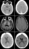
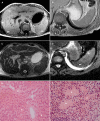
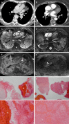
Total-body CT and MR features of postmortem change in in-hospital deaths
- PMID: 28953923
- PMCID: PMC5617178
- DOI: 10.1371/journal.pone.0185115
Total-body CT and MR features of postmortem change in in-hospital deaths
Abstract
Objectives: To evaluate the frequency of total-body CT and MR features of postmortem change in in-hospital deaths.
Materials and methods: In this prospective blinded cross-sectional study, in-hospital deceased adult patients underwent total-body postmortem CT and MR followed by image-guided biopsies. The presence of PMCT and PMMR features related to postmortem change was scored retrospectively and correlated with postmortem time interval, post-resuscitation status and intensive care unit (ICU) admittance.
Results: Intravascular air, pleural effusion, periportal edema, and distended intestines occurred more frequently in patients who were resuscitated compared to those who were not. Postmortem clotting was seen less often in resuscitated patients (p = 0.002). Distended intestines and loss of grey-white matter differentiation in the brain showed a significant correlation with postmortem time interval (p = 0.001, p<0.001). Hyperdense cerebral vessels, intravenous clotting, subcutaneous edema, fluid in the abdomen and internal livores of the liver were seen more in ICU patients. Longer postmortem time interval led to a significant increase in decomposition related changes (p = 0.026).
Conclusions: There is a wide variety of imaging features of postmortem change in in-hospital deaths. These imaging features vary among clinical conditions, increase with longer postmortem time interval and must be distinguished from pathologic changes.
Conflict of interest statement
Figures


Similar articles
-
Intravascular gas distribution in the upper abdomen of non-traumatic in-hospital death cases on postmortem computed tomography.Leg Med (Tokyo). 2011 Jul;13(4):174-9. doi: 10.1016/j.legalmed.2011.03.002. Epub 2011 May 11. Leg Med (Tokyo). 2011. PMID: 21561795
-
Normal cranial postmortem CT findings in children.Forensic Sci Int. 2015 Jan;246:43-9. doi: 10.1016/j.forsciint.2014.10.036. Epub 2014 Oct 31. Forensic Sci Int. 2015. PMID: 25437903
-
Time-related course of pleural space fluid collection and pulmonary aeration on postmortem computed tomography (PMCT).Leg Med (Tokyo). 2015 Jul;17(4):221-5. doi: 10.1016/j.legalmed.2015.01.002. Epub 2015 Jan 21. Leg Med (Tokyo). 2015. PMID: 25657038
-
Essence of postmortem computed tomography for in-hospital deaths: what clinical radiologists should know.Jpn J Radiol. 2023 Oct;41(10):1039-1050. doi: 10.1007/s11604-023-01443-w. Epub 2023 May 17. Jpn J Radiol. 2023. PMID: 37193920 Free PMC article. Review.
-
Postmortem imaging: MDCT features of postmortem change and decomposition.Am J Forensic Med Pathol. 2010 Mar;31(1):12-7. doi: 10.1097/PAF.0b013e3181c65e1a. Am J Forensic Med Pathol. 2010. PMID: 20010292 Review.
Cited by
-
Efficient cascaded V-net optimization for lower extremity CT segmentation validated using bone morphology assessment.J Orthop Res. 2022 Dec;40(12):2894-2907. doi: 10.1002/jor.25314. Epub 2022 Mar 15. J Orthop Res. 2022. PMID: 35239226 Free PMC article.
-
Postmortem changes in brain cell structure: a review.Free Neuropathol. 2023 May 31;4:10. doi: 10.17879/freeneuropathology-2023-4790. eCollection 2023 Jan. Free Neuropathol. 2023. PMID: 37384330 Free PMC article.
-
Post-mortem computed tomography (PMCT) radiological findings and assessment in advanced decomposed bodies.Radiol Med. 2019 Oct;124(10):1018-1027. doi: 10.1007/s11547-019-01052-6. Epub 2019 Jun 28. Radiol Med. 2019. PMID: 31254219
-
Optimization of Postprocessing parameters for abdominal Forensic CT scans.Forensic Sci Int Synerg. 2024 May 14;8:100478. doi: 10.1016/j.fsisyn.2024.100478. eCollection 2024. Forensic Sci Int Synerg. 2024. PMID: 38779309 Free PMC article.
-
The current state of forensic imaging - post mortem imaging.Int J Legal Med. 2025 May;139(3):1141-1159. doi: 10.1007/s00414-025-03461-x. Epub 2025 Mar 24. Int J Legal Med. 2025. PMID: 40126650 Free PMC article. Review.
References
-
- Burton JL, Underwood J. Clinical, educational, and epidemiological value of autopsy. Lancet. 2007;369(9571):1471–80. doi: 10.1016/S0140-6736(07)60376-6 . - DOI - PubMed
-
- Gaensbacher S, Waldhoer T, Berzlanovich A. The slow death of autopsies: a retrospective analysis of the autopsy prevalence rate in Austria from 1990 to 2009. Eur J Epidemiol. 2012;27(7):577–80. doi: 10.1007/s10654-012-9709-3 . - DOI - PubMed
-
- Turnbull A, Martin J, Osborn M. The death of autopsy? Lancet. 2015;386(10009):2141 doi: 10.1016/S0140-6736(15)01049-1 . - DOI - PubMed
-
- Shojania KG, Burton EC, McDonald KM, Goldman L. Changes in rates of autopsy-detected diagnostic errors over time: a systematic review. JAMA. 2003;289(21):2849–56. doi: 10.1001/jama.289.21.2849 . - DOI - PubMed
-
- Roberts IS, Benamore RE, Benbow EW, Lee SH, Harris JN, Jackson A, et al. Post-mortem imaging as an alternative to autopsy in the diagnosis of adult deaths: a validation study. Lancet. 2012;379(9811):136–42. doi: 10.1016/S0140-6736(11)61483-9 ; PubMed Central PMCID: PMCPMC3262166. - DOI - PMC - PubMed
MeSH terms
LinkOut - more resources
Full Text Sources
Other Literature Sources
Medical

