. 2017 Sep 26;20(13):3025-3033.
doi: 10.1016/j.celrep.2017.09.002.

Augmentation of Antitumor Immunity by Human and Mouse CAR T Cells Secreting IL-18

Affiliations

Augmentation of Antitumor Immunity by Human and Mouse CAR T Cells Secreting IL-18

Biliang Hu et al. Cell Rep. .

Abstract

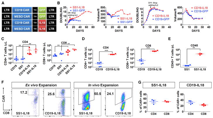
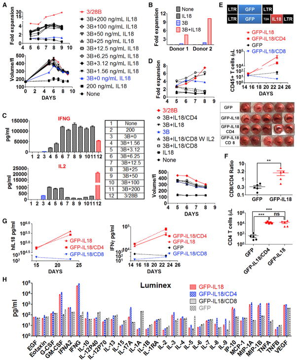
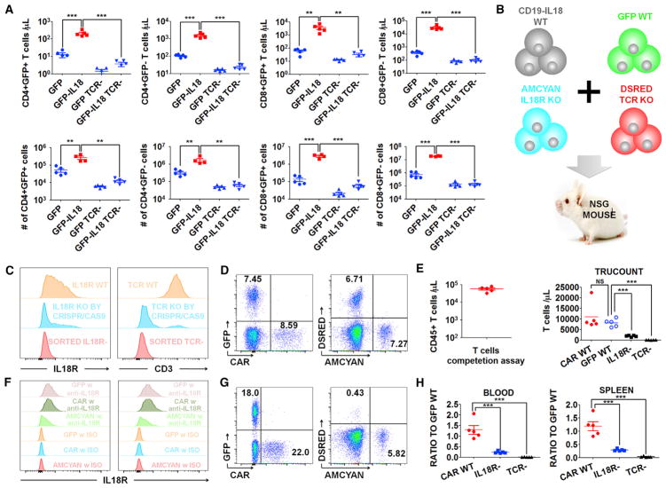
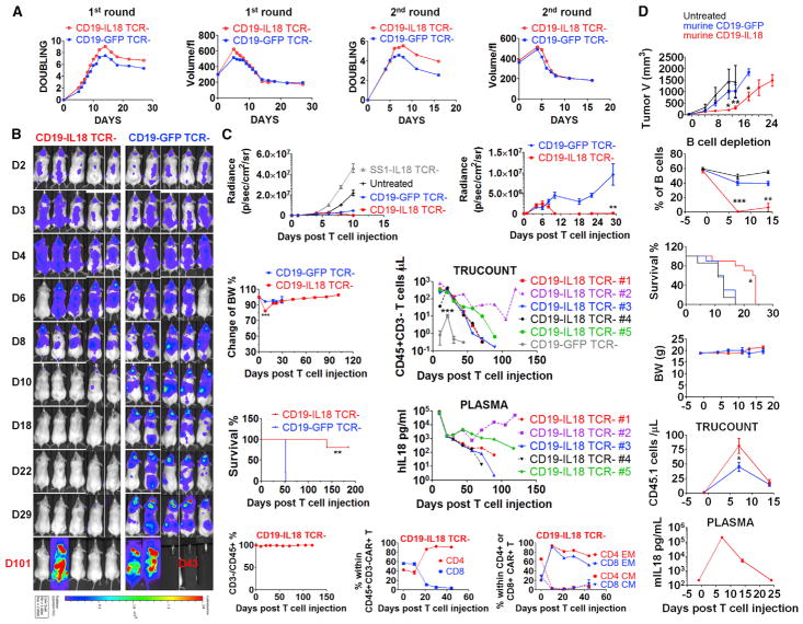
The effects of transgenically encoded human and mouse IL-18 on T cell proliferation and its application in boosting chimeric antigen receptor (CAR) T cells are presented. Robust enhancement of proliferation of IL-18-secreting human T cells occurred in a xenograft model, and this was dependent on TCR and IL-18R signaling. IL-18 augmented IFN-γ secretion and proliferation of T cells activated by the endogenous TCR. TCR-deficient, human IL-18-expressing CD19 CAR T cells exhibited enhanced proliferation and antitumor activity in the xenograft model. Antigen-propelled activation of cytokine helper ensemble (APACHE) CAR T cells displayed inducible expression of IL-18 and enhanced antitumor immunity. In an intact mouse tumor model, CD19-IL-18 CAR T cells induced deeper B cell aplasia, significantly enhanced CAR T cell proliferation, and effectively augmented antitumor effects in mice with B16F10 melanoma. These findings point to a strategy to develop universal CAR T cells for patients with solid tumors.

Keywords: IL-18; T cell; adoptive transfer; cancer; chimeric antigen receptor; immunotherapy.


Figures

Figure 1
Figure 1. IL18-CAR T Cells Have Enhanced Proliferation In Vitro and In Vivo
(A) The construct designs. (B) Population doubling and cell volume of SS1 anti-mesothelin or anti-CD19 CAR T cells following the first restimulation with irradiated K562-Meso or K562-CD19 with exogenous IL-2. (C) NSG mice (n = 5) bearing an AsPC1 pancreatic flank tumor received CD19 or SS1 CAR T cells (2e6), and 3 weeks later, peripheral blood was analyzed by Trucount. (D) NSG mice (n = 5) bearing a systemic Nalm6 acute lymphoblastic leukemia (ALL) tumor received 1e6 CD19 or CD19-IL-18 CAR T cells. After 18 days, circulating T cells were assessed. (E) Tumor-free NSG mice (n = 5) were inoculated with 5e6 SS1 or SS1-IL-18 CAR T cells and, 3 weeks later, analyzed for circulating T cells. (F and G) CAR T cells were analyzed on day 9 of ex vivo expansion. In vivo expansion of SS1-IL-18 and CD19-IL-18 CAR T cells was determined by harvesting spleens from the mice described in (C) and (D), respectively. (F) Representative fluorescence-activated cell sorting (FACS) plots of CD8+CAR+ cells. (G) The percentage of CD4+CAR+ or CD8+CAR+ T cells in spleens from (F). All data with error bars are presented as mean ± SEM. Student’s t test: **p < 0.01, ***p < 0.001.
Figure 2
Figure 2. Combined TCR and IL-18 Signaling Promotes CD4+ T Cell Expansion in the Absence of CAR Signaling
(A–C) Primary T cells from a healthy donor were activated with anti-CD3 beads and varying concentrations (1.56 doubled up to 200 ng/mL) of rIL-18 added on days 0 and 3. (A) Changes in T cell volume and cell numbers. (B) IL-18-dependent expansion of T cells from two additional donors stimulated with rIL-18, anti-CD3 beads, and anti-CD3 beads plus rIL-18. (C) Luminex analysis of secreted IFN-γ and IL-2 with 12 culture conditions indicated. Additional cytokine data are shown in Figure S2A. (D) Normal donor T cells expanded with anti-CD3/CD28 beads for 10 days were selected by MACS columns to purify CD8+ and CD4+ T cells. Purified CD4+ T cells, CD8+T cells, or bulk T cells were restimulated with anti-CD3 beads and 100 ng/mL rIL-18, added on days 0 and 3, as indicated in the legend. (E–H) T cells transduced with lentivectors encoding GFP orGFP-IL-18 were selected using MACS columns to purify CD8+ and CD4+ T cells. Tumor-free NSG mice (n = 5) were injected with 2e6 T cells as indicated. The data shown are representative of two independent experiments. (E) Circulating CD45+ T cells and pictures of spleens from various groups. (F) Circulating CD45+CD4+ T cells on day 23 following T cell injection and the ratio of CD8+ to CD4+ T cells. (G) Serum levels of IL-18 and IFN-γ. (H) Serum from mice on day 23 was analyzed by Luminex to assess the cytokine profile. All data with error bars are presented as mean ± SEM. Student’s t test: **p < 0.01, ***p < 0.001.
Figure 3
Figure 3. IL-18-Enhanced T Cell Proliferation Requires TCR and IL-18R Signaling
(A) Tumor-free NSG mice (n = 5) were injected with 2e6 TCR KO or TCR WT GFP-IL-18 T cells or the indicated controls. After 18 days, T cells in the blood (top) or spleens (bottom) were assessed. (B) Cell-competitive repopulation design. CD19 CAR-IL-18 WT, GFP WT, AmCyan IL-18R KO and DsRed TCR KO T cells were mixed together and injected into tumor-free NSG mice (n = 5). (C) Purity of TCR KO and IL-18R KO cells after CRISPR/Cas9 gene editing and MACS sorting. (D) Initial frequency of each cell population shown in (B). (E–H) The mice were analyzed 3 weeks after T cell injection. (E) Left: total circulating CD45+ cells, indicating robust expansion. Right: individual populations of T cells. (F) Expression levels of IL-18Rα on T cells in the blood (left) and spleen (right). (G) Representative FACS plot of each cell population in the blood. (H) The ratios of CD19 CAR-IL-18 WT, IL-18R KO AmCyan, and TCR KO DsRed to GFP WT cells in the blood (left) and spleen (right). All data with error bars are presented as mean ± SEM. Student’s t test: **p < 0.01, ***p < 0.001.
Figure 4
Figure 4. IL-18 Costimulation Significantly Enhances Proliferation and Antitumor Activity of Human TCR-Deficient and Syngeneic Murine CD19 CAR T Cells
(A) Expansion kinetics and volume of TCR KO CAR T cells following the first (with IL-2) and second (without IL-2) restimulation with irradiated K562-CD19cells. (B and C) NSG mice were injected intravenously with 1e6 Nalm6 ALL cells expressing CBG-GFP. On day 7 after tumor injection, mice (n = 5) received the indicated TCR KO CAR T cells (1e6). The data shown are representative of two independent experiments. (B) Live animal imaging of tumor growth. (C) Tumor growth curves of four groups (left) and a magnified plot of TCR KO CD19-GFP and TCR KOCD19-IL-18 CAR T cohorts (right). Additional data include body weight, survival, number of CD45+CD3 KO T cells, IL-18 plasma concentration, percentage of CD3 KO cells among total CD45+ cells, distribution of CD4+ and CD8+ T cells within CD19 CAR T cells, and memory phenotype in CD4+ and CD8+ CAR T cells. (D) Study of murine CD19-IL-18 CAR T cells in the B16F10 melanoma tumor model that was derived from the B6 mouse in 1954. Lymphocyte-replete C57BL/6 (CD45.2 strain) mice were inoculated subcutaneously (s.c.) with 2e6 B16F10 tumor cells expressing murine CD19 (B16-mCD19). One week later, mice were randomized and treated with 5e6 murine CD19-GFP (n = 10) or CD19-IL18 (n = 10) CAR T cells (CD45.1 origin) or left untreated (n = 7). The mice were monitored for tumor growth, B cell frequency, survival, body weight, CD45.1 T cells, and murine IL-18 plasma concentration. All data with error bars are presented as mean ± SEM. In some cases, the magnitude of the SEM is less that the size of the symbol. Student’s t test: *p < 0.05, **p < 0.01, ***p < 0.001.

References

    1. Brown CE, Alizadeh D, Starr R, Weng L, Wagner JR, Naranjo A, Ostberg JR, Blanchard MS, Kilpatrick J, Simpson J, et al. Regression of Glioblastoma after Chimeric Antigen Receptor T-Cell Therapy. N Engl J Med. 2016;375:2561–2569. - PMC - PubMed
    1. Carroll RG, Carpenito C, Shan X, Danet-Desnoyers G, Liu R, Jiang S, Albelda SM, Golovina T, Coukos G, Riley JL, et al. Distinct effects of IL-18 on the engraftment and function of human effector CD8 T cells and regulatory T cells. PLoS ONE. 2008;3:e3289. - PMC - PubMed
    1. Chmielewski M, Kopecky C, Hombach AA, Abken H. IL-12 release by engineered T cells expressing chimeric antigen receptors can effectively Muster an antigen-independent macrophage response on tumor cells that have shut down tumor antigen expression. Cancer Res. 2011;71:5697–5706. - PubMed
    1. Chmielewski M, Hombach AA, Abken H. Of CARs and TRUCKs: chimeric antigen receptor (CAR) T cells engineered with an inducible cytokine to modulate the tumor stroma. Immunol Rev. 2014;257:83–90. - PubMed
    1. Cohen J. IL-12 deaths: explanation and a puzzle. Science. 1995;270:908. - PubMed