. 2017 Sep 26;20(13):3123-3134.
doi: 10.1016/j.celrep.2017.09.010.

Microprocessor Recruitment to Elongating RNA Polymerase II Is Required for Differential Expression of MicroRNAs

Affiliations

Microprocessor Recruitment to Elongating RNA Polymerase II Is Required for Differential Expression of MicroRNAs

Victoria A Church et al. Cell Rep. .

Abstract

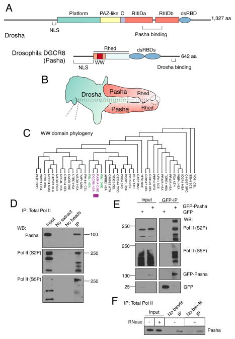
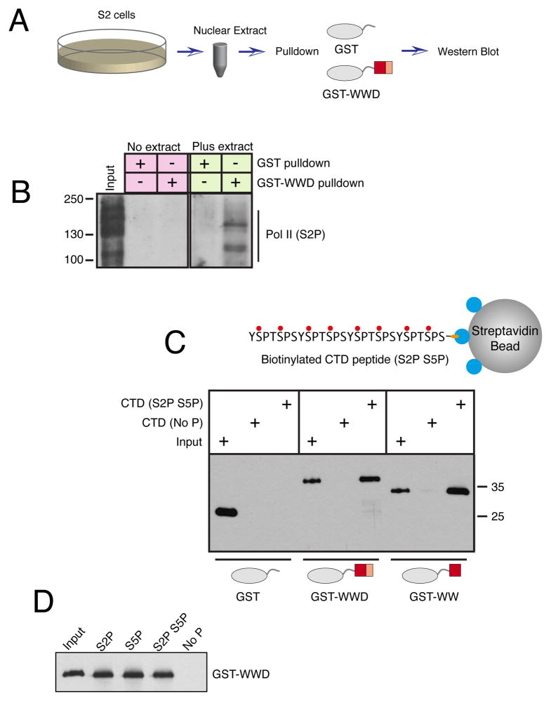
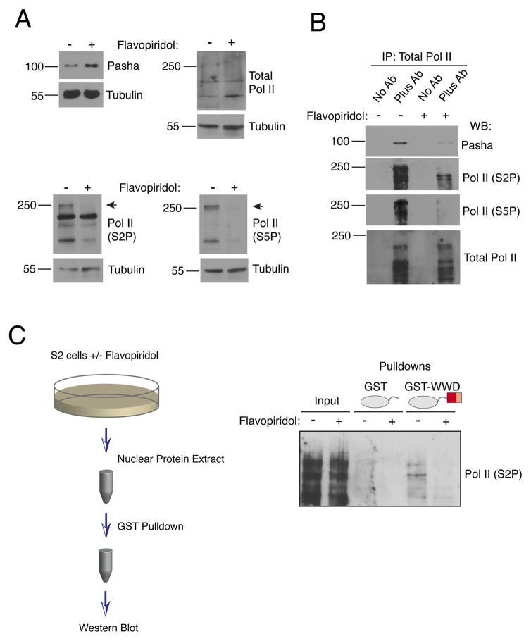
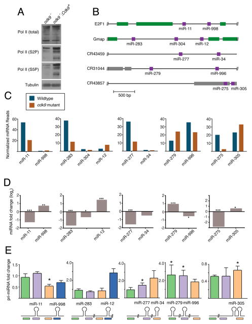
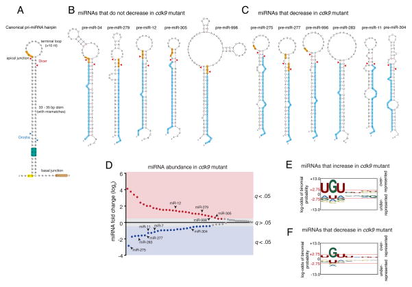
The cellular abundance of mature microRNAs (miRNAs) is dictated by the efficiency of nuclear processing of primary miRNA transcripts (pri-miRNAs) into pre-miRNA intermediates. The Microprocessor complex of Drosha and DGCR8 carries this out, but it has been unclear what controls Microprocessor's differential processing of various pri-miRNAs. Here, we show that Drosophila DGCR8 (Pasha) directly associates with the C-terminal domain of the RNA polymerase II elongation complex when it is phosphorylated by the Cdk9 kinase (pTEFb). When association is blocked by loss of Cdk9 activity, a global change in pri-miRNA processing is detected. Processing of pri-miRNAs with a UGU sequence motif in their apical junction domain increases, while processing of pri-miRNAs lacking this motif decreases. Therefore, phosphorylation of RNA polymerase II recruits Microprocessor for co-transcriptional processing of non-UGU pri-miRNAs that would otherwise be poorly processed. In contrast, UGU-positive pri-miRNAs are robustly processed by Microprocessor independent of RNA polymerase association.

Keywords: DGCR8; Drosophila; RNA polymerase II; microRNA.


Figures

Figure 1
Figure 1. The Pasha subunit of Microprocessor associates with phosphorylated Pol II
See also Figure S1. (A) Schematic structures of the Drosophila Microprocessor subunits, Drosha and Pasha. Both proteins have nuclear localization sequences (NLS). Drosha has two RNase III domains, RIIIDa and RIIIDb, and a double-strand RNA binding domain (dsRBD). Pasha has two dsRBDs, and a Rhed domain, which drives homotypic dimerization and also binds heme and pri-miRNA hairpins. The WW domain is located within a region (orange) of Rhed that is sufficient for heme binding and dimerization. (B) Model of Microprocessor structure when bound to a pri-miRNA hairpin. The cleavage site in the hairpin is marked by blue arrowheads. The model is adapted from Kim and colleagues (Kwon et al., 2016; Nguyen et al., 2015). (C) Phylogenetic tree of a subset of eukaryotic WW domains. The sub-lineage containing DGCR8 and Pin1 is highlighted. (D) Immunoprecipitation from S2 cell lysate using 4H8 antibody, which recognizes all CTD isoforms of Pol II (Brodsky et al., 2005; Schroder et al., 2013). Molecular weights of standards are shown on the right. 0.3% input was loaded for Pasha and 10% input for Pol II. (E) Immunoprecipitation from S2 cell lysate using anti-GFP to purify GFP-Pasha or GFP. Molecular weights of standards are shown on the left. 2% input was loaded for Pol II and 8% input was loaded for GFP and GFP-Pasha. (F) Immunoprecipitation from S2 cell lysate using an antibody recognizing all Pol II isoforms (4H8), in which some samples were treated with a mixture of RNases. Precipitates were probed for Pasha as shown. 0.3% input was loaded.
Figure 2
Figure 2. The WW domain of Pasha associates with the phosphorylated CTD of Pol II
See also Figure S1. (A) Scheme of assay for binding between GST proteins and phospho-Pol II present in nuclear extracts. (B) Nuclear extract was incubated with GST proteins, as indicated, and pulled down material was probed for S2-phosphorylated Pol II. Input represents 4% of the total pulldown reaction. (C) Binding between GST proteins and biotinylated peptides containing four CTD heptad repeats. Peptide-associated GST proteins were visualized by Western blot. Inputs shown represent 5% of total binding reactions. (D) Biotinylated peptides with different phospho-modifications were assayed for binding to GST-WWD. Input is 5% of the binding reactions.
Figure 3
Figure 3. Pasha binding to Pol II requires Cdk9 activity
See also Figure S1. (A) S2 cells were treated with flavopiridol for 20 hours. Whole cell extracts prepared from cells were blotted for Pasha, total Pol II, S2P-modified Pol II, S5P-modified Pol II, and tubulin. (B) Extracts analyzed in (A) were subjected to immunoprecipitation with antibody specific for all isoforms of Pol II. Immunoprecipitates were probed for Pasha, total Pol II, S2P-modified Pol II and S5P-modified Pol II. (C) S2 cells were treated with flavopiridol for 2 hours. Nuclear extracts were subjected to pulldown reactions with GST proteins, as indicated, and pulled down material was probed for S2P-modified Pol II. Inputs represent 5% of total pulldown reactions.
Figure 4
Figure 4. Cdk9 is required for differential miRNA processing
See also Figure S2. (A) Western blot of total Pol II and specific phospho-isoforms of Pol II from cdk9 mutant tissue. (B) Schematic structures of five polycistronic genes that were analyzed for miRNA expression. (C) Levels of mature miRNAs expressed from polycistronic genes. Shown are normalized RNA-seq read levels (1000s) from animals that are wildtype and cdk9 mutant. (D) Differential expression of mature miRNAs between mutant and wildtype animals as determined by RNA-seq. (*, p ≤ .01; **, p ≤ .001; ***, p ≤ .0001; edgeR exact test). (E) Differential expression of pri-miRNAs between mutant and wildtype animals as determined by RT-qPCR. Shown below are positions of the various RT-qPCR products being assayed. Error bars are standard deviations. (*, p ≤ .01; t-test)
Figure 5
Figure 5. An apical junction sequence is related to Cdk9-dependent processing
See also Figures S3 and S4. (A) Generalized structural and sequence features of pri-miRNA hairpin. Sequence motifs that affect in vitro processing specificity and efficiency are highlighted. Cleavage sites for Drosha and Dicer are shown with arrowheads. (B) Predicted pre-miRNA structures for those miRNAs whose abundance did not decrease in cdk9 mutants. Highlighted are the UGU motif (yellow) and mature miRNA (blue). (C) Predicted pre-miRNA structures for those miRNAs whose abundance decreased in cdk9 mutants. For B and C, mFold and RNAfold were independently used to predict the pre-miRNA structures. (D) Differential expression of all miRNAs detected by RNA-seq between mutant and wildtype samples. miRNAs are ranked by fold-change, and those whose fold-change is considered significant (a FDR below 5%) are colored. Those miRNAs that are derived from polycistronic genes are noted. (E,F) Logo graphs of nucleotide sequence bias within the apical junctions of pri-miRNA hairpins. Contrasted are the 30 miRNAs whose expression is greater in cdk9 mutants (E) versus the 26 miRNAs whose expression is reduced in cdk9 mutants (F). The y-axes correspond to the binomial probability of residue frequencies, with respect to the background of all Drosophila pre-miRNA sequences. Threshold values of p < 0.05 significance (2.75) are shown in red and marked with red horizontal lines.
Figure 6
Figure 6. Differential processing has a significant effect on gene silencing in vivo
See also Figure S5. (A,B) Expression of the Brd reporter gene in the developing eye. Each image contains approximately 800 cells, some of which are genetically wildtype and some of which are mutant for the cdk9 gene. Cell genotypes have been marked by the presence or absence of an RFP marker, as indicated. RFP and GFP channels are shown separately along with a merged image. Since cdk9 mutant cells in (B) also expressed the Cdk9 rescue transgene, silencing of the Brd reporter is restored within these cells. (C) When rbf mutant eye discs are stained for activated caspase protein, they show a zone of prevalent cell apoptosis within the morphogenetic furrow. (D) This zone is absent if eye cells overexpress miR-998 via the GAL4/UAS system. (E) When rbf mutant cells are also mutant for cdk9, then there is greatly reduced apoptosis. (F) A summary of the genetic experiments showing the pathways of gene regulation downstream of Cdk9. Scale bars for A–E, 10 μm.

Similar articles

Cited by

References

    1. Alarcon CR, Lee H, Goodarzi H, Halberg N, Tavazoie SF. N6-methyladenosine marks primary microRNAs for processing. Nature. 2015;519:482–485. - PMC - PubMed
    1. Auyeung VC, Ulitsky I, McGeary SE, Bartel DP. Beyond secondary structure: primary-sequence determinants license pri-miRNA hairpins for processing. Cell. 2013;152:844–858. - PMC - PubMed
    1. Bartel DP. MicroRNAs: target recognition and regulatory functions. Cell. 2009;136:215–233. - PMC - PubMed
    1. Bartkowiak B, Liu P, Phatnani HP, Fuda NJ, Cooper JJ, Price DH, Adelman K, Lis JT, Greenleaf AL. CDK12 is a transcription elongation-associated CTD kinase, the metazoan ortholog of yeast Ctk1. Genes Dev. 2010;24:2303–2316. - PMC - PubMed
    1. Brodsky AS, Meyer CA, Swinburne IA, Hall G, Keenan BJ, Liu XS, Fox EA, Silver PA. Genomic mapping of RNA polymerase II reveals sites of co-transcriptional regulation in human cells. Genome Biol. 2005;6:R64. - PMC - PubMed