. 2017 Sep 26;20(13):3176-3187.
doi: 10.1016/j.celrep.2017.09.006.

Lymphatic Vessels Balance Viral Dissemination and Immune Activation following Cutaneous Viral Infection

Affiliations

Lymphatic Vessels Balance Viral Dissemination and Immune Activation following Cutaneous Viral Infection

Christopher P Loo et al. Cell Rep. .

Abstract

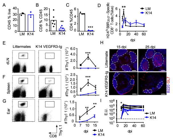
Lymphatic vessels lie at the interface between peripheral sites of pathogen entry, adaptive immunity, and the systemic host. Though the paradigm is that their open structure allows for passive flow of infectious particles from peripheral tissues to lymphoid organs, virus applied to skin by scarification does not spread to draining lymph nodes. Using cutaneous infection by scarification, we analyzed the effect of viral infection on lymphatic transport and evaluated its role at the host-pathogen interface. We found that, in the absence of lymphatic vessels, canonical lymph-node-dependent immune induction was impaired, resulting in exacerbated pathology and compensatory, systemic priming. Furthermore, lymphatic vessels decouple fluid and cellular transport in an interferon-dependent manner, leading to viral sequestration while maintaining dendritic cell transport for immune induction. In conclusion, we found that lymphatic vessels balance immune activation and viral dissemination and act as an "innate-like" component of tissue host viral defense.

Keywords: cutaneous infection; dissemination; lymphatic vessels; tissue immunity; tissue microenvironment; type I interferons; vaccinia virus.


Figures

Figure 1
Figure 1. Infection by scarification induces lymphatic vessel remodeling but not lymphangiogenesis
C57Bl/6 mice infected by skin scarification with 2x10⁶ PFU VACV. (A) Representative images of lymphatic vessels (LYVE1) (Scale bar=200μm) and quantification of (B) vessels per length of skin and (C) vessel distension (width/length). (D) Representative images of blood vessels (CD31) (Scale bar=200μm) and (E) quantification of vessels per length of skin. (F) Whole mount images (maximal projection) of skin (scale bar=50μm; LYVE1). (G) Distribution of lymphatic vessel diameters in skin (n=4; >10 measurements per image). (H) Whole mount images (maximal projection) of lymphatic capillary blunt (scale bar=10μm; LYVE1). (I) VEGF-A (pg per ear). (J and K) Mice were infected and pulsed with BrdU (day 0–7) and percent incorporation in (J) blood endothelial cells (BECs;CD45CD31+gp38) and (K) lymphatic endothelial cells (LECs; CD45CD31+gp38+). Naïve (N) and sterile infected (M) controls. (Each point represents an individual animal). (L) Images of VACV dLNs. (Top) Whole lymph nodes and (Bottom) inset of subcapsular and follicular zones. Lymphatic vessels (LYVE1, green), T cells (CD3ε, red), and B cells (B220, blue). (Scale bar = 200μm.) Statistical significance determined by one-way ANOVA. *p<0.05, **p<0.01, ***p<0.001. See also Figure S1.
Figure 2
Figure 2. Viral persistence and exacerbated pathology in the absence of lymphatic vessels
K14 VEGFR3-Ig (K14) mice and littermate controls (LM) were infected with VACV by skin scarification. (A) Virus quantified in infected skin (plaque forming units; PFU). (B) Quantification of ear thickness (n>10). (C) Mile’s Assay for vascular permeability ten days post infection (dpi) in LM and K14 mice. (D) Tissue histology (representative images; left) and quantification (right) in infected skin 10 dpi. Total leukocytes (CD45), neutrophils (Ly6G), B cells (B220), and degranulating mast cells (toluidine blue). Statistics determined by students unpaired t-test or Fisher’s exact test. *p<0.05, **p<0.01, ***p<0.001. Each point represents an individual mouse.
Figure 3
Figure 3. Delayed anti-viral adaptive immune responses in mice lacking dermal lymphatic vessels following scarification
K14 VEGFR3-Ig (K14) mice or littermate controls (LM) were infected by skin scarification with VACV-GP33 (n=4). Quantification of (A) total leukocytes (CD45+ %live) and (B) CD8+ and (C) CD4+ T cells (%CD45) in skin 7 days post infection (dpi). (D) Endogenous H2-Kd B8R20-27-specific CD8+ T cells in blood. (E and F) 25K TCR-tg P14 Thy1.1+CD8+ T cells transferred in K14 or LM mice one day prior to infection. (E–G) Representative flow plots of Thy1.1+CD8+ P14 T cells 7 dpi (left; gated on live lymphocytes) and (right) absolute cell numbers on 7, 10 and 15 dpi in (E) draining lymph nodes (dLN), (F) spleen, and (G) infected skin (n≥4). (H) DLNs from K14 VEGFR3-Ig mice and littermate controls imaged by immunofluorescence at 15 and 25 dpi (Scale bar = 200μm, B220, blue; GL7, red). (I) Serum VACV-specific IgG from K14 VEGFR3-Ig mice and littermate controls (Each line is an animal). Dotted line indicates limit of detection. Statistics determined by unpaired students t-test and two-way ANOVA at day 10. *p<0.05, **p<0.01, ***p<0.001. See also Figure S2 and 3.
Figure 4
Figure 4. Lymph node independent adaptive immune priming in the absence of dermal lymphatic drainage
Either wildtype (WT) or CD62L knockout (LKO) TCR-tg P14 CD8+ T cells transferred into K14 VEGFR3-Ig (K14) mice or littermate (LM) controls one day prior to infection by scarification with VACV-GP33. (A) Representative plots of circulating Thy1.1+ P14 T cells 10 days post infection (dpi) and (B) quantification in blood (Thy1.1+/live) (n≥4). (C) Spleens from K14 and LM mice evaluated 10 days dpi for reactive germinal centers (Scale bar = 200μm, B220, blue; GL7, green; CD33, red). (D) Acquisition of a central memory phenotype (CD44+CD62L+KLRG1) by circulating H2-Kd B8R20-27-specific endogenous CD8+ T cells in K14 and LM mice. (E–I) LCMV immune mice were infected with VACV-GP33. (E) Number of splenic and (F) lymph node (LN) TCR-tg P14 CD8+ T cells. Number of (G) total endogenous CD8+ T cells or (H) TCR-tg P14 CD8+ T cells in skin 3 dpi. (I) Plaque forming units (PFU) from skin 3 dpi of naïve or LCMV-immune K14 and LM. Data from representative experiment. Statistics determined by unpaired students t-test or one-way ANOVA. *p<0.05, **p<0.01, ***p<0.001. Each point indicates an individual animal. See also Figure S4.
Figure 5
Figure 5. Cutaneous viral infection reduces lymphatic fluid transport
C57Bl/6 mice were infected by skin scarification with VACV. (A) Fluorescence microlymphangiography (155kDa FITC dextran) following scarification (Scale bar=500μm). Arrows indicate draining lymphatic vessels. (B) Quantification of lymphatic drainage following intradermal injection of Evan’s Blue and quantification in draining lymph nodes (dLN). (C) Miles’ assay for vascular leakiness. (D–I) Intradermal injections of 100ng (1μl) VEGF- C(Cys156Ser) daily starting on the first day of scarification with VACV. (D) Quantification of virus in skin 5 days post infection (dpi) (plaque forming units; PFU). (E) Measurement of ear thickness (n=6). (F) Representative images (left) and quantification (right) of cutaneous lymphatic vessels (purple; LYVE1) following intradermal VEGF-C or PBS (Scale bar=200μm). (I) PFU in dLNs 5 dpi. Statistics determined by one-way ANOVA or Fisher’s exact test. *p<0.05, **p<0.01, ***p<0.001. See also Figure S5.
Figure 6
Figure 6. Lymphatic endothelial cells are activated following infection and allow for dendritic cell migration to draining lymph nodes
Wildtype C57BL/6 mice were infected by cutaneous scarification with VACV-GP33. (A) Representative flow cytometry (gated on CD45CD31+) of cutaneous endothelial cells at 0, 3 and 7 days post infection. (B) Quantification of activation markers on blood endothelial cells (BEC; CD45CD31+gp38) and lymphatic endothelial cells (LEC;CD45CD31+gp38+) (n=6). (C) Quantification of CCL21 in draining lymph nodes (dLN) (n=4). (D) Quantification of migratory DCs present in dLNs following cutaneous VACV infection at the indicated time points or 24 hours following administration of dibutyl phthalate (DBP). (E) Quantification of FITC+ migratory DCs present in dLNs. 5% FITC dissolved in DMSO:Acetone painted on skin 24 hours prior to each time point. FITC dissolved in the skin irritant dibutyl phthalate (DBP):acetone used as a positive control. Statistics determined by students unpaired t test. *p<0.05, **p<0.01, ***p<0.001. Each point is an individual mouse.
Figure 7
Figure 7. Type I IFN signaling regulates lymphatic remodeling, transport, and viral spread following cutaneous infection
Antibodies blocking the interferon-alpha/beta receptor alpha chain (i.p. 500μg; MAR1-5A3) or isotype control were administered 0 and 2 days post infection (dpi). (A) Quantification of virus in skin 5 dpi (plaque forming unit; PFU). (B) Representative images of lymphatic vessels in infected skin with MAR1-5A3 (αIFNAR1) or isotype control (LYVE1; Scale bar = 200μm). (C) Lymphatic vessel density (vessels per mm length) and (D) lymphatic vessel distension (width/length) (n=7). (E) Whole mount images (maximal projection) of skin 5 dpi (scale bar=50μm; LYVE1, green). (F) Evan’s blue lymphangiography (3 dpi). Representative images and (G) incidence of drainage. (H) Quantification of Evan’s blue dye in draining lymph nodes (dLN) following intradermal injection (2 dpi). (I) PFU in dLNs (5 dpi). Dotted line indicates limit of detection. (J) Fluorescent imaging of subcapsular GFP in draining lymph nodes 5 dpi with VACV-GFP. (LYVE1, red; CD169, purple; GFP, green; scale bar = 200μm). Statistical significance determined by students unpaired t test or Fishers exact test. *p<0.05, **p<0.01, ***p<0.001. Each point represents an individual animal.

References

    1. Allan RS, Waithman J, Bedoui S, Jones CM, Villadangos JA, Zhan Y, Lew AM, Shortman K, Heath WR, Carbone FR. Migratory Dendritic Cells Transfer Antigen to a Lymph Node-Resident Dendritic Cell Population for Efficient CTL Priming. Immunity. 2006;25:153–162. - PubMed
    1. Baluk P, Tammela T, Ator E, Lyubynska N, Achen MG, Hicklin DJ, Jeltsch M, Petrova TV, Pytowski B, Stacker SA, et al. Pathogenesis of persistent lymphatic vessel hyperplasia in chronic airway inflammation. J Clin Invest. 2005;115:247–257. - PMC - PubMed
    1. Baluk P, Fuxe J, Hashizume H, Romano T, Lashnits E, Butz S, Vestweber D, Corada M, Molendini C, Dejana E, et al. Functionally specialized junctions between endothelial cells of lymphatic vessels. J Exp Med. 2007;204:2349–2362. - PMC - PubMed
    1. Bedoui S, Whitney PG, Waithman J, Eidsmo L, Wakim L, Caminschi I, Allan RS, Wojtasiak M, Shortman K, Carbone FR, et al. Cross-presentation of viral and self antigens by skin-derived CD103+ dendritic cells. Nat Immunol. 2009;10:488–495. - PubMed
    1. Bruehl RE, Bertozzi CR, Rosen SD. Minimal sulfated carbohydrates for recognition by L-selectin and the MECA-79 antibody. J Biol Chem. 2000;275:32642–32648. - PubMed