. 2017 Sep 26;22(10):1618.
doi: 10.3390/molecules22101618.

A Fluorescent Coumarin-Based Probe for the Fast Detection of Cysteine with Live Cell Application

Affiliations

A Fluorescent Coumarin-Based Probe for the Fast Detection of Cysteine with Live Cell Application

Rui-Feng Zeng et al. Molecules. .

Abstract

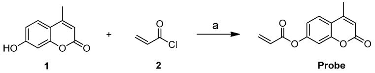
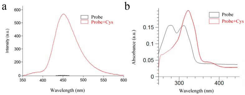
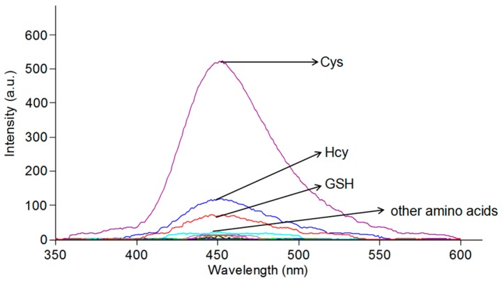
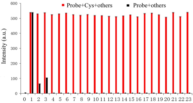
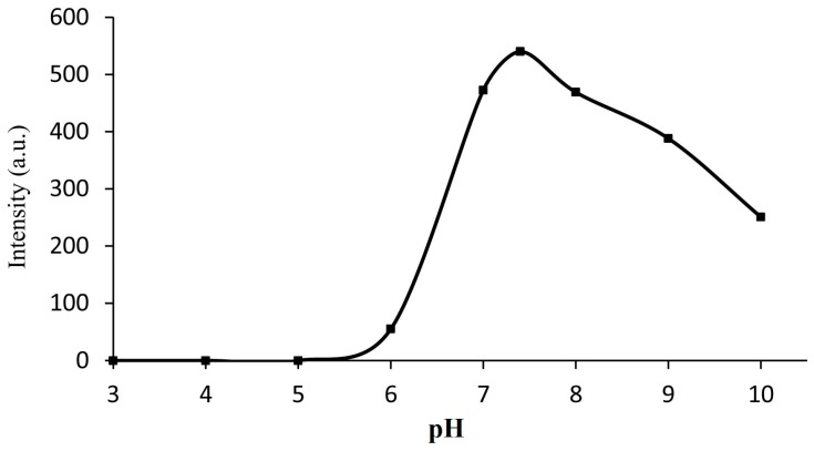
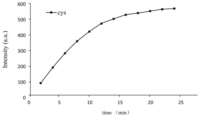
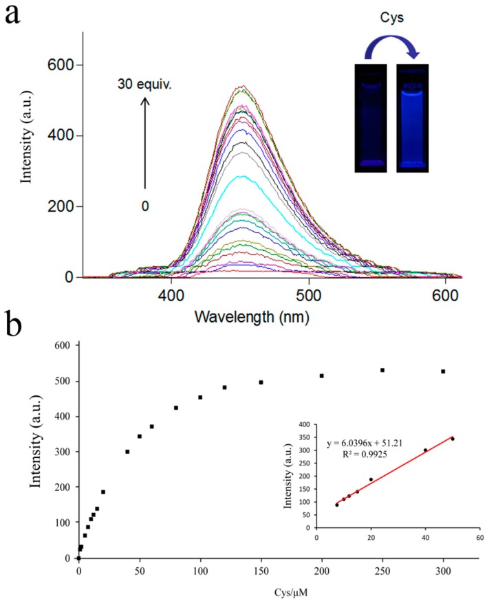
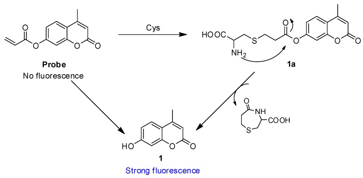
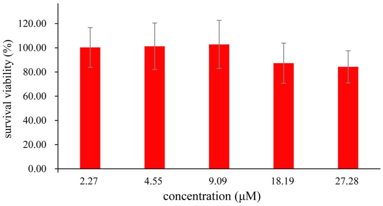
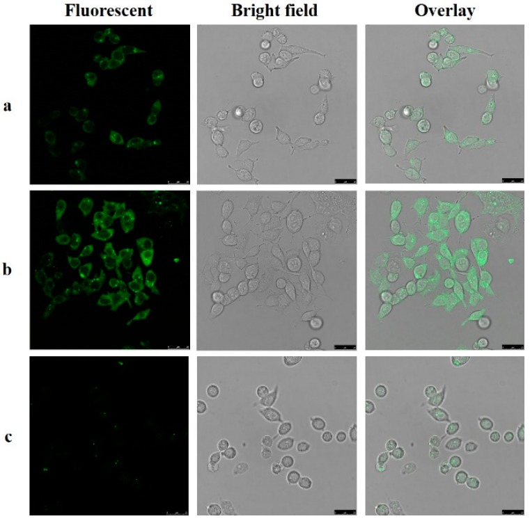
A new coumarin-based fluorescent probe, containing an allylic esters group, has been designed and synthesized for sensing cysteine in physiological pH. In this fluorescent probe, the coumarin was applied as the fluorophore and an allylic esters group was combined as both a fluorescence quencher and a recognition unit. The probe can selectively and sensitively detect cysteine (Cys) over homocysteine, glutathione, and other amino acids, and has a rapid response time of 30 min and a low detection limit of 47.7 nM. In addition, the probe could be applied for cell imaging with low cytotoxicity.

Keywords: bioimaging; coumarin; cysteine; fluorescent probe.


Conflict of interest statement

The authors declare no conflict of interest.

Figures

Scheme 1
Scheme 1
Synthesis of the probe. Reagents and conditions: (a) trimethylamine, CH2Cl2, r.t.
Figure 1
Figure 1
(a) The fluorescence intensity of the probe and the probe added to Cys-buffered solution (PBS (phosphate buffered solution):DMSO (dimethyl sulfoxide) = 6:4, pH = 7.4) at room temperature; (b) The absorption spectra of the probe and the probe added to Cys in buffered solution (PBS:DMSO = 6:4, pH = 7.4) at room temperature. λex = 325 nm.
Figure 2
Figure 2
Fluorescence intensity of the probe (10 μM) in the presence of different compounds (50 equiv.); each reaction was in buffered solution (PBS:DMSO = 6:4, pH = 7.4) at room temperature. λex = 325 nm.
Figure 3
Figure 3
Black bar represents the fluorescence response of the probe (10 μM) to various compounds (50 equiv.). Red bar represents the fluorescence response of the probe (10 μM) to Cys (50 equiv.) in the presence of other compounds (50 equiv.). Each reaction was in buffered solution (PBS:DMSO = 6:4, pH = 7.4) at room temperature. The numbers represent analytes: 0. Blank; 1. Cys; 2. Hcy; 3. GSH; 4. Asp; 5. Val; 6. Glu; 7. Pro; 8. Gly; 9. Phe; 10. Met; 11. Thr; 12. Ser; 13. Ile; 14. His; 15. Phe; 16. Lys; 17. Try; 18. Leu; 19. Cys C; 20. CN; 21. SCN; 22. HS; 23. SO42−. λex = 325 nm, λem = 450 nm.
Figure 4
Figure 4
The effect of pH on the fluorescence response of the probe (10 μM) in the presence of Cys (500 μM) in buffered solution (PBS:DMSO = 6:4, pH = 3–10), pH was adjusted by NaOH and HCl. λex = 325 nm, λem = 450 nm.
Figure 5
Figure 5
Kinetic analysis of the probe towards Cys (10 μM probe with 50 equiv. of Cys) in buffer solution (DMSO:PBS = 4:6, pH = 7.4). λex = 325 nm, λem = 450 nm.
Figure 6
Figure 6
(a) Fluorescent spectral changes (b) The fluorescence intensity at 450 nm of probe (10 μM) upon addition of Cys (0–300 μM) in buffer solution (DMSO:PBS = 4:6, PH 7.4), incubated 30 min before detected. λex = 325 nm, λem = 450 nm.
Scheme 2
Scheme 2
Proposed reaction mechanism of the probe with Cys.
Figure 7
Figure 7
MTS assay of HepG2 cells incubated in the presence of the probe (0–27 μM) at 37 °C for 24 h.
Figure 8
Figure 8
Fluorescent imaging of HepG2 cells: (Left) fluorescent image; (Middle) bright field image; and (Right) overlay image. λex = 405 nm, λem = 420–480 nm. (a) Images of cells incubated with the probe (25 μM) for 2 h at 37 °C; (b) Images of cells preincubated with Cys (100 μM) for 1 h and then incubated with the probe (25 μM) for 2 h at 37 °C; (c) Images of cells preincubated with N-ethylmaleimide (NEM) (500 μM) for 1 h and then incubated with the probe (25 μM) for 2 h at 37 °C.

References

    1. Bragoszewski P., Wasilewski M., Sakowska P., Gornicka A., Bottinger L., Qiu J., Wiedemann N., Chacinska A. Retro-translocation of mitochondrial intermembrane space proteins. Proc. Natl. Acad. Sci. USA. 2015;112:7713–7718. doi: 10.1073/pnas.1504615112. - DOI - PMC - PubMed
    1. Abby S.L., Harris I.M., Harris K.M. Homocysteine and cardiovascular disease. Curr. Atheroscler. Rep. 2004;6:101–106. doi: 10.3122/15572625-11-5-391. - DOI - PubMed
    1. Chen X., Zhou Y., Peng X., Yoon J. Fluorescent and colorimetric probes for detection of thiols. Chem. Soc. Rev. 2010;39:2120–2135. doi: 10.1039/b925092a. - DOI - PubMed
    1. Hirai T., Kanda T., Sato K., Takaishi M., Nakajima K., Yamamoto M., Kamijima R., Digiovanni J., Sano S. Cathepsin K is involved in development of psoriasis-like skin lesions through TLR-dependent Th17 activation. J. Immunol. 2013;190:4805–4811. doi: 10.4049/jimmunol.1200901. - DOI - PubMed
    1. Shahrokhian S. Lead phthalocyanine as a selective carrier for preparation of a cysteine-selective electrode. Anal. Chem. 2001;73:5972–5978. doi: 10.1021/ac010541m. - DOI - PubMed