. 2017 Sep 26;5(4):31.
doi: 10.3390/vaccines5040031.

Newcastle Disease Virus Vectored Bivalent Vaccine against Virulent Infectious Bursal Disease and Newcastle Disease of Chickens

Affiliations

Newcastle Disease Virus Vectored Bivalent Vaccine against Virulent Infectious Bursal Disease and Newcastle Disease of Chickens

Sohini Dey et al. Vaccines (Basel). .

Abstract

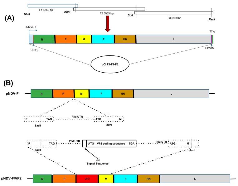
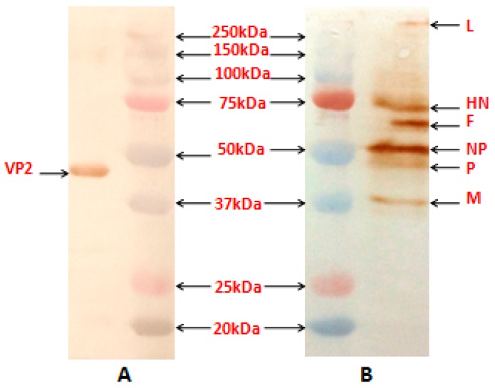
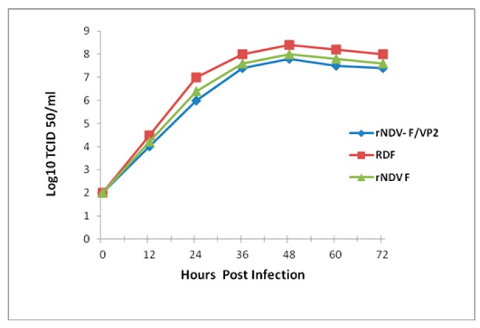
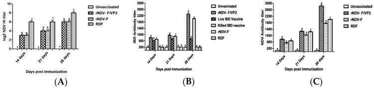
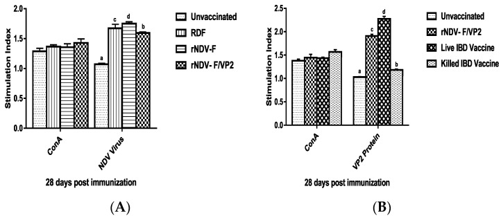
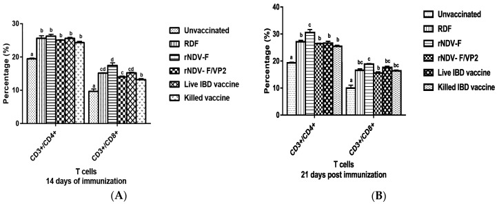
Newcastle disease virus (NDV) strain F is a lentogenic vaccine strain used for primary vaccination in day-old chickens against Newcastle disease (ND) in India and Southeast Asian countries. Recombinant NDV-F virus and another recombinant NDV harboring the major capsid protein VP2 gene of a very virulent infectious bursal disease virus (IBDV); namely rNDV-F and rNDV-F/VP2, respectively, were generated using the NDV F strain. The rNDV-F/VP2 virus was slightly attenuated, as compared to the rNDV-F virus, as evidenced from the mean death time and intracerebral pathogenicity index analysis. This result indicates that rNDV-F/VP2 behaves as a lentogenic virus and it is stable even after 10 serial passages in embryonated chicken eggs. When chickens were vaccinated with the rNDV F/VP2, it induced both humoral and cell mediated immunity, and was able to confer complete protection against very virulent IBDV challenge and 80% protection against virulent NDV challenge. These results suggest that rNDV-F could be an effective and inherently safe vaccine vector. Here, we demonstrate that a bivalent NDV-IBDV vaccine candidate generated by reverse genetics method is safe, efficacious and cost-effective, which will greatly aid the poultry industry in developing countries.

Keywords: IBDV-VP2 protein; Newcastle disease virus vector; bivalent vaccine candidate; humoral and cell mediated immune responses; protection efficacy.


Conflict of interest statement

The authors declare that they have no conflict of interest.

Figures

Figure 1
Figure 1
Generation of full-length clone of strain F with and without VP2 gene cassette. (A) Schematic representation of the construction of full-length clone pCI F1-F2-F3 of Newcastle disease virus (NDV) strain F. Assembly was carried out by sequential cloning of F1, F2 and F3 fragments into pCI vector. Fragment F1 contains hammerhead ribozyme (HHRz) at the 5′-end and fragment F3 contains hepatitis delta ribozyme (HdvRz) at the 3′-end. Restriction sites used for cloning are indicated. Abbreviations: CMV/T7, cytomegalovirus immediate-early enhancer and promoter/T7 RNA polymerase promoter; T7ɸ, T7 transcription termination. (B) Schematic representation of the NDV genome with the SacII and AvrII sites between the P and M genes and insertion of VP2 gene with HN signal between these sites.
Figure 2
Figure 2
Western blot analysis of the proteins produced by purified recombinant NDV containing VP2 gene of infectious bursal disease virus (IBDV). Purified virus samples were separated on 12.5% sodium dodecyl sulfate polyacrylamide gel electrophoresis (SDS-PAGE). Proteins were then transferred to nitrocellulose membrane and blocked in 1% BSA in TBST. Membranes were incubated with either mouse anti-VP2 monoclonal antibody or chicken anti-NDV polyclonal antibody at 4 °C for 1 h. Membranes were washed in TBST and then incubated with secondary antibody conjugated with HRP in 1% BSA in TBST for 1 h at room temperature. Membranes were washed in TBST and incubated with Sigma Fast DAB (Sigma, USA) color development reagent for 15 min and then visualized. (A) The position of the VP2 protein of about 48-kDa (right) and marker proteins (Precision Plus Protein Standards, Bio-Rad, USA) are as indicated. The blot was probed with anti-mouse IBDV-VP2 monoclonal antibody (Abcam, USA). (B) The positions of the NDV L, HN, F, NP, P and M (right) and marker proteins are as indicated. The blot was probed with anti-chicken NDV polyclonal antibody.
Figure 3
Figure 3
Multicycle growth kinetics of parent NDV strain F and recombinant viruses rNDV-F/VP2 and rNDVF in Vero cells. Monolayer of Vero cells were infected with each of the viruses at a multiplicity of infection (MOI) of 0.01 and supernatant collected and replaced with an equal volume of fresh medium at 12 h interval until 72 h, and viral titers were determined by limiting dilution assay and calculated as TCID50 by Reed and Muench method.
Figure 4
Figure 4
Immunofluorescence analysis of VP2 protein expression in rNDV-F/VP2 virus. Immunofluorescence analysis of VP2 protein expression in rNDV-F/VP2 virus. Confluent monolayers of Vero cells were infected with recombinant viruses rNDV-F/VP2 and rNDV-F at a multiplicity of infection (MOI) of 0.01. The cells were examined by immunofluorescence assay (IFA) with IBDV specific mAb against the VP2 protein (Abcam, USA) and an NDV-specific polyclonal antibody (Abcam, USA), followed by a mixture of the FITC labeled sheep anti-mouse IgG (B,F) and Alexa Fluor 568-labeled goat anti-chicken IgG (C,G). Fluorescence was monitored and photographed using a confocal microscope (Olympus FV 10, USA) at a magnification of 60× under UV light of matching excitation filters for FITC and Alexa Fluor 568 respectively.
Figure 5
Figure 5
NDV and IBDV specific serum antibody responses in the experimental chickens. (A) HI assay was carried out 14 and 28 days post-immunization. All HI titers are expressed as mean reciprocal log2 titre ± SEM (standard error of the mean) (n = 10). Statistical differences were calculated by one-way ANOVA with p < 0.01 and Waller-Duncan as post-hoc test. The antibody titers as determined on 14, 21 and 28 days post-immunization by ELISA, (B) higher than 500 were considered positive for IBDV antibody and (C) higher than 200 were considered positive for NDV. Data represent the mean ± standard error. Statistical analysis was done by one-way ANOVA (p < 0.01 and Waller-Duncan as post-hoc test. Level not connected by same letter are significantly different (p < 0.01).
Figure 6
Figure 6
Antigen specific lymphocyte proliferative response in chickens on 28 days post immunization. Chicken PBMCs from immunized and control groups (n = 6) were stimulated with NDV virus (A), recombinant VP2 protein expressed in Saccharomyces cerevisiae (B), and concanavalin A (conA) as positive control. Lymphocyte proliferative response was measured and expressed as stimulation index. All the data presented as mean value ± standard error and Waller-Duncan as post-hoc test. Level not connected by same letter are significantly different (p < 0.01).
Figure 7
Figure 7
Flow cytometric analysis of CD4+ and CD8+ T cells in peripheral blood mono-nuclear cells (PBMCs) of vaccinated and unvaccinated birds at 14 (A) and 21 (B) dpi. PBMCs were collected from the birds and 1 × 10⁵ cells were analyzed after addition of chicken specific Mabs conjugated with fluorescent dyes. Data represent the percentage of cells ± SEM. The values depicted in different lowercase superscripts differ significantly (p < 0.01) and Waller-Duncan as post-hoc test.

Similar articles

Cited by

References

    1. Alexander D.J., Aldous E.W., Fuller C.M. The long view: A selective review of 40 years of Newcastle disease research. Avian Pathol. 2012;41:329–335. doi: 10.1080/03079457.2012.697991. - DOI - PubMed
    1. Van den Berg T.P., Eterradossi N., Toquin D., Meulemans G. Infectious bursal disease (Gumboro disease) Rev. Sci. Tech. 2000;19:509–543. doi: 10.20506/rst.19.2.1227. - DOI - PubMed
    1. Delmas B., Mundt E., Vakharia V.N., Wu J.L. Family Birnaviridae. In: Adams M.J., Carstens E.B., Lefkowitz E.J., editors. King AMQ. Academic Press; London, UK: 2012. pp. 499–507. Virus Taxonomy: Ninth Report of the International Committee on Taxonomy of Viruses.
    1. Sanchez A.B., Rodriguez J.F. Proteolytic processing in infectious bursal disease virus: Identification of the polyprotein cleavage sites by site directed mutagenesis. Virology. 1999;262:190–199. doi: 10.1006/viro.1999.9910. - DOI - PubMed
    1. Fahey K.J., Erny K., Crooks J. A conformational immunogen on VP-2 of infectious bursal disease virus that induces virus-neutralizing antibodies that passively protect chickens. J. Gen. Virol. 1989;70:1473–1481. doi: 10.1099/0022-1317-70-6-1473. - DOI - PubMed

LinkOut - more resources