Review
. 2017 Dec;52(6):696-714.
doi: 10.1080/10409238.2017.1380597. Epub 2017 Sep 28.

Functions of SMARCAL1, ZRANB3, and HLTF in maintaining genome stability

Affiliations
Review

Functions of SMARCAL1, ZRANB3, and HLTF in maintaining genome stability

Lisa A Poole et al. Crit Rev Biochem Mol Biol. 2017 Dec.

Abstract

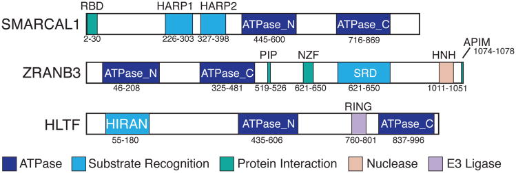
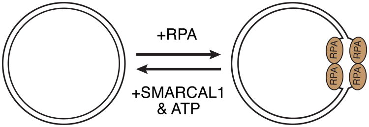
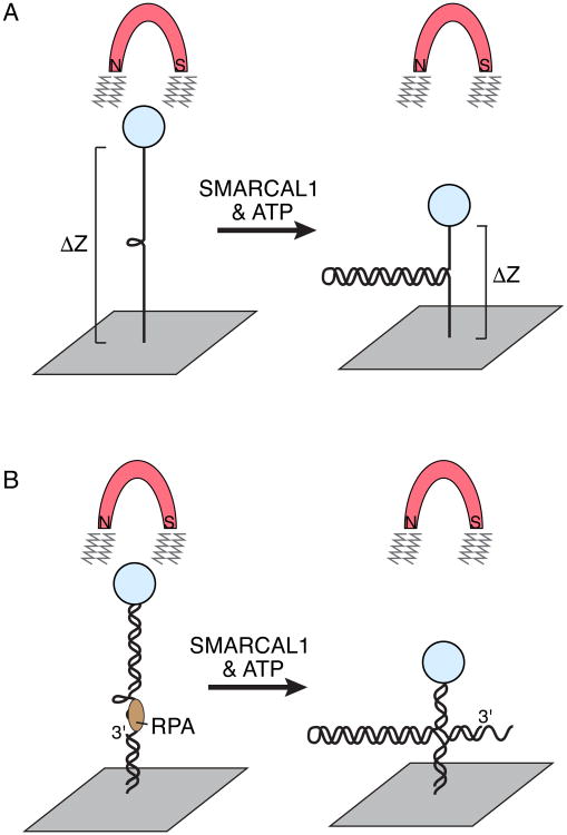
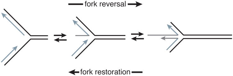
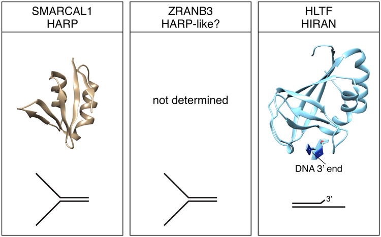
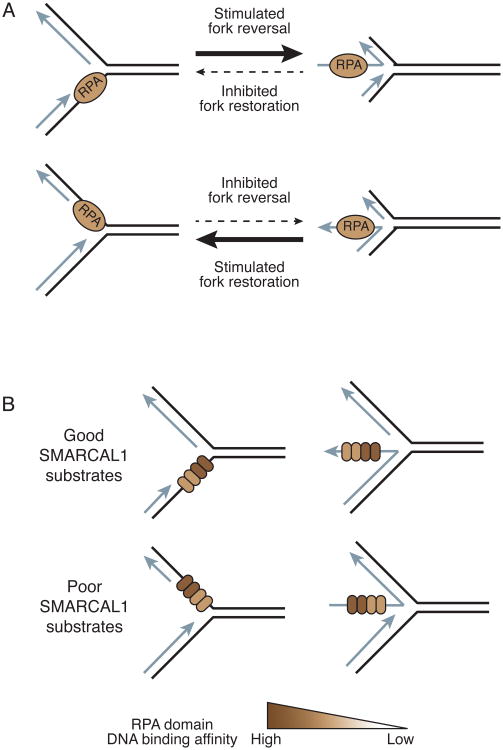
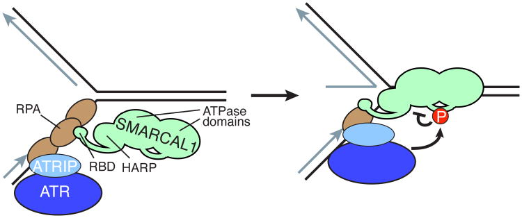
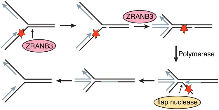
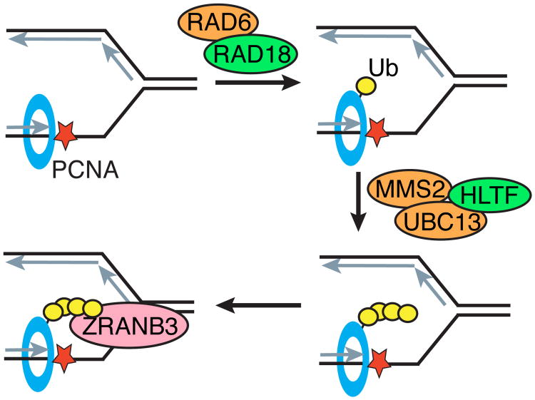
A large number of SNF2 family, DNA and ATP-dependent motor proteins are needed during transcription, DNA replication, and DNA repair to manipulate protein-DNA interactions and change DNA structure. SMARCAL1, ZRANB3, and HLTF are three related members of this family with specialized functions that maintain genome stability during DNA replication. These proteins are recruited to replication forks through protein-protein interactions and bind DNA using both their motor and substrate recognition domains (SRDs). The SRD provides specificity to DNA structures like forks and junctions and confers DNA remodeling activity to the motor domains. Remodeling reactions include fork reversal and branch migration to promote fork stabilization, template switching, and repair. Regulation ensures these powerful activities remain controlled and restricted to damaged replication forks. Inherited mutations in SMARCAL1 cause a severe developmental disorder and mutations in ZRANB3 and HLTF are linked to cancer illustrating the importance of these enzymes in ensuring complete and accurate DNA replication. In this review, we examine how these proteins function, concentrating on their common and unique attributes and regulatory mechanisms.

Keywords: DNA repair; Fork reversal; PCNA; RPA; checkpoint; replication stress.


Figures

Figure 1
Figure 1
Domain structures of SMARCAL1, ZRANB3, and HLTF. The ATPase, substrate recognition, protein interaction, and other enzymatic domains are depicted. RBD, RPA binding domain; SRD, substrate recognition domain.
Figure 2
Figure 2
Annealing helicase activity assay. At high concentrations, RPA will induce and stabilize a single-stranded DNA bubble in supercoiled plasmid DNA. SMARCAL1 uses the energy of ATP hydrolysis to re-anneal the complementary DNA strands and displace RPA.
Figure 3
Figure 3
Single molecule magnetic tweezer experiment to monitor SMARCAL1 annealing and fork reversal activities. (A) A 1.2kb DNA hairpin substrate is attached to a glass slide on one end and a magnetic bead on the other. Application of a magnetic field will stretch the DNA and unwind the duplex except for the last 20-30 base pairs because of their high GC content. MARCAL1 catalyzes re-annealing of the duplex DNA against the applied force, which is measured as a change in the distance of the bead from the glass slide. This experimental setup revealed that a single molecule of SMARCAL1 catalyzes bursts of repetitive annealing. (B) Addition of oligonucleotides to the stretched DNA allows the creation of substrates that mimic replication forks with ssDNA gaps on either the leading strand (depicted) or lagging template strands. RPA can be added to bind the ssDNA. This experimental set up revealed that RPA increases the distance SMARCAL1 moves per annealing event when it is bound to the leading template strand.
Figure 4
Figure 4
Diagram depicting fork reversal and fork restoration. Fork reversal anneals the parental (black) and nascent (silver) DNA strands in a concerted reaction generating a chicken foot structure. Migrating the four-way junction in the opposite direction yields fork restoration. A ssDNA gap is diagramed on the leading strand template in this example.
Figure 5
Figure 5
Substrate recognition domains of SMARCAL1, ZRANB3, and HLTF. Structures of the SMARCAL1 HARP and HLTF HIRAN domains were derived from PDB 4S0N and 4XZF respectively. The DNA binding specificity of the domains is illustrated. The structure of the ZRANB3 SRD domain has not been determined.
Figure 6
Figure 6
The orientation of RPA binding to the replication fork substrate differentially regulates SMARCAL1. (A) Fork reversal is stimulated when RPA is bound to the leading strand template and is inhibited when it is bound to the lagging strand template. Fork restoration is activated when RPA is bound to a longer nascent leading strand and inhibited when it is bound to the nascent lagging strand. (B) RPA binds asymmetrically to ssDNA using four DNA binding domains with varying affinities. RPA stimulates SMARCAL1 when the two highest affinity DNA binding domains are located next to the fork junction and inhibits SMARCAL1 when the low affinity binding domains are located next to the junction.
Figure 7
Figure 7
ATR inhibits SMARCAL1. ATRIP binding to the 70N domain of RPA recruits ATR to replication forks. The SMARCAL1 RBD binds RPA32C to bring SMARCAL1 to replication forks. Once SMARCAL1 binds to the forked DNA via its ATPase and HARP domains, it becomes a substrate for ATR phosphorylation. Phosphorylation within the linker between the ATPase domains inhibits SMARCAL1 activity.
Figure 8
Figure 8
Model for how ZRANB3 nuclease and fork remodeling activities could be coordinated to repair a leading strand template lesion (red star). ZRANB3 endonuclease cuts two nucleotides into the parental duplex. Fork reversal could then stabilize the fork and allow for strand displacement DNA synthesis. A flap endonuclease could then remove the damaged DNA and permit fork restoration to restart the fork. This model is adapted from Weston et al., Genes and Development (Weston et al., 2012).
Figure 9
Figure 9
Model for cooperativity between HLTF and ZRANB3. PCNA is monoubiquitinated by the RAD6/RAD18 complex after the replication fork stalls. Polyubiquitin chains are added by MMS2/UBC13 with the ubiquitin ligase HLTF. ZRANB3 then binds polyubiquitinated PCNA and remodels or cleaves the stalled replication fork. Ub, ubiquitin.

References

    1. Achar YJ, Balogh D, Haracska L. Coordinated protein and DNA remodeling by human HLTF on stalled replication fork. Proc Natl Acad Sci U S A. 2011;108:14073–8. - PMC - PubMed
    1. Achar YJ, Balogh D, Neculai D, Juhasz S, Morocz M, Gali H, Dhe-Paganon S, Venclovas C, Haracska L. Human HLTF mediates postreplication repair by its HIRAN domain-dependent replication fork remodelling. Nucleic Acids Res. 2015;43:10277–91. - PMC - PubMed
    1. Badu-Nkansah A, Mason AC, Eichman BF, Cortez D. Identification of a Substrate Recognition Domain in the Replication Stress Response Protein Zinc Finger Ran-binding Domain Containing Protein 3 (ZRANB3) J Biol Chem 2016 - PMC - PubMed
    1. Bansbach CE, Betous R, Lovejoy CA, Glick GG, Cortez D. The annealing helicase SMARCAL1 maintains genome integrity at stalled replication forks. Genes Dev. 2009;23:2405–14. - PMC - PubMed
    1. Bansbach CE, Boerkoel CF, Cortez D. SMARCAL1 and replication stress: an explanation for SIOD? Nucleus. 2010;1:245–8. - PMC - PubMed

Publication types

LinkOut - more resources