. 2017 Nov 1;37(44):10554-10566.
doi: 10.1523/JNEUROSCI.1313-17.2017. Epub 2017 Sep 27.

The Long 3'UTR mRNA of CaMKII Is Essential for Translation-Dependent Plasticity of Spontaneous Release in Drosophila melanogaster

Affiliations

The Long 3'UTR mRNA of CaMKII Is Essential for Translation-Dependent Plasticity of Spontaneous Release in Drosophila melanogaster

Elena A Kuklin et al. J Neurosci. .

Abstract

A null mutation of the Drosophila calcium/calmodulin-dependent protein kinase II gene (CaMKII) was generated using homologous recombination. Null animals survive to larval and pupal stages due to a large maternal contribution of CaMKII mRNA, which consists of a short 3'-untranslated region (UTR) form lacking regulatory elements that guide local translation. The selective loss of the long 3'UTR mRNA in CaMKII-null larvae allows us to test its role in plasticity. Development and evoked function of the larval neuromuscular junction are surprisingly normal, but the resting rate of miniature excitatory junctional potentials (mEJPs) is significantly lower in CaMKII mutants. Mutants also lack the ability to increase mEJP rate in response to spaced depolarization, a type of activity-dependent plasticity shown to require both transcription and translation. Consistent with this, overexpression of miR-289 in wild-type animals blocks plasticity of spontaneous release. In addition to the defects in regulation of mEJP rate, CaMKII protein is largely lost from synapses in the mutant. All phenotypes are non-sex-specific and rescued by a fosmid containing the entire wild-type CaMKII locus, but only viability and CaMKII localization are rescued by genomic fosmids lacking the long 3'UTR. This suggests that synaptic CaMKII accumulates by two distinct mechanisms: local synthesis requiring the long 3'UTR form of CaMKII mRNA and a process that requires zygotic transcription of CaMKII mRNA. The origin of synaptic CaMKII also dictates its functionality. Locally translated CaMKII has a privileged role in regulation of spontaneous release, which cannot be fulfilled by synaptic CaMKII from the other pool.SIGNIFICANCE STATEMENT As a regulator of synaptic development and plasticity, CaMKII has important roles in both normal and pathological function of the nervous system. CaMKII shows high conservation between Drosophila and humans, underscoring the usefulness of Drosophila in modeling its function. Drosophila CaMKII-null mutants remain viable throughout development, enabling morphological and electrophysiological characterization. Although the structure of the synapse is normal, maternally contributed CaMKII does not localize to synapses. Zygotic production of CaMKII mRNA with a long 3'-untranslated region is necessary for modulating spontaneous neurotransmission in an activity-dependent manner, but not for viability. These data argue that regulation of CaMKII localization and levels by local transcriptional processes is conserved. This is the first demonstration of distinct functions for Drosophila CaMKII mRNA variants.

Keywords: Drosophila; activity-dependent plasticity; calcium/calmodulin-dependent protein kinase II; local translation; microRNA; synaptic localization.


Figures

Figure 1.
Figure 1.
Generation of CaMKIIstop null alleles used in this study. A, Strategy for generation of a null mutant. Top, Diagram of the exon organization of the CaMKII locus and the location of recombination arms (gold boxes) and point mutation targets (indicated by red arrow). Green represents coding exons. White represents noncoding. Bottom, Sequence of locus and point mutations induced by homologous recombination. Two stop codons were introduced at Met1 and Cys7. B, Alleles used in this study. The CaMKIIstopw+ allele was generated after homologous recombination and retains the mini-white selection cassette, which can be used to follow the engineered chromosome in adults. Induced stop codons are denoted by a red “X.” The CaMKIIWTw+ control allele was generated by recombination with arms containing the fully WT CaMKII sequence and also contains the mini-white cassette. The CaMKIIstop fully recombined null allele was generated by removing the mini-white cassette using Cre recombinase. It retains a 79 bp loxP site between exons 4 and 5.
Figure 2.
Figure 2.
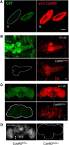
CaMKII is supplied maternally, and levels decline through development. A, Embryonic CaMKII levels are not reduced in the null mutant. Late-stage embryos from a w1118;;;PBac{EGFP-IV}eIF4GKM0133/CaMKIlstopw+ stock were stained for CaMKII with rabbit anti-CaMKII 88 at 1:10,000. The presence of the GFP chromosome allows identification of homozygous null, heterozygous, and WT animals from the same cross. Left, GFP visualization with a null animal (*) on the left and a WT animal (sibling control) on the right. Right, The same embryos have similar levels of CaMKII. Scale bar, 100 μm. B, Third instar larval CaMKII levels are reduced in null mutants and have an abnormal distribution. Top panels, WT sibling control animal with two copies of the GFP chromosome. Bottom panels, Homozygous null animal. GFP visualization (left) and anti-CaMKII 88 staining (right) are shown for both. Samples were dissected, processed, and imaged concurrently. Scale bar, 50 μm. Levels of CaMKII were variable between individual third instar larvae, likely due to differences in maternal mRNA levels. Images shown are for animals with relatively high levels of CaMKII. C, Stage P6–P7 pupal brains from null animals have a small amount of abnormally distributed CaMKII and appear to be structurally disrupted compared with GFP sibling control. Samples were dissected, processed, and imaged concurrently. Scale bar, 50 μm. D, Stage P13–P15 pupal brains from CaMKIIstopw+-null animals (right) have no detectable CaMKII compared with CaMKIIWTw+ sibling controls (left). Brains were stained with mouse monoclonal anti-CaMKII 18 at 1:10,000. Samples were dissected, processed, and imaged concurrently. Scale bar, 50 μm.
Figure 3.
Figure 3.
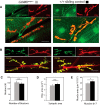
Maternal CaMKII does not localize normally in CaMKIIstopw+ null mutants, but overall morphology is normal. Levels of CaMKII were variable between individual third instar larvae, likely due to differences in maternal mRNA levels. A, B, Images are for animals with relatively high levels of CaMKII. A, CaMKII mutants lack synaptic CaMKII. Confocal stacks of mutant (left) and WT (right) third instar NMJs stained with anti-CaMKII (1:500 88) and anti-Dlg (1:50 mouse monoclonal 4F3). Samples were dissected, processed, and imaged concurrently with identical settings. Scale bar, 20 μm. Mutant animals have normal axonal levels of CaMKII but have no synaptic kinase. B, NMJ morphology is normal in CaMKIIstopw+-null mutants. Third instar larval NMJs were stained with anti-Dlg and anti-HRP, and muscles 6/7 in segment A2 were visualized using a confocal microscope. Left panels, Mutant NMJ. Right panels, Sibling control animal homozygous for the GFP marker chromosome. Scale bar, 25 μm. C, Bouton number is not altered in CaMKII-null animals. D, Postsynaptic area, as assessed by the measuring the extent of Dlg immunoreactivity, is unchanged in CaMKII-null animals. E, Muscle area is slightly decreased in CaMKII-null animals. The areas of muscles 6 and 7 were assessed using ImageJ Fiji. N = 18 muscles for CaMKIIstopw+ and 16 for WT sibling controls. *p < 0.05 (Student's t test).
Figure 4.
Figure 4.
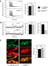
CaMKIIstopw+-null mutants have decreased mEJP frequency. A, EJPs are normal in the CaMKIIstopw+ mutant. A 1 s 10 Hz stimulus delivered to the segmental nerve produces a train of EJPs in both WT and CaMKIIstopw+ larvae. Left, Data are average of 50 traces per genotype. Facilitation/depression indices were not affected by genotype (data not shown). Right, Amplitude of the first EJP in the train was not affected by genotype. Data are mean ± SEM and were analyzed using Student's t test (p > 0.05); N = 10. B, mEJP frequency is decreased in the CaMKII mutant. Left, Example traces of mEJPs recorded from muscle 6. Middle, mEJP amplitude was not affected by genotype. Right, mEJP frequency is significantly decreased in CaMKII-null larvae. Average RMP: −62.37 (for sibling control animals); −61.18 (for CaMKIIstopw+ animals). Data are mean ± SEM. *p < 0.05 (Student's t test). N = 10. C, Release site number is not changed in the CaMKII mutant. Third instar larval NMJs were stained with mAb nc82, which recognizes Brp, and anti-Dlg to visualize synapses. Left panels, Sibling control NMJ. Right, CaMKIIstopw+ mutant NMJ. Scale bar, 5 μm. The number of nc82+ puncta per μm2 of Dlg staining was assessed and data tested for significance using Student's t test (p > 0.05). N = 16 for CaMKIIstopw+ and 14 for WT sibling controls.
Figure 5.
Figure 5.
CaMKIIstopw+-null mutants fail to show activity-dependent increases in mEJP frequency. A, Spaced depolarization of WT sibling control animals increases mEJP frequency in a transcription- and translation-dependent manner. Inclusion of 5 mm actinomycin (ACT) to block transcription or 100 mm cycloheximide (CHX) to block translation completely prevented activity-dependent increases in mEJP frequency in WT animals. N ≥ 5 per condition. B, CaMKIIstopw+ animals do not exhibit increased mEJP frequency in response to depolarization. N ≥ 5 per condition. C, ala2 animals, which globally express a CaMKII inhibitor peptide, show normal mEJP plasticity. N = 5. D, Schematic of experimental procedure, as modified from Ataman et al. (2008). Five pulses of high K+ were delivered over the course of 1.5 h with rest intervals of 15 min between pulses (5× K+). The control treatment (Con) consisted of the same protocol but with pulses of normal HL3. Drugs were delivered in normal HL3 with the control protocol. RMP for +/+ sibling animals (Con, 5× K+, ACT, ACT 5× K+, CHX, CHX 5× K+) in mV as follows: −61.25, −63.58, −62.16, −65.85, −64.24, −62.37, respectively. Average RMP for CaMKIIstopw+ animals (Con, 5× K+, ACT, ACT 5× K+, CHX, CHX 5×) in mV as follows: −66.68, −64.52, −62.88, −61.33, −62.08, −65.19, respectively. Data were analyzed by two-way ANOVA with genotype and treatment as main effects and pairwise comparisons made using Bonferroni and Holm–Sidak post hoc tests with α = 0.05. Data are mean ± SEM. * p < 0.05, significant difference from all other conditions for that genotype. mEJP amplitude was unchanged by genotype or condition (data not shown).
Figure 6.
Figure 6.
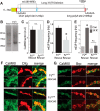
The long 3′UTR is required for normal mEJP rates but is not required for CaMKII synaptic localization. A, Diagram of the 3′ end of the CaMKII gene. B, 3′RACE of the CaMKII 3′UTR from 0–2 h embryonic and adult head mRNA. Both a long and a short 3′UTR are found in adult (arrows), whereas the form containing the long 3′UTR is not expressed in maternal RNA. Bands were cloned and sequenced to confirm identity. C, CaMKII levels from FsWT and FsΔUTR larvae are not significantly different. Blots from extracts of 10 independent biological replicates were probed with anti-CaMKII (1:1000, rabbit 88) and anti-actin (1:1000 mouse mAb C4). Total CaMKII signal was normalized to actin and compared. p > 0.5 (Student's t test). D, Spontaneous activity was recorded from homozygous CaMKIIstopw+ mutants with chromosome 3 rescue fosmid transgenes containing either the WT CaMKII gene (FsWT) or a CaMKII gene lacking long 3′UTR sequences (FsΔUTR). FsΔUTR fails to rescue the CaMKIIstopw+ mEJP phenotype (Fig. 4B). n = 15 for both FsWT and FsΔUTR. Data are mean ± SEM. *p < 0.05, significant difference (Student's t test). mEJP amplitude was not significantly different (data not shown). Average RMPs for FsWT and FsΔUTR were not significantly different, in mV as follows: −63.52, −62.92, respectively. E, A spaced depolarization protocol was applied to FsWT and FsΔUTR lines. FsΔUTR fails to rescue mEJP plasticity. n = 6 for each genotype. Data are mean ± SEM. *p < 0.05, significant difference from all other conditions for that genotype (Student's t test). Basal and spaced depolarization-induced mEJP amplitudes were not significantly changed by genotype or condition (data not shown). Average RMPs were not significantly different between control and 5× K+ conditions for either genotype. In mV after protocol: −64.17, −63.84 for FsWT and FsΔUTR, respectively. F, Postsynaptic CaMKII localizes normally in animals rescued with a fosmid transgene lacking long 3′UTR sequences. Single confocal sections of mutant animals rescued with a WT fosmid transgene (FsWT, top) and a fosmid lacking long UTR sequences (FsΔUTR, bottom). Two examples of each genotype are shown. Third instar NMJs were stained with anti-CaMKII and anti-Dlg. Samples were dissected, processed, and imaged concurrently with identical settings. Scale bar, 10 μm. G, Presynaptic CaMKII localizes normally in animals rescued with a fosmid transgene lacking long 3′UTR sequences. Single confocal sections of mutant animals rescued with a WT fosmid transgene (FsWT, top) and a fosmid lacking long UTR sequences (FsΔUTR, bottom). Two examples of each genotype are shown. Third instar NMJs were stained with anti-CaMKII and anti-Brp. Samples were dissected, processed, and imaged concurrently with identical settings. Scale bar, 10 μm.
Figure 7.
Figure 7.
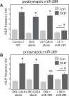
miR-289 overexpression attenuates activity-dependent increases in mEJP frequency. Spaced depolarization protocol was performed with Canton S control animals, and animals overexpressing miR-289 presynaptically (B) in motor neurons (OK6- and C380-GAL4) and postsynaptically (A) in muscles (C57-GAL4). RMPs for Canton S Con, Canton S 5× K+, Pre GAL4 Con, Pre GAL4 5× K+, Post GAL4 Con, Post GAL4 5× K+, UAS Con, UAS 5× K+, Pre miR-289 Con, Pre miR-289 5× K+, Post miR-289 Con, Post miR-289 5× K+, in mV as follows: −63.87, −62.39, −65.41, −64.68, −63.93, −64.45, −66.46, −64.94, −62.78, −64.56, −63.77, −66.31, respectively. Data were analyzed by two-way ANOVA with genotype and treatment as main effects and pairwise comparisons made using Bonferroni post hoc test with α = 0.05. Data are mean ± SEM. Control condition was not significantly different across genotypes. *Significant difference from the other condition for that genotype. #Significant difference from WT, UAS, and GAL4 controls for that condition. mEJP amplitude was not significantly changed by genotype or condition (data not shown). RMPs were not significantly different between control and 5× K+ conditions.

References

    1. Aakalu G, Smith WB, Nguyen N, Jiang C, Schuman EM (2001) Dynamic visualization of local protein synthesis in hippocampal neurons. Neuron 30:489–502. 10.1016/S0896-6273(01)00295-1 - DOI - PubMed
    1. Ashraf SI, McLoon AL, Sclarsic SM, Kunes S (2006) Synaptic protein synthesis associated with memory is regulated by the RISC pathway in Drosophila. Cell 124:191–205. 10.1016/j.cell.2005.12.017 - DOI - PubMed
    1. Ataman B, Ashley J, Gorczyca M, Ramachandran P, Fouquet W, Sigrist SJ, Budnik V (2008) Rapid activity-dependent modifications in synaptic structure and function require bidirectional Wnt signaling. Neuron 57:705–718. 10.1016/j.neuron.2008.01.026 - DOI - PMC - PubMed
    1. Attrill H, Falls K, Goodman JL, Millburn GH, Antonazzo G, Rey AJ, Marygold SJ (2016) FlyBase: establishing a Gene Group resource for Drosophila melanogaster. Nucleic Acids Res 44:D786–D792. 10.1093/nar/gkv1046 - DOI - PMC - PubMed
    1. Barber CF, Jorquera RA, Melom JE, Littleton JT (2009) Postsynaptic regulation of synaptic plasticity by synaptotagmin 4 requires both C2 domains. J Cell Biol 187:295–310. 10.1083/jcb.200903098 - DOI - PMC - PubMed

Publication types

MeSH terms

Substances

LinkOut - more resources