. 2017 Nov 1;37(44):10567-10586.
doi: 10.1523/JNEUROSCI.0758-17.2017. Epub 2017 Sep 27.

Sex- and Estrus-Dependent Differences in Rat Basolateral Amygdala

Affiliations

Sex- and Estrus-Dependent Differences in Rat Basolateral Amygdala

Shannon R Blume et al. J Neurosci. .

Abstract

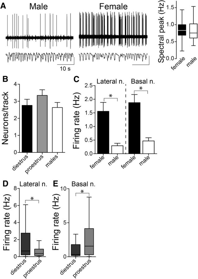
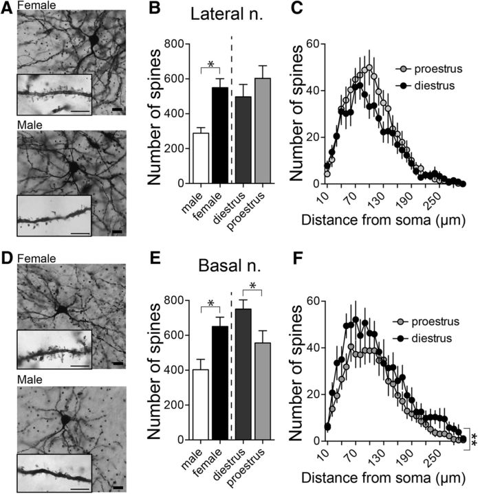
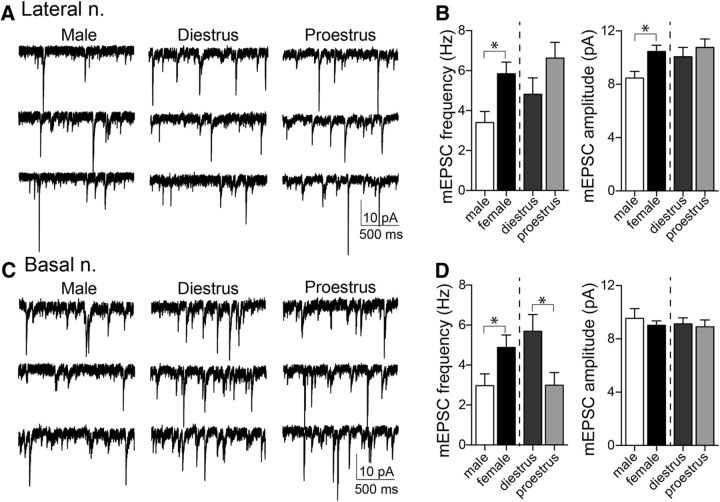
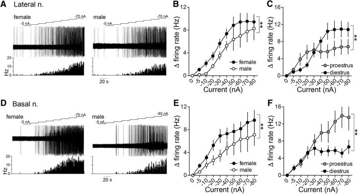
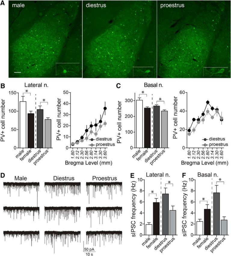
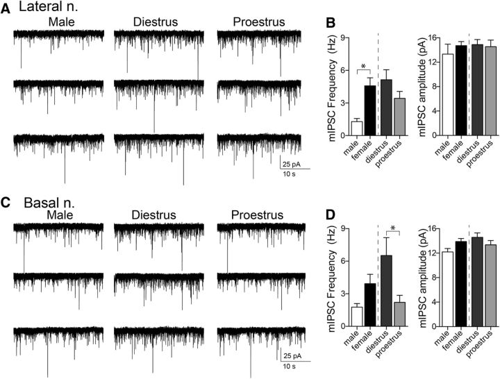
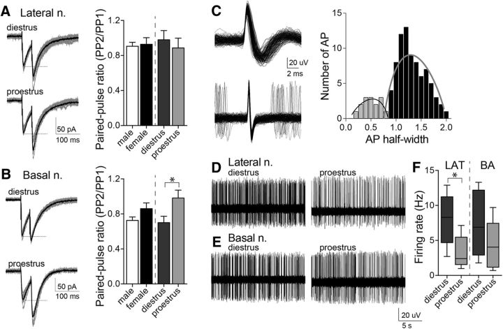
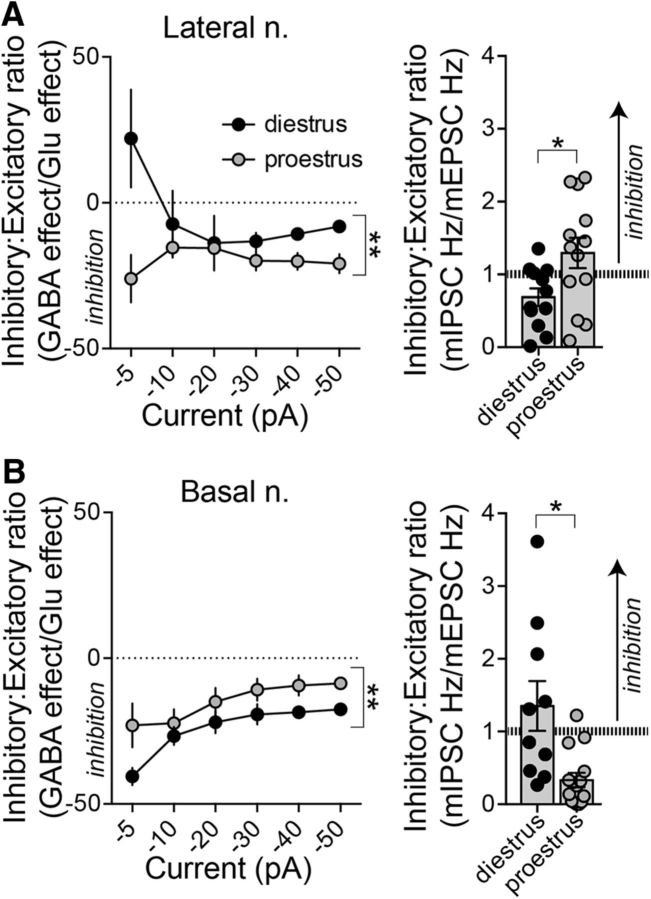
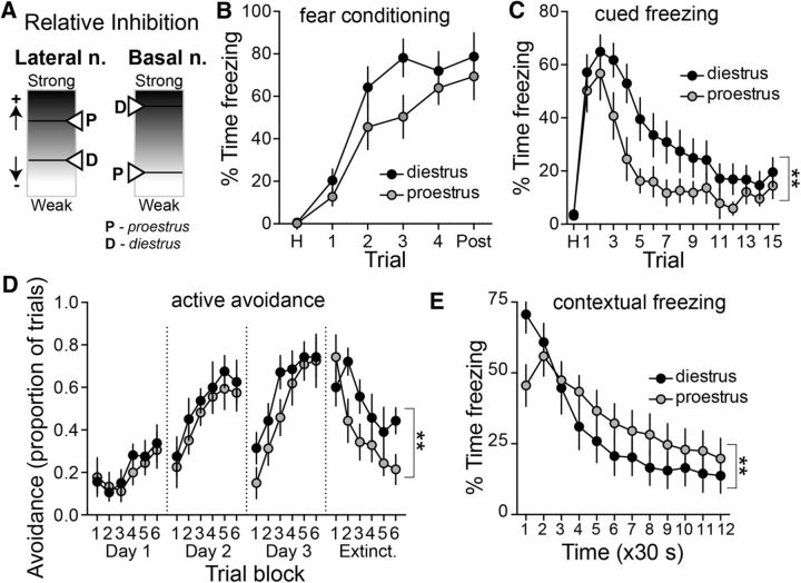
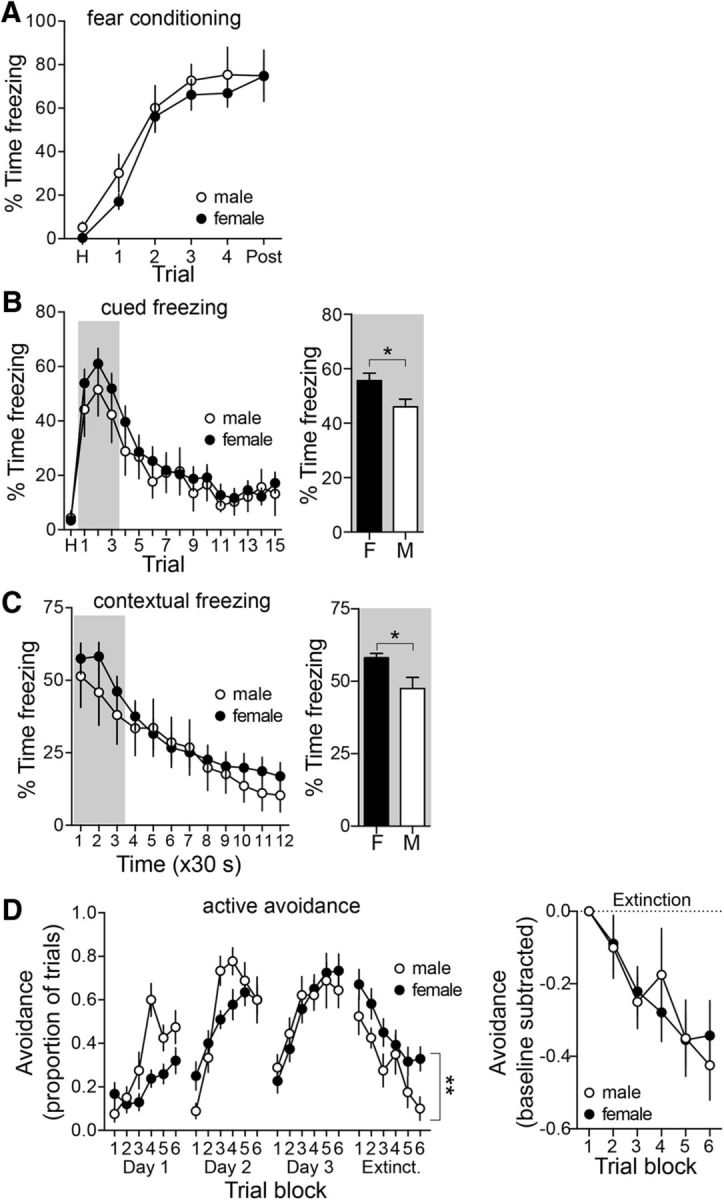
Depression and anxiety are diagnosed almost twice as often in women, and the symptomology differs in men and women and is sensitive to sex hormones. The basolateral amygdala (BLA) contributes to emotion-related behaviors that differ between males and females and across the reproductive cycle. This hints at sex- or estrus-dependent features of BLA function, about which very little is known. The purpose of this study was to test whether there are sex differences or estrous cyclicity in rat BLA physiology and to determine their mechanistic correlates. We found substantial sex differences in the activity of neurons in lateral nuclei (LAT) and basal nuclei (BA) of the BLA that were associated with greater excitatory synaptic input in females. We also found strong differences in the activity of LAT and BA neurons across the estrous cycle. These differences were associated with a shift in the inhibition-excitation balance such that LAT had relatively greater inhibition during proestrus which paralleled more rapid cued fear extinction. In contrast, BA had relatively greater inhibition during diestrus that paralleled more rapid contextual fear extinction. These results are the first to demonstrate sex differences in BLA neuronal activity and the impact of estrous cyclicity on these measures. The shift between LAT and BA predominance across the estrous cycle provides a simple construct for understanding the effects of the estrous cycle on BLA-dependent behaviors. These results provide a novel framework to understand the cyclicity of emotional memory and highlight the importance of considering ovarian cycle when studying the BLA of females.SIGNIFICANCE STATEMENT There are differences in emotional responses and many psychiatric symptoms between males and females. This may point to sex differences in limbic brain regions. Here we demonstrate sex differences in neuronal activity in one key limbic region, the basolateral amygdala (BLA), whose activity fluctuates across the estrous cycle due to a shift in the balance of inhibition and excitation across two BLA regions, the lateral and basal nuclei. By uncovering this push-pull shift between lateral and basal nuclei, these results help to explain disparate findings about the effects of biological sex and estrous cyclicity on emotion and provide a framework for understanding fluctuations in emotional memory and psychiatric symptoms.

Keywords: amygdala; electrophysiology; estrous; fear extinction; female.


Figures

Figure 1.
Figure 1.
Sex differences in BLA neuron firing rate. A, BLA neurons in females exhibited a higher firing rate compared with males, demonstrated as the number of action potentials per second in these representative recording traces. Depth of anesthesia could contribute to the differences in BLA neuron firing rate and was monitored by cortical local field potential. There was no significant sex difference in the primary component of the cortical oscillation (p = 0.248, two-tailed unpaired t test; shown here is box plot with or without Tukey's test). B, The number of spontaneously firing BLA neurons per electrode track were similar across female group (diestrus and proestrus) and male group (p = 0.347, one-way ANOVA). C, LAT and BA neurons recorded in female rats had significantly higher firing rates than male rats (LAT: p < 0.05; BA: p < 0.05; Mann–Whitney U tests). D, LAT neurons recorded in diestrous and proestrous females were compared to examine the influence of the estrous cycle on neuronal firing rate. Females in diestrus had a higher LAT neuronal firing rate compared with females in proestrus (p < 0.05, Mann–Whitney U test; shown here is box plot with or without Tukey's test). E, Females in diestrus had a lower BA neuronal firing rate compared with proestrus (p < 0.05, Mann–Whitney U test; shown here is the box plot with or without Tukey's test). *p < 0.05 Mann–Whitney U test. Calibration y-axis: top traces, 20 μV; bottom traces, 10 μV.
Figure 2.
Figure 2.
Sex differences in LAT and BA neuron morphology. A, Representative pictures of Golgi-Cox-stained LAT neurons in a female (top) and a male (bottom) rat. Scale bars: 20 μm; inset, 5 μm. B, There were significantly more spines in LAT neurons in females compared with males (p < 0.05, two-tailed unpaired t test) and no significant difference in spine number between diestrous and proestrous females (p = 0.282, two-tailed unpaired t test). C, Across the dendritic tree there were no differences in spine number (p = 0.294, two-way RM-ANOVA) between diestrous and proestrous females. D, Representative pictures of Golgi-Cox-stained BA neurons in a female (top) and male (bottom) rat. Scale bars: 20 μm; inset, 5 μm. E, The number of spines on BA neurons was higher in females compared with males (p < 0.05, two-tailed unpaired t test) and was dependent on the estrous cycle where BA neurons in diestrous females had more spines compared with BA neurons in proestrous females (right; p = 0.050, two-tailed unpaired t test). F, A greater number of spines was observed across the dendritic tree in BA neurons from diestrous females (p < 0.05, two-way RM-ANOVA). *p < 0.05 two-tailed unpaired t test; **p < 0.05 two-way RM-ANOVA.
Figure 3.
Figure 3.
Greater excitatory synaptic input in LAT and BA neurons in females. mEPSCs were measured from LAT principal neurons in vitro in female and male animals to determine whether the observed anatomical differences had a functional correlate. A, Displayed here are representative traces of mEPSC recordings in male and female groups. B, Females had a significantly higher frequency of mEPSCs in LAT neurons (left: p < 0.05, two-tailed unpaired t test) and greater amplitude of mEPSCs compared with males (right: p < 0.05, two-tailed unpaired t test). mEPSC frequency (left: p = 0.124, two-tailed unpaired t test) and amplitude (right: p = 0.757, two-tailed unpaired t test) did not differ across the estrous cycle. C, Representative traces of mEPSC recordings in BA neurons in male and female groups. D, The frequency of mEPSCs in BA neurons was greater in females compared with males (left: p < 0.05, two-tailed unpaired t test); however, there was no difference in mEPSC amplitude between sexes (right: p = 0.463, two-tailed unpaired t test). The frequency of mEPSCs was significantly higher in diestrous females compared with proestrous females (left: p < 0.05, two-tailed unpaired t test); however, mEPSC amplitudes were similar between the cycle stages (right: p = 0.757, two-tailed unpaired t test). *p < 0.05, two-tailed unpaired t test.
Figure 4.
Figure 4.
Glutamate iontophoresis drives LAT and BA neuronal firing rate more effectively in females than males. A, Representative traces of iontophoretic current applied to eject glutamate (top), the neuronal response to glutamate (middle), and a time histogram of the firing rate (Hz) of the neuron across time (bottom) in LAT neurons recorded in a female (left) and male (right) rat. B, Glutamate drove LAT neuron firing more effectively in females compared with males (p < 0.05, two-way ANOVA). C, The effectiveness of glutamate to drive LAT neurons was dependent on the estrous cycle stage, where glutamate was more effective at lower current applications in proestrous females and higher current applications were more effective in diestrous females (p < 0.05, two-way ANOVA). D, Representative traces of iontophoretic current applied to eject glutamate (top), the neuronal response to glutamate (middle), and a time histogram of the firing rate (Hz) of the neuron across time (bottom) in BA neurons recorded in a female (left) and male (right) rat. E, Glutamate drove BA neuron firing more effectively in females compared with males (p < 0.05, two-way ANOVA). F, BA neurons in proestrous females were more responsive to glutamate iontophoresis compared with diestrous females (p < 0.05, two-way ANOVA). **p < 0.05, two-way ANOVA.
Figure 5.
Figure 5.
PV-immunopositive cell numbers are decreased in the BLA of female rats in proestrus. A, Representative images of PV staining of LAT and BA nuclei, in diestrous, proestrous, and male groups (−2.56 mm from bregma). Inset depicts higher-magnification image of single cells. B, The number of PV+ neurons in the LAT nucleus was significantly higher in male compared with female rats (p = 0.0321, two-tailed unpaired t test). Females in proestrus had lower numbers of PV+ neurons in the LAT nucleus compared with females in diestrus (p = 0.0288, two-tailed unpaired t test), and this was observed across rostral-caudal regions. C, Males had a higher number of PV+ neurons in the BA nucleus compared with females (p = 0.0045, two-tailed unpaired t test), and proestrous females had lower numbers of PV+ neurons than diestrous females (p = 0.0210, two-tailed unpaired t test). D, sIPSCs were measured in LAT neurons and BA neurons to examine GABAergic synaptic input. Shown here are examples of sIPSCs recorded from BA slices obtained from a male, a diestrus female, and a proestrus female rat. E, The sIPSC frequency in LAT neurons was higher in females compared with males (p = 0.0015, two-tailed unpaired t test). Across the estrous cycle, there was a lower frequency of sIPSCs in LAT neurons during proestrus (p = 0.0291, two-tailed unpaired t test). F, In a similar manner, the sIPSC frequency in BA neurons was higher in females compared with males (p = 0.0254, two-tailed unpaired t test). Across the estrous cycle, there was a lower frequency of sIPSCs in BA neurons during proestrus (p = 0.0002, two-tailed unpaired t test). *p < 0.05, two-tailed unpaired t test. Scale bar, 100 μm.
Figure 6.
Figure 6.
mIPSC frequency in BA neurons is higher in diestrous females. To examine the effect of sex/estrous cycle on GABA release probability and number of synapses, mIPSCs were measured in female and male groups. A, Representative traces of mIPSCs recorded from LAT neurons in male and female groups. B, Females had greater mIPSC frequencies in LAT neurons compared with males (left; p < 0.05, two-tailed unpaired t test); however, the frequency of mIPSCs did not differ between diestrous and proestrous groups (left; p = 0.135, two-tailed unpaired t test). There were no differences in mIPSC amplitudes between males and females (right; p = 0.3547, two-tailed unpaired t test), or diestrous and proestrous females (right; p = 0.8010, two-tailed unpaired t test). C, Representative traces of mIPSCs recorded from BA neurons in male and female groups. D, In BA neurons, males and females had similar mIPSC frequencies (left; p = 0.129, two-tailed unpaired t test) and no differences in mIPSC amplitudes (right; p = 0.0576, two-tailed unpaired t test). Diestrous females had greater mIPSC frequencies compared with proestrous females (left; p < 0.05, two-tailed unpaired t test), with no differences in mIPSC amplitudes observed (right; p = 0.2401, two-tailed unpaired t test). *p < 0.05, two-tailed unpaired t test.
Figure 7.
Figure 7.
Paired-pulse stimulation and interneuron firing activity indicate different sources for estrous cyclicity in LAT and BA. Paired-pulse stimulation of GABAergic input was measured. A, Representative examples of paired-pulse responses recorded from a LAT neuron during diestrus or proestrus (gray: 10 consecutive responses; black: average). There was no difference in the paired-pulse ratio of GABAergic input across the estrous cycle in LAT neurons (right; p = 0.5550, two-tailed unpaired t test). B, Representative examples of paired-pulse responses recorded from a BA neuron during diestrus or proestrus (gray: 10 consecutive responses; black: average). There was a significant shift in the paired-pulse ratio of GABAergic input across the estrous cycle in BA neurons (right; p < 0.05, two-tailed unpaired t test). C, Presumptive interneurons displayed a short action potential half-width compared with principal neurons (left). Based on the distribution of half-widths, a cutoff of <0.9 ms was used to identify presumptive interneurons (right). D, Representative example of a presumptive LAT interneuron firing recorded in a diestrous or proestrous female. E, Representative example of a presumptive BA interneuron firing recorded in a diestrous or proestrous female. F, The average firing rate of presumptive LAT interneurons was significantly higher in diestrous compared with proestrous females (p < 0.05, two-tailed unpaired t test). In contrast, the average firing rate of presumptive BA interneurons was not significantly different across the estrous cycle (p = 0.2547, two-tailed unpaired t test; shown here is the box plot with or without Tukey's test). *p < 0.05, two-tailed unpaired t test.
Figure 8.
Figure 8.
Opposite in vivo sensitivity to GABA across estrous females between LAT and BA. GABA was iontophoresed in ascending current, while neuronal firing was measured. A, Representative traces of iontophoretic current applied to eject GABA (top), the neuronal response to GABA (middle), and a time histogram of the firing rate of LAT neurons recorded from a male (left) and female rat (right). B, In LAT neurons, GABA was more effective in suppressing neuron firing in males compared with females (p < 0.05, two-way ANOVA). C, GABA was also more effective in suppressing LAT neuron firing in proestrous females compared with diestrous females (p < 0.05, two-way ANOVA). D, Representative traces of iontophoretic current applied to eject GABA (top), the neuronal response to GABA (middle) and a time histogram of the firing rate of BA neurons recorded from a male (left) and a female (right) rat. E, In contrast to LAT neurons, GABA was more effective at suppressing the firing of BA neurons in females compared with males (p < 0.05, two-way ANOVA). F, Also in contrast to LAT neurons, GABA was more effective in suppressing BA neuron firing in diestrous compared with proestrous females (p < 0.05, two-way ANOVA). **p < 0.05, two-way ANOVA.
Figure 9.
Figure 9.
Opposite inhibition–excitation balance in LAT and BA across the estrous cycle. To examine the balance of glutamatergic and GABAergic function in our in vivo and in vitro measures, we compared the ratio of GABAergic inhibition to glutamatergic excitation. A, In the LAT, the balance was shifted toward greater in vivo inhibition during proestrus compared with diestrus. Proestrous females had a higher inhibitory/excitatory ratio in LAT neurons both in vivo (left; p < 0.05, two-way ANOVA) and in vitro (right; p < 0.05, two-tailed unpaired t test). B, In the BA nucleus, diestrous females had a greater inhibitory/excitatory ratio both in vivo (left; p < 0.05, two-way ANOVA) and in vitro (right; p < 0.05, two-tailed unpaired t test). *p < 0.05, two-tailed unpaired t test; **p < 0.05, two-way ANOVA.
Figure 10.
Figure 10.
Shift between LAT and BA function across the estrous cycle. A, Schematic illustration of the opposite effects of estrous cyclicity on relative inhibition in the LAT and BA. B, Cued fear conditioning of a tone paired with a footshock led to gradual acquisition of freezing. There was no significant difference between diestrus (n = 17) and proestrus (n = 15) in the freezing during repeated tone–footshock conditioning trials (p > 0.05, two-way RM-ANOVA). C, Acquisition of extinction of conditioned freezing to a cue was measured as the reduced conditioned freezing across nonreinforced trials on the testing day. The acquisition of extinction to the cue was significantly slower in diestrous compared with proestrous females (p < 0.05, two-way RM-ANOVA). D, Extinction of active avoidance was measured as the reduced avoidance response to a cue. Diestrous and proestrous females displayed similar learning behavior (days 1–3); however, diestrous females displayed slower extinction compared with proestrous females (p < 0.05, two-way RM-ANOVA). E, Acquisition of the extinction of contextual fear was measured as reduced conditioned freezing in a footshock-paired context over time. Proestrous females displayed slower acquisition of extinction compared with diestrous females (p < 0.05, two-way RM-ANOVA). **p < 0.05, two-way RM-ANOVA.
Figure 11.
Figure 11.
Sex differences in the expression of conditioned fear. A, Fear conditioning was measured during acquisition in male and female rats. Acquisition of conditioned fear was similar in male and female rats (p > 0.05, two-way RM-ANOVA). B, Conditioned freezing to a cue was measured in a novel context over the course of repeated trials. There were no sex differences overall in conditioned freezing (left; p > 0.05, two-way RM-ANOVA; male n = 11; female n = 32). However, when assessing the initial expression of conditioned cued freezing, before significant extinction of the conditioned response, females displayed greater conditioned freezing (freezing during the first three trials collapsed; p < 0.05, two-tailed unpaired t test). C, Conditioned freezing to a context was measured in the conditioned context over time. There were no sex differences overall in conditioned freezing (left; p > 0.05, two-way RM-ANOVA; males, n = 11; females, n = 38). However, when assessing the initial expression of conditioned contextual freezing, before significant extinction of the conditioned response, females displayed greater conditioned freezing (freezing during the first two segments collapsed; p < 0.05, two-tailed unpaired t test. D, The expression of active avoidance during the extinction session was significantly greater in female rats (p < 0.05, two-way RM-ANOVA). The greater expression in female rats throughout the extinction session could be due to higher initial expression or slower extinction. However, when normalized by the initial expression (trial block 1), there was no significant difference between males and females across extinction blocks (p > 0.05, two-way RM-ANOVA). *p < 0.05, two-tailed unpaired t test; **p < 0.05, two-way RM-ANOVA.

Similar articles

Cited by

References

    1. Aguilar R, Gil L, Gray JA, Driscoll P, Flint J, Dawson GR, Giménez-Llort L, Escorihuela RM, Fernández-Teruel A, Tobeña A (2003) Fearfulness and sex in f2 roman rats: males display more fear though both sexes share the same fearfulness traits. Physiol Behav 78:723–732. 10.1016/S0031-9384(03)00043-X - DOI - PubMed
    1. Akirav I, Raizel H, Maroun M (2006) Enhancement of conditioned fear extinction by infusion of the GABA(A) agonist muscimol into the rat prefrontal cortex and amygdala. Eur J Neurosci 23:758–764. 10.1111/j.1460-9568.2006.04603.x - DOI - PubMed
    1. Andreano JM, Cahill L (2009) Sex influences on the neurobiology of learning and memory. Learn Mem 16:248–266. 10.1101/lm.918309 - DOI - PubMed
    1. Arruda-Carvalho M, Clem RL (2014) Pathway-selective adjustment of prefrontal-amygdala transmission during fear encoding. J Neurosci 34:15601–15609. 10.1523/JNEUROSCI.2664-14.2014 - DOI - PMC - PubMed
    1. Baran SE, Armstrong CE, Niren DC, Hanna JJ, Conrad CD (2009) Chronic stress and sex differences on the recall of fear conditioning and extinction. Neurobiol Learn Mem 91:323–332. 10.1016/j.nlm.2008.11.005 - DOI - PMC - PubMed

Publication types

LinkOut - more resources