Clinical Trial
. 2017 Sep 27;9(409):eaan0241.
doi: 10.1126/scitranslmed.aan0241.

RNAi-based treatment of chronically infected patients and chimpanzees reveals that integrated hepatitis B virus DNA is a source of HBsAg

Affiliations
Clinical Trial

RNAi-based treatment of chronically infected patients and chimpanzees reveals that integrated hepatitis B virus DNA is a source of HBsAg

Christine I Wooddell et al. Sci Transl Med. .

Abstract

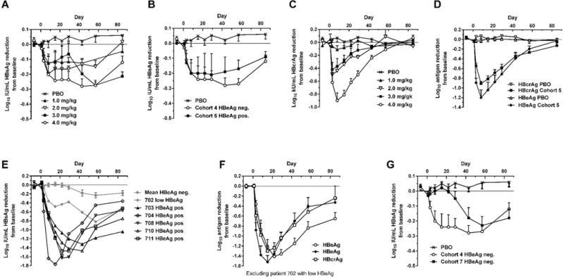
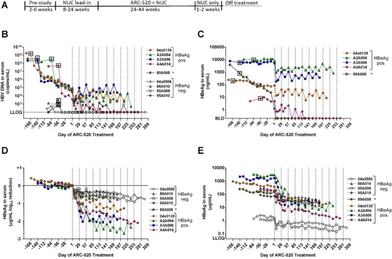
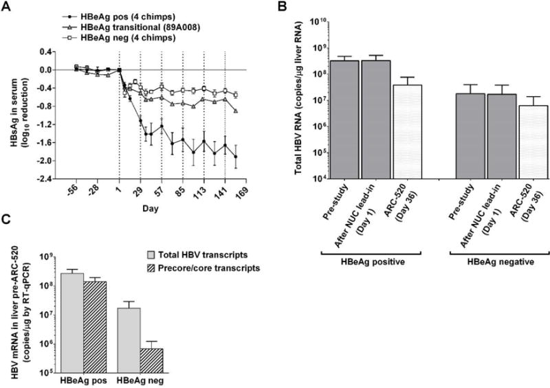
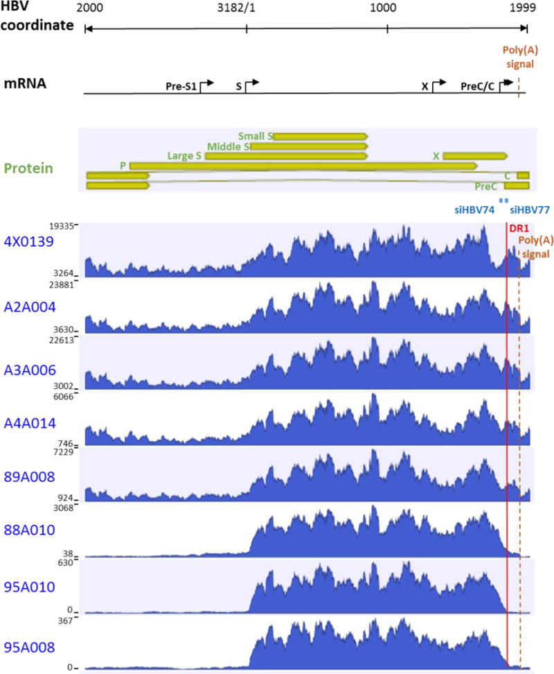
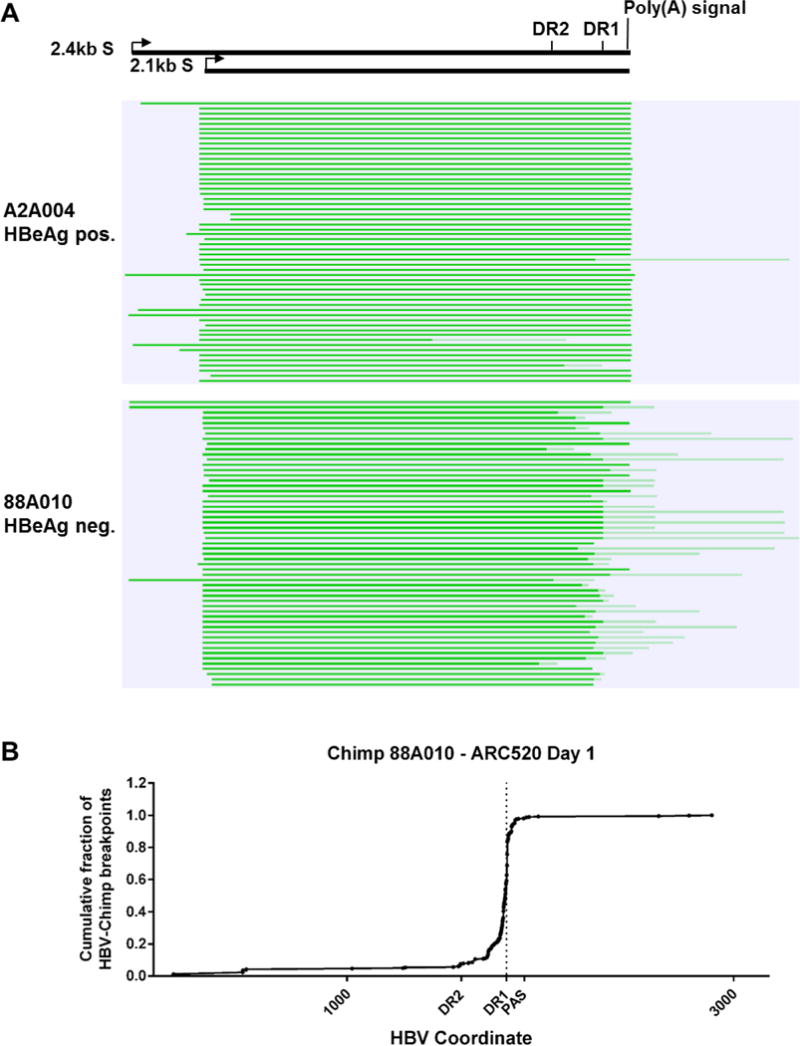
Chronic hepatitis B virus (HBV) infection is a major health concern worldwide, frequently leading to liver cirrhosis, liver failure, and hepatocellular carcinoma. Evidence suggests that high viral antigen load may play a role in chronicity. Production of viral proteins is thought to depend on transcription of viral covalently closed circular DNA (cccDNA). In a human clinical trial with an RNA interference (RNAi)-based therapeutic targeting HBV transcripts, ARC-520, HBV S antigen (HBsAg) was strongly reduced in treatment-naïve patients positive for HBV e antigen (HBeAg) but was reduced significantly less in patients who were HBeAg-negative or had received long-term therapy with nucleos(t)ide viral replication inhibitors (NUCs). HBeAg positivity is associated with greater disease risk that may be moderately reduced upon HBeAg loss. The molecular basis for this unexpected differential response was investigated in chimpanzees chronically infected with HBV. Several lines of evidence demonstrated that HBsAg was expressed not only from the episomal cccDNA minichromosome but also from transcripts arising from HBV DNA integrated into the host genome, which was the dominant source in HBeAg-negative chimpanzees. Many of the integrants detected in chimpanzees lacked target sites for the small interfering RNAs in ARC-520, explaining the reduced response in HBeAg-negative chimpanzees and, by extension, in HBeAg-negative patients. Our results uncover a heretofore underrecognized source of HBsAg that may represent a strategy adopted by HBV to maintain chronicity in the presence of host immunosurveillance. These results could alter trial design and endpoint expectations of new therapies for chronic HBV.


Conflict of interest statement

Competing interests

C.I.W., D.L.L, Z.X., B.D.G., T.S., J.H., and S.B.K. are or were employees of Arrowhead Pharmaceuticals. R.G.G., S.A.L., C.L.L., C.F. and J.Y.N.L. receive compensation from Arrowhead. M.F.Y., H.Y.C., R.E.L. and D. C. performed work under contract with Arrowhead.

Figures

Fig. 1
Fig. 1. Serum HBsAg, HBcrAg and HBeAg reduction in human patients treated with a single dose of ARC-520
CHB patients were given a single intravenous dose of 1 to 4 mg/kg ARC-520 on a background of daily oral NUCs. HBsAg (A) or HBcrAg (C) reduction in CHB patients that were HBeAg negative, NUC-experienced and received single doses of 1 to 4 mg/kg (cohorts 1 to 4). (B) HBsAg reduction in CHB patients that were HBeAg negative and NUC-experienced (cohort 4) or HBeAg positive and NUC-experienced (cohort 5) that received a single dose of 4 mg/kg. (D) HBcrAg and HBeAg reduction in CHB patients that were HBeAg positive, NUC-experienced and received a single 4 mg/kg dose (cohort 5). (E) HBsAg reduction for individual CHB patients that were HBeAg positive in cohort 7. (F) HBsAg, HBeAg and HBcrAg reductions in CHB patients that were HBeAg positive, treatment naïve, and received a single 4 mg/kg dose (cohort 7). (G) HBsAg reduction in CHB patients that were HBeAg negative and treatment naïve (cohort 7) or NUC experienced (cohort 4). PBO, patients on NUC therapy given placebo injection; HBcrAg, HBV core-related antigen. Error bars show SEM.
Fig. 2
Fig. 2. Response to repeat dosing of chimpanzees with ARC-520
(A) Following a pre-study evaluation, nine chimpanzees began daily oral NUC dosing. After a variable NUC lead-in period to reduce viremia, NUC treatment continued concomitant with Q4W dosing of ARC-520 that began on Day 1. Dosing days are indicated by vertical dashed lines. Blood samples were collected periodically throughout the study and serum HBV DNA (B), HBeAg (C) and HBsAg (D, E) were measured. LLOQ, lower limit of quantitation. BLD, below the limit of detection. Asterisk, chimpanzee 89A008 transitioned from HBeAg positive to negative during the NUC lead-in period.
Fig. 3
Fig. 3. Serum HBsAg and liver HBV mRNA reduction in chimpanzees dosed with ARC-520
Following pre-study evaluation, nine chimpanzees began NUC dosing for a lead-in period of 8–24 weeks. Q4W dosing with ARC-520 began on Day 1. (A) HBsAg levels in serum. The mean log10 change in HBsAg is shown for the 8 weeks preceding the first dose of ARC-520 and during ARC-520 treatment of four HBeAg positive (●), four HBeAg negative (□), and in the HBeAg transitional chimpanzee (▲). (B, C) Liver mRNA from biopsies of HBeAg positive (A2A004, A3A006 and A4A014) and HBeAg negative (88A010, 95A008 and 95A010) chimpanzees was evaluated for HBV gene expression by RT-qPCR with probe sets in core to measure the levels of precore mRNA/pgRNA and in X to measure total HBV mRNA. (B) Mean total HBV mRNA levels were compared between HBeAg positive and negative chimpanzees pre-study, after the NUC lead-in (Day 1), and one week after the second ARC-520 injection (Day 36). (C) The numbers of total HBV transcripts and precore/pgRNA transcripts on Day 1 are compared for the HBeAg positive and negative chimpanzees. Error bars show SEM.
Fig. 4
Fig. 4. Histogram of liver HBV mRNA paired-end sequencing reads in HBeAg positive and negative chimpanzees
The HBV mRNA and HBV protein open reading frames are positioned relative to the coordinates of the HBV genome. The mRNA sequencing read histograms are shown for HBeAg positive chimpanzees 4x0139, A2A004, A3A006 and A4A014; for HBeAg transitional chimpanzee 89A008; and for HBeAg negative chimpanzees 88A010, 95A010 and 95A008. Locations of the DR1 sequence (red line), HBV PAS (brown dashed line) and binding sites for the siRNAs in ARC-520 (siHBV-74 and siHBV-77) are indicated.
Fig. 5
Fig. 5. Mapping of HBV S transcripts from an HBeAg positive and an HBeAg negative chimpanzee
Total RNA isolated from the liver biopsies after the NUC lead-in and before ARC-520 dosing on Day 1 from HBeAg positive chimpanzee A2A004 and HBeAg negative chimpanzee 88A010 was reverse transcribed, size-selected and sequenced with single molecule real-time (SMRT) sequencing. (A) Full-length nonconcatemer reads (see methods) were aligned to each chimpanzee’s consensus HBV DNA sequence with CLC Genomics Workbench. Precore/pgRNA transcripts are not shown in this view. These were detected in A2A004 but were not in 88A010 at this sequencing depth. Green lines represent HBV-containing transcripts. Dark green represents sequences aligning to HBV and light green represents those not aligning to HBV. The HBV coordinates are shown with elements DR2, DR1 and the HBV polyadenylation signal. (B) Cumulative fraction of HBV-chimpanzee breakpoints were plotted against the HBV coordinate.
Fig. 6
Fig. 6. Treatment of HBeAg negative chimpanzees with siRNA targeted outside the DR1-DR2 region
Following a NUC lead-in, all 4 HBeAg negative chimpanzees were given seven Q4W doses of ARC-520. Chimpanzees 4x0506 and 95A008 were then given an additional three doses of ARC-520 (4 mg/kg) while 95A010 and 88A010 were given three doses of 4 mg/kg siHBV-75 plus 4 mg/kg ARC-EX1 delivery reagent.

Comment in

References

    1. G.B.D. Mortality. C. Causes of Death, Global, regional, and national age-sex specific all-cause and cause-specific mortality for 240 causes of death, 1990–2013: a systematic analysis for the Global Burden of Disease Study 2013. Lancet. 2015;385:117–171. - PMC - PubMed
    1. Yuen MF, Ahn SH, Chen DS, Chen PJ, Dusheiko GM, Hou JL, Maddrey WC, Mizokami M, Seto WK, Zoulim F, Lai CL. Chronic Hepatitis B Virus Infection: Disease Revisit and Management Recommendations. J Clin Gastroenterol. 2016;50:286–294. - PubMed
    1. Beasley RP, Hwang LY, Lin CC, Chien CS. Hepatocellular carcinoma and hepatitis B virus. A prospective study of 22 707 men in Taiwan. Lancet. 1981;2:1129–1133. - PubMed
    1. Bock CT, Schwinn S, Locarnini S, Fyfe J, Manns MP, Trautwein C, Zentgraf H. Structural organization of the hepatitis B virus minichromosome. J Mol Biol. 2001;307:183–196. - PubMed
    1. Jiang Z, Jhunjhunwala S, Liu J, Haverty PM, Kennemer MI, Guan Y, Lee W, Carnevali P, Stinson J, Johnson S, Diao J, Yeung S, Jubb A, Ye W, Wu TD, Kapadia SB, de Sauvage FJ, Gentleman RC, Stern HM, Seshagiri S, Pant KP, Modrusan Z, Ballinger DG, Zhang Z. The effects of hepatitis B virus integration into the genomes of hepatocellular carcinoma patients. Genome Res. 2012;22:593–601. - PMC - PubMed

Publication types

MeSH terms