. 2017 Sep 27;8(1):707.
doi: 10.1038/s41467-017-00786-z.

Clinical responses to adoptive T-cell transfer can be modeled in an autologous immune-humanized mouse model

Affiliations

Clinical responses to adoptive T-cell transfer can be modeled in an autologous immune-humanized mouse model

Henrik Jespersen et al. Nat Commun. .

Abstract

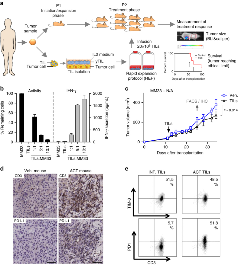
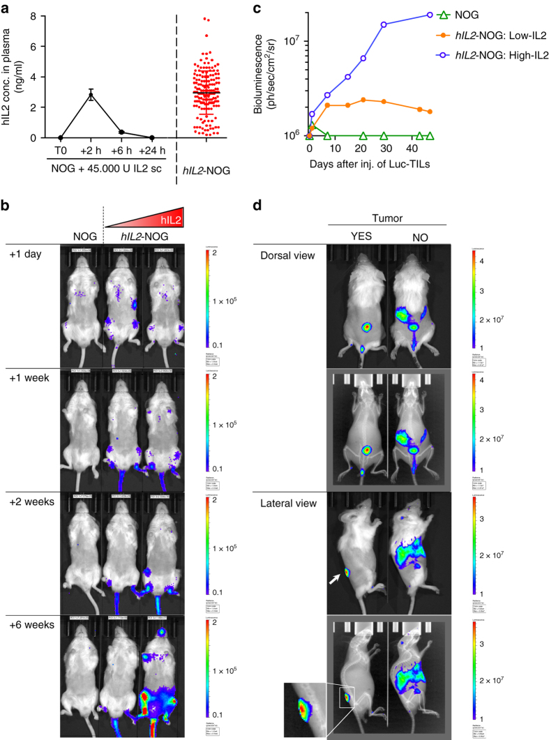
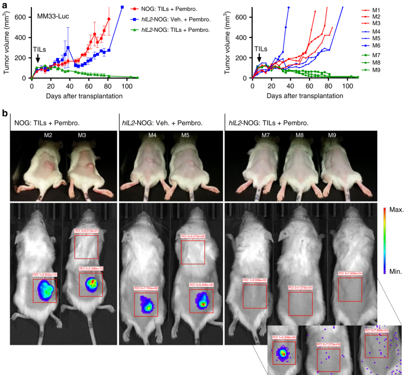
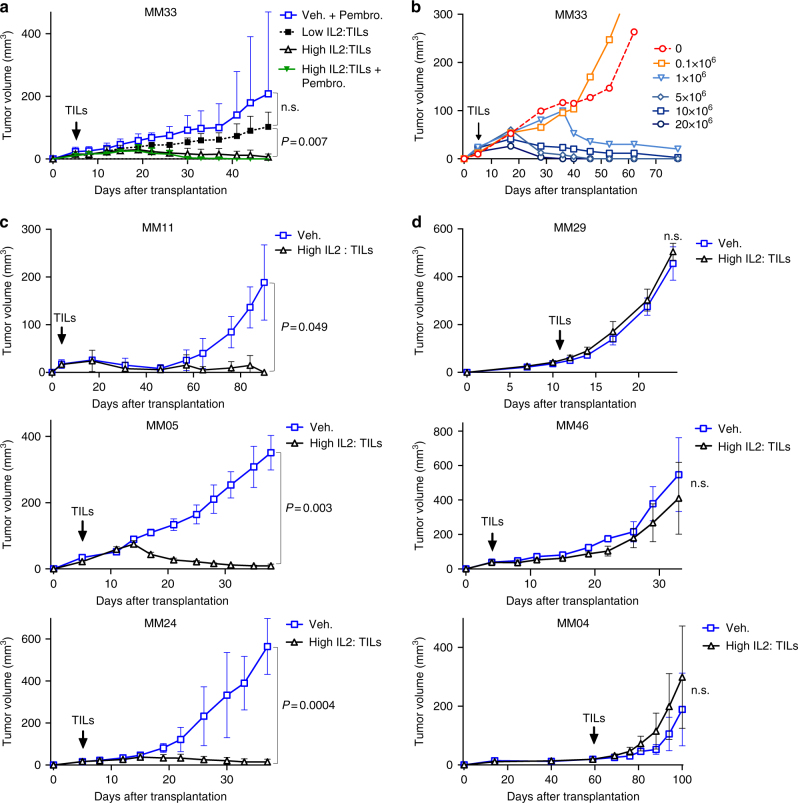
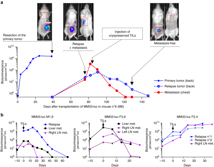
Immune checkpoint inhibitors and adoptive cell transfer (ACT) of autologous tumor-infiltrating T cells have shown durable responses in patients with melanoma. To study ACT and immunotherapies in a humanized model, we have developed PDXv2.0 - a melanoma PDX model where tumor cells and tumor-infiltrating T cells from the same patient are transplanted sequentially in non-obese diabetic/severe combined immune-deficient/common gamma chain (NOG/NSG) knockout mouse. Key to T-cell survival/effect in this model is the continuous presence of interleukin-2 (IL-2). Tumors that grow in PDXv2.0 are eradicated if the autologous tumor cells and T cells come from a patient that exhibited an objective response to ACT in the clinic. However, T cells from patients that are non-responders to ACT cannot kill tumor cells in PDXv2.0. Taken together, PDXv2.0 provides the potential framework to further model genetically diverse human cancers for assessing the efficacy of immunotherapies as well as combination therapies.Combining different types of immune therapies might benefit certain patients. Here, the authors develop an autologous immune-humanized melanoma mouse model that allows the preclinical assessment of cancer cell-T cell interactions from each individual patient and the benefits of immunotherapies combinations.


Conflict of interest statement

The authors declare no competing financial interests

Figures

Fig. 1
Fig. 1
Effective in vitro cytotoxicity does not result in effective anti-tumoral activity in NOG mice. a Schematic representation of the humanization process (PDXv2.0). b Melanoma cells from patient #33 (MM33) were transduced with a luciferase lentivirus. Cells were plated in a 96-well plate and were mixed with post-REP TILs from the original tumor. Luciferin was added to the media and the viability of the luciferase-expressing tumor cells was measured in a luminometer. The experiment was done in triplicates and the error bar represents ± SEM. c MM33 cells were transplanted into ten NOG mice. 1 week after transplantation, mice were randomized into two groups, one of which received a tail vein injection with autologous TILs. 45,000 U IL-2 was given daily for 3 days following TIL injection and thereafter twice weekly during 3 weeks. Tumor growth was measured using calipers. P-values are from a multiple t-test analysis. d, e When robust progression was noted in all mice, the mice with the slowest and fastest growing tumors in each group were sacrificed and the tumors were analyzed by immunohistochemistry (d, bar = 50 µm) or flow cytometry e for indicated markers. When PD1 expression was observed, anti-PD1 antibody pembroluzimab was given to the mice twice weekly. The experiments shown were performed once
Fig. 2
Fig. 2
Transgenic expression of human IL-2 enables NOG mice to support viability of TILs. a Changes in IL-2 plasma levels in NOG mice injected with 45,000 U IL-2 over time and steady-state levels of IL-2 in hIL2-NOG mice. b In vivo bioluminescence images over time of two hIL2-NOG transgenic mice with different serum levels of hIL-2 injected with MM33 TILs labeled with a luciferase lentivirus. c Quantification of radiance of luciferase-expressing MM33 TILs growing in mice (from b) with different serum levels of IL-2. d In vivo bioluminescence images from different angles of tumor-bearing or non-tumor-bearing hIL2-NOG transgenic mice injected with luciferase-expressing MM33 TILs. The experiments shown here were performed once
Fig. 3
Fig. 3
Transgenic expression of human IL2 enables TIL-mediated tumor eradication in NOG mice. a Six hIL2-NOG transgenic mice and three NOG mice were transplanted with luciferase labeled MM33 tumor cells. After tumor growth had been confirmed by IVIS imaging, all NOG mice, and three of the hIL2-NOG mice, were injected with autologous REP TILs and tumor growth was measured with calipers. Shown to the right are individual mice. b IVIS imaging to investigate if mice are disease free. Inset, very prolonged exposure yielding mostly spark signals from static electricity. The experiment described here was performed once, follow-up experiments confirming the data are in Fig. 4 and Supplementary Fig. 5
Fig. 4
Fig. 4
ACT in mice with high levels of plasma hIL-2 correlates with responses in patients. a Tumor growth curves of MM33 in hIL2-NOG mice expressing high (>2 ng/ml) or low levels of hIL-2 (< 1.5 ng/ml), treated either with PBS or TILs in the presence or absence of pembrolizumab. b Dose titration response of MM33 to different amounts of injected TILs (from 0.1×10⁶ to 20×106 TILs) in hIL2-NOG mice with high levels of hIL2. c, d Tumor growth curves of cells from three responders in the clinic (c: MM11, MM24 and MM05) and from two non-responders (d: MM04, MM46 and MM29) in hIL2-NOG mice with high levels of hIL-2 treated either with PBS or TILs. Data are caliper measurements (MM33/MM24/MM05/MM04, n = 4 per group; MM11/MM29/MM46, n = 3 per group). P-values are from multiple t-test (with Sidak corrections) for tumor growth curves. Experiment in a was performed twice, a replicate shown in Supplementary Fig. 5. Experiments in b, c were performed once except for MM24 where a replicate experiment is shown in Supplementary Fig. 5
Fig. 5
Fig. 5
Transgenic expression of human IL2 enables TIL-mediated tumor eradication of metastases in NOG mice. Transgenic hIL2-NOG mice transplanted with luciferase labeled MM33 tumor cells were allowed to develop subcutaneous tumors until tumors reached >500 mm3. The tumors were surgically removed and relapse was followed using the IVIS imager. When metastases had appeared TILs were injected and responses were measured by IVIS imaging. a A representative mouse showing the disease progression. b IVIS imaging of mice with additional sites of metastasis of melanoma. Graphs are labeled with gender of mouse (F or M) and corresponding plasma level of hIL-2 (ng/ml)

References

    1. Balch CM, et al. Final version of 2009 AJCC melanoma staging and classification. J. Clin. Oncol. 2009;27:6199–6206. doi: 10.1200/JCO.2009.23.4799. - DOI - PMC - PubMed
    1. Berger MF, et al. Melanoma genome sequencing reveals frequent PREX2 mutations. Nature. 2012;485:502–506. - PMC - PubMed
    1. Lawrence MS, et al. Mutational heterogeneity in cancer and the search for new cancer-associated genes. Nature. 2013;499:214–218. doi: 10.1038/nature12213. - DOI - PMC - PubMed
    1. Bald T, et al. Ultraviolet-radiation-induced inflammation promotes angiotropism and metastasis in melanoma. Nature. 2014;507:109–113. doi: 10.1038/nature13111. - DOI - PubMed
    1. Fritsch EF, et al. HLA-binding properties of tumor neoepitopes in humans. Cancer Immunol. Res. 2014;2:522–529. doi: 10.1158/2326-6066.CIR-13-0227. - DOI - PMC - PubMed

Publication types

MeSH terms