Review
. 2017 Sep;34(3):272-279.
doi: 10.1055/s-0037-1605368. Epub 2017 Sep 11.

Special Consideration for Intraosseous Arteriovenous Malformations

Affiliations
Review

Special Consideration for Intraosseous Arteriovenous Malformations

Young Soo Do et al. Semin Intervent Radiol. 2017 Sep.

Abstract

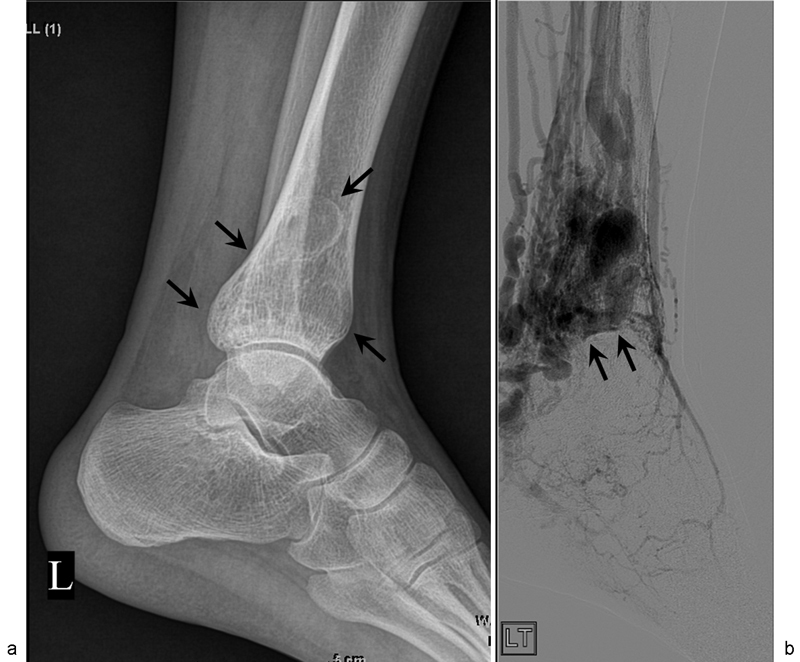
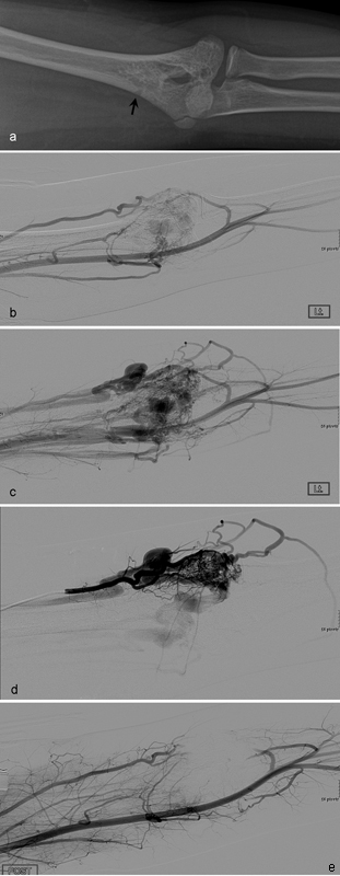
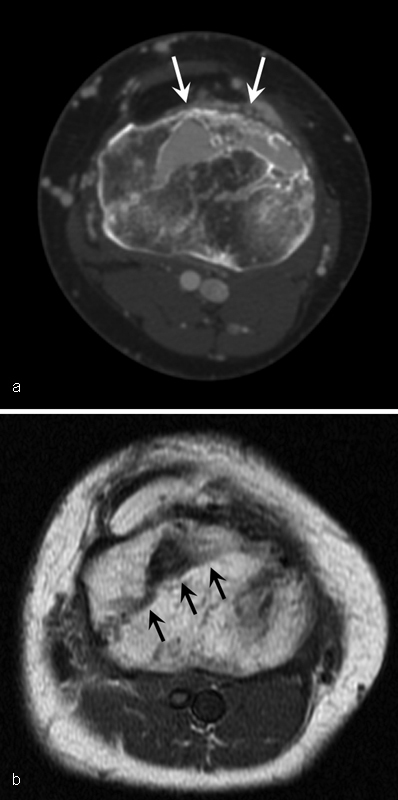
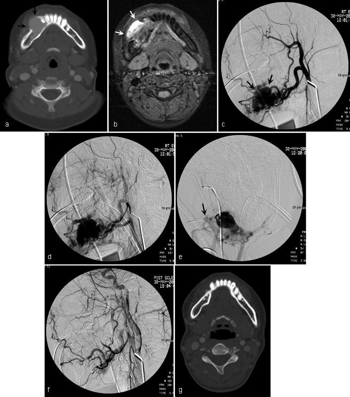
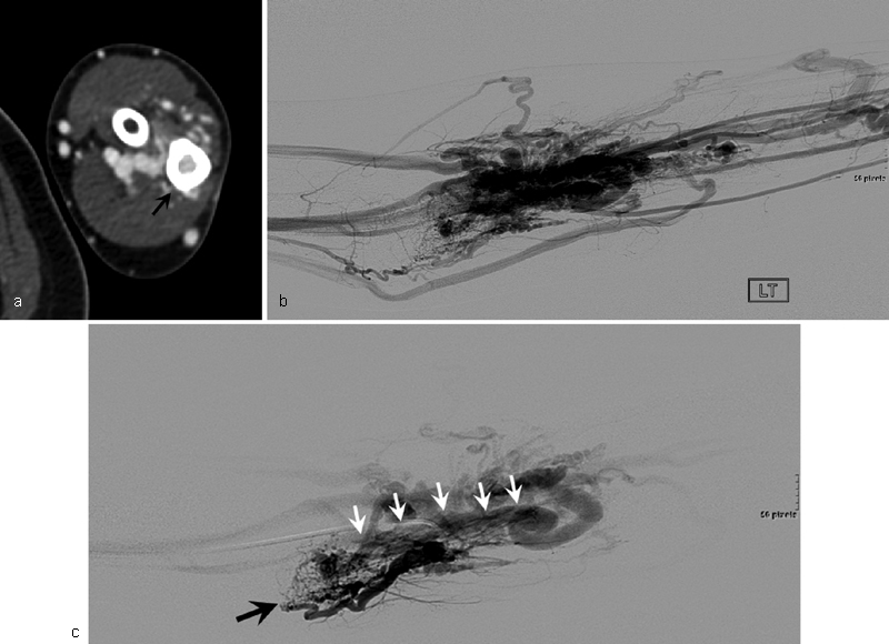
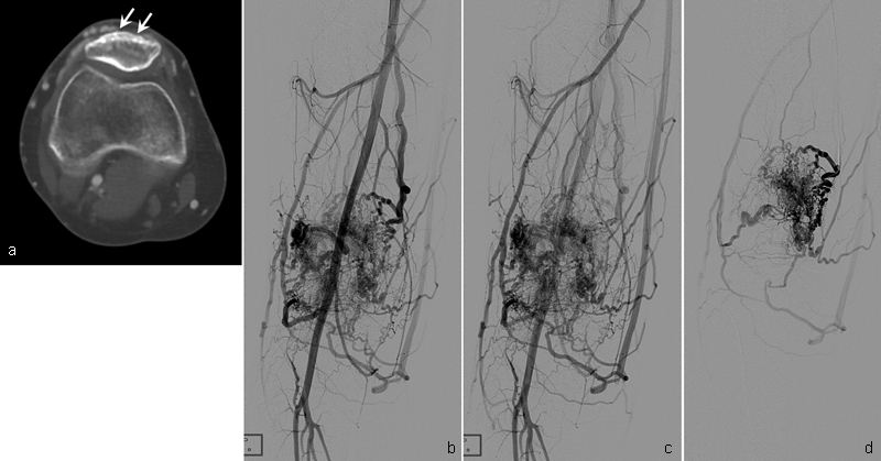
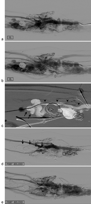
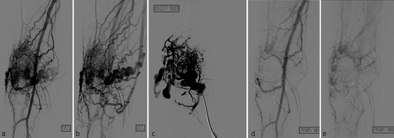
Intraosseous arteriovenous malformations (AVMs) have been associated with distortion, hypertrophy, osteolytic skeletal changes, bleeding, leg length discrepancy, and pathologic fracture. Computed tomography or magnetic resonance imaging is helpful in the evaluation of the extent and depth of intraosseous AVMs and associated soft-tissue AVMs. Treatment approaches can differ, depending on the angiographic classification. Embolotherapy with ethanol, coils, or n-butyl cyanoacrylate is the primary treatment for symptomatic intraosseous AVMs, and the goal of treatment is symptom improvement with few complications.

Keywords: embolization; embolotherapy; interventional radiology; intraosseous AVM.


Figures

Fig. 1
Fig. 1
A 21-year-old man with intraosseous arteriovenous malformations (AVMs) at the distal tibia. ( a ) Simple radiography shows large osteolytic lesion in the medulla without cortical expansion (arrows). ( b ) Oblique angiography shows intraosseous AVMs at the distal tibia (arrows).
Fig. 2
Fig. 2
A 12-year-old boy with type III intraosseous arteriovenous malformations (AVMs) at the distal humerus. ( a ) Radiography demonstrates a lucent serpiginous lesion involving the cortex and medulla of the distal humerus (arrow). Pretreatment angiographies in arterial ( b ) and venous ( c ) phases show type III intraosseous AVMs at the distal humerus. ( d ) Selective angiography shows more details of type III intraosseous AVMs. ( e ) Angiography after transarterial injection of 30 mL of absolute ethanol shows complete obliteration of intraosseous AVMs.
Fig. 3
Fig. 3
A 46-year-old woman with intraosseous arteriovenous malformations at the proximal tibia. ( a ) Axial CT shows multiple cortical defect and osteolytic lesion (arrows) with contrast enhancement at the proximal tibia. ( b ) Axial T1-weighted MR image shows signal voiding lesion (arrows) at the proximal tibia.
Fig. 4
Fig. 4
A 15-year-old girl with type II intraosseous arteriovenous malformations (AVMs) of the mandible. ( a ) Axial CT shows contrast-enhanced osteolytic lesion (arrows) with cortical erosion of the mandible. ( b ) Axial fat-suppressed gradient-echo MR image shows high signal lesion (arrows) at the mandible. Pretreatment lateral angiographies in arterial ( c ) and venous ( d ) phases show type II intraosseous AVMs (arrows). ( e ) Embolotherapy was performed with 10 mL of absolute ethanol through a direct-punctured needle (arrow). ( f ) Completion angiography shows complete obliteration of intraosseous AVMs. ( g ) Follow-up axial CT image 4 years later shows no residual intraosseous AVMs.
Fig. 5
Fig. 5
A 32-year-old woman with type II intraosseous arteriovenous malformations (AVMs) of the proximal ulna. ( a ) Axial CT shows contrast-enhanced osteolytic lesion (arrow) in the medulla of the ulna. ( b ) Angiography shows AVMs at the forearm. ( c ) Selective angiography shows the feeding artery (black arrow) of AVMs and intraosseous type II AVMs (white arrows) at the proximal ulna.
Fig. 6
Fig. 6
A 21-year-old woman with type III intraosseous arteriovenous malformations (AVMs) at the patella. ( a ) Axial CT shows multiple small enhancing osteolytic lesions (arrows) in the cortex of the patella. Angiography in early arterial ( b ) and late arterial ( c ) phases show type III intraosseous AVMs at the patella. ( d ) Selective angiography shows more details of type III intraosseous AVMs.
Fig. 7
Fig. 7
A 24-year-old woman with type II intraosseous arteriovenous malformations (AVMs) at the forearm and upper arm. She underwent four sessions of embolotherapy in the past 3 years. Pretreatment angiographies in arterial ( a ) and venous ( b ) phases show AVMs at the forearm and upper arm. ( c ) The draining vein (arrows) of the intraosseous AVMs (arrow heads) was punctured with a 21-gauge needle and a microwire was inserted into the intraosseous AVMs. After insertion of a microcatheter in the intraosseous AVMs, embolotherapy was performed with multiple microcoils and 20 mL of absolute ethanol. Posttreatment angiography in arterial ( d ) and venous ( e ) phases shows complete obliteration of the forearm AVMs. Note multiple coils (arrows) in the medulla of the proximal radius. Most dilated vessels of Fig. b ) in the forearm are the dilated draining veins of intraosseous type II AVMs.
Fig. 8
Fig. 8
A 15-year-old boy with type III intraosseous arteriovenous malformations (AVMs) at the patella. Pretreatment angiographies in arterial ( a ) and venous ( b ) phases show type III intraosseous AVMs at the patella. ( c ) Angiography after direct puncture of the intraosseous AVMs with a needle shows more details of type III intraosseous AVMs. During angiography, BP cuff at the thigh was inflated to 200 mm Hg to slow down the blood flow. Embolotherapy was performed with 10 mL of absolute ethanol under BP cuff inflation. Completion angiographies in arterial ( d ) and venous ( e ) phases show markedly reduced intraosseous AVMs.

References

    1. Do Y S, Park K B, Park H S et al.Extremity arteriovenous malformations involving the bone: therapeutic outcomes of ethanol embolotherapy. J Vasc Interv Radiol. 2010;21(06):807–816. - PubMed
    1. Savader S J, Savader B L, Otero R R. Intraosseous arteriovenous malformations mimicking malignant disease. Acta Radiol. 1988;29(01):109–114. - PubMed
    1. Boyd J B, Mulliken J B, Kaban L B, Upton J, III, Murray J E. Skeletal changes associated with vascular malformations. Plast Reconstr Surg. 1984;74(06):789–797. - PubMed
    1. Szilagyi D E, Smith R F, Elliott J P, Hageman J H. Congenital arteriovenous anomalies of the limbs. Arch Surg. 1976;111(04):423–429. - PubMed
    1. Flye M W, Jordan B P, Schwartz M Z. Management of congenital arteriovenous malformations. Surgery. 1983;94(05):740–747. - PubMed