Signal changes in standing magnetic resonance imaging of osseous injury at the origin of the suspensory ligament in four Thoroughbred racehorses under tiludronic acid treatment
- PMID: 28955160
- PMCID: PMC5608961
- DOI: 10.1294/jes.28.87
Signal changes in standing magnetic resonance imaging of osseous injury at the origin of the suspensory ligament in four Thoroughbred racehorses under tiludronic acid treatment
Abstract
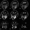
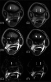
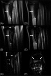
Problems associated with the proximal metacarpal region, such as an osseous injury associated with tearing of Sharpey's fibers or an avulsion fracture of the origin of the suspensory ligament (OISL), are important causes of lameness in racehorses. In the present study, four Thoroughbred racehorses (age range, 2-4 years) were diagnosed as having forelimb OISL and assessed over time by using standing magnetic resonance imaging (sMRI). At the first sMRI examination, all horses had 3 characteristic findings, including low signal intensity within the trabecular bone of the third metacarpus on T1-weighted images, intermediate-to-high signal intensity surrounded by a hypointense rim on T2*-weighted images, and high signal intensity on fat-suppressed images. Following the sMRI examination, all horses received 50 mg of tiludronic acid by intravenous regional limb perfusion once weekly for 3 weeks. Attenuation of the high signal intensity on T2*-weighted and fat-suppressed images was observed on follow-up sMRI in 3 horses. Following rest and rehabilitation, these 3 horses successfully returned to racing. In contrast, the other horse that did not show attenuation of the high signal intensity failed to return to racing. To our knowledge, this is the first report of OISL in Thoroughbred racehorses assessed over time by sMRI under tiludronic acid treatment. Our findings support the use of sMRI for examining lameness originating from the proximal metacarpal region to refine the timing of returning to exercise based on follow-up examinations during the recuperation period.
Keywords: magnetic resonance imaging; osseous injury; proximal metacarpus; racehorse; tiludronic acid.
Figures


Similar articles
-
Use of standing low-field magnetic resonance imaging to assess oblique distal sesamoidean ligament desmitis in three Thoroughbred racehorses.J Vet Med Sci. 2016 Oct 1;78(9):1475-1480. doi: 10.1292/jvms.15-0656. Epub 2016 Jun 16. J Vet Med Sci. 2016. PMID: 27320360 Free PMC article.
-
Standing magnetic resonance imaging of distal phalanx fractures in 6 cases of Thoroughbred racehorse.J Vet Med Sci. 2019 May 11;81(5):689-693. doi: 10.1292/jvms.18-0183. Epub 2019 Mar 21. J Vet Med Sci. 2019. PMID: 30905907 Free PMC article.
-
Standing magnetic resonance imaging detection of bone marrow oedema-type signal pattern associated with subcarpal pain in 8 racehorses: a prospective study.Equine Vet J. 2010 Jan;42(1):10-7. doi: 10.2746/042516409X471467. Equine Vet J. 2010. PMID: 20121907
-
[Disorders of the origin of the suspensory ligament in the horse: a diagnostic challenge].Schweiz Arch Tierheilkd. 2006 Feb;148(2):86-97. doi: 10.1024/0036-7281.148.2.86. Schweiz Arch Tierheilkd. 2006. PMID: 16509170 Review. German.
-
Musculoskeletal injury in thoroughbred racehorses: correlation of findings using multiple imaging modalities.Vet Clin North Am Equine Pract. 2012 Dec;28(3):539-61. doi: 10.1016/j.cveq.2012.09.005. Epub 2012 Oct 18. Vet Clin North Am Equine Pract. 2012. PMID: 23177131 Review.
Cited by
-
The Evolution of Lesions on Follow-Up Magnetic Resonance Imaging of the Proximal Metacarpal Region in Non-Racing Sport Horses That Returned to Work (2015-2023).Animals (Basel). 2024 Jun 8;14(12):1731. doi: 10.3390/ani14121731. Animals (Basel). 2024. PMID: 38929351 Free PMC article.
References
-
- Bertone A.L. 2011. Suspensory ligament desmitis. pp. 644–648. In: Adams and Stashak’s Lameness in Horses, 6th ed. (Baxter, G.M. ed.), Blackwell Publishing, Ltd., West Sussex.
-
- Bischofberger A.S., Konar M., Ohlerth S., Geyer H., Lang J., Ueltschi G., Lischer C.J. 2006. Magnetic resonance imaging, ultrasonography and histology of the suspensory ligament origin: a comparative study of normal anatomy of warmblood horses. Equine Vet. J. 38: 508–516. - PubMed
-
- Brokken M.T., Schneider R.K., Sampson S.N., Tucker R.L., Gavin P.R., Ho C.P. 2007. Magnetic resonance imaging features of proximal metacarpal and metatarsal injuries in the horse. Vet. Radiol. Ultrasound 48: 507–517. - PubMed
-
- Butler J. 2000. Clinical radiology of the horse. pp. 158–165. In: Clinical Radiology of the Horse, 2nd ed. (Butler, J.A., Colles, C.M., Dyson, S.J., Kold, S.E. and Poulos, P.W. eds.), Blackwell Science, Ltd., Oxford.
-
- Carpenter R.S. 2012. How to treat dorsal metacarpal disease with regional tiludronate and extracorporeal shock wave therapies in thoroughbred racehorses. Proceedings of the 58th annual convention of the American Association of Equine Practitioners 58: 546–549.
LinkOut - more resources
Full Text Sources
Other Literature Sources
