. 2017 Sep 25;90(3):351-360.
eCollection 2017 Sep.

Changes in Muscle Metabolism are Associated with Phenotypic Variability in Golden Retriever Muscular Dystrophy

Affiliations

Changes in Muscle Metabolism are Associated with Phenotypic Variability in Golden Retriever Muscular Dystrophy

Peter P Nghiem et al. Yale J Biol Med. .

Abstract

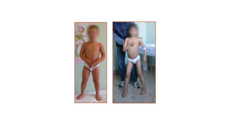
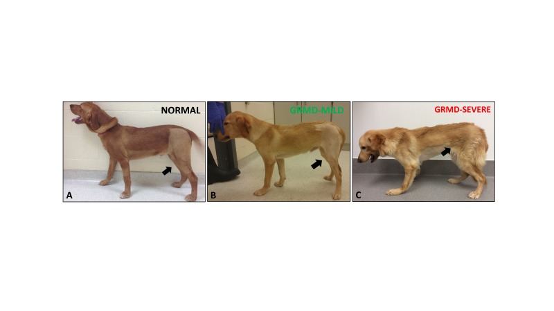
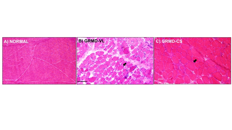
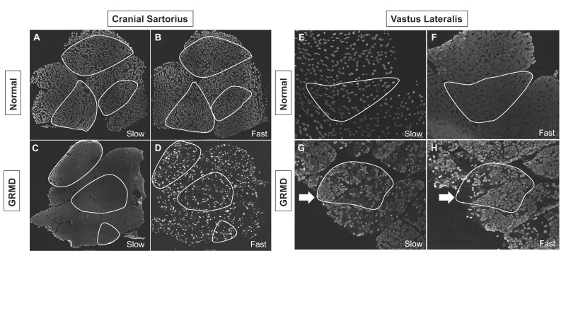
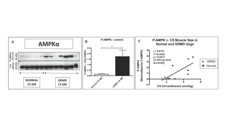
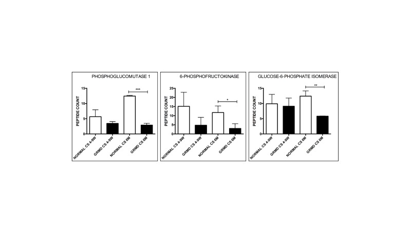
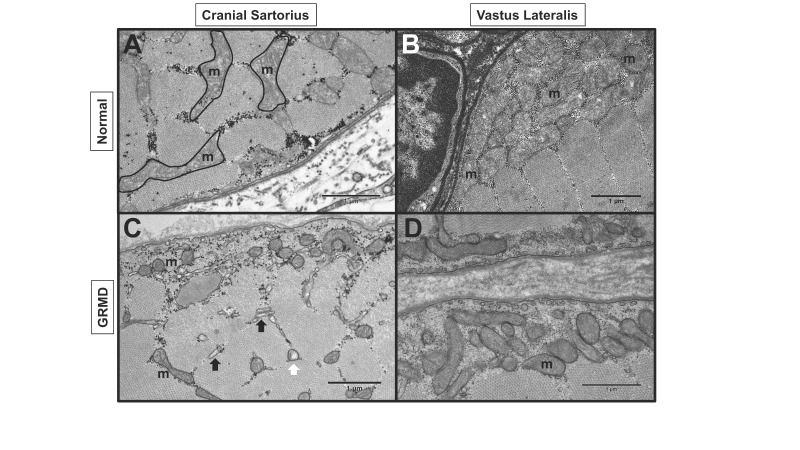
Duchenne muscular dystrophy (DMD) is an X-chromosome-linked disorder and the most common monogenic disease in people. Affected boys are diagnosed at a young age, become non-ambulatory by their early teens, and succumb to cardiorespiratory failure by their thirties. Despite being a monogenic condition resulting from mutations in the DMD gene, affected boys have noteworthy phenotypic variability. Efforts have identified genetic modifiers that could modify disease progression and be pharmacologic targets. Dogs affected with golden retriever muscular dystrophy (GRMD) have absent dystrophin and demonstrate phenotypic variability at the functional, histopathological, and molecular level. Our laboratory is particularly interested in muscle metabolism changes in dystrophin-deficient muscle. We identified several metabolic alterations, including myofiber type switching from fast (type II) to slow (type I), reduced glycolytic enzyme expression, reduced and morphologically abnormal mitochondria, and differential AMP-kinase phosphorylation (activation) between hypertrophied and wasted muscle. We hypothesize that muscle metabolism changes are, in part, responsible for phenotypic variability in GRMD. Pharmacological therapies aimed at modulating muscle metabolism can be tested in GRMD dogs for efficacy.

Keywords: AMPK; Duchenne; dystrophin; golden retriever muscular dystrophy; metabolism; muscle; phenotype.


Figures

Figure 1
Figure 1
Phenotypic variation was observed in DMD boys. Left: Mildly affected DMD patient. Note the apparent leg musculature enlargement. Right: Severely affected DMD boy with muscle wasting and weakness in the arms and legs. Note the boy needed assistance to stand. Photos courtesy of L.B.
Figure 2
Figure 2
GRMD dogs showed significant variation in phenotype. A) Normal dog (Winthrop) at 6 months of age. B) Mildly affected GRMD dog (Lunes) at 6 months of age. Note the mild muscle atrophy of the pelvic limbs (black arrows) and mild hyperextension of the carpal joints. C) Severely affected GRMD dog (Kermit) at 6 months. Note the prominent joint angle changes and postural deficits, particularly at the carpal, tibiotarsal, stifle, and hip joints, which were due to severe muscle weakness/wasting and joint contractures.
Figure 3
Figure 3
H&E staining: Muscles from GRMD dogs were variably affected. A) Normal muscle. Note the similar-sized myofibers, minimal connective tissue, and lack of immune cell infiltration. B) VL muscle of a GRMD dog showed degeneration and regeneration (myofiber size variation, black arrow), increased fibrosis and connective tissue, and increased immune cell infiltration. C) The CS muscle and its myofibers in GRMD were hypertrophied (black arrow), as noted by their increased myofiber diameter compared to GRMD VL and normal muscle. Marker = 50 microns.
Figure 4
Figure 4
Dystrophin-deficient GRMD dog muscle underwent fiber type switching. In GRMD cranial sartorius (CS) and vastus lateralis (VL) muscle, there was reduction in the number of fast staining fibers, while nearly every fiber stained with the myosin heavy chain (MHC) slow twitch antibody (gray colors). A: Normal CS: Slow twitch MHC staining. B: Normal CS: Fast twitch MHC staining. C: GRMD CS: Slow twitch MHC staining. D: GRMD CS: Fast twitch MHC staining. E: Normal VL: Slow twitch MHC staining. F: Normal VL: Fast twitch MHC staining. G: GRMD VL: Slow twitch MHC staining. H: GRMD VL: Fast twitch MHC staining. All dogs shown at ~6 months of age. Images A-D were 3X3 stitched 4X objective images displaying the entire muscle biopsy sections. Images E-H were single images collected with a 4X objective. Outlined white areas highlight representative serially sectioned muscle fascicles used for identifying slow and fast twitch fiber type variation. White arrows indicate serially sectioned area in the GRMD VL that had increased slow twitch staining.
Figure 5
Figure 5
The hypertrophied CS muscle had increased phosphorylated (P) AMPKα compared to normal. A) Note the variability in western blot expression of P-AMPKα in GRMD dog samples. B) P-AMPKα was normalized to Total AMPKα protein and increased compared to normal samples. * = p < 0.05. C) P-AMPKα was positively correlated with CS circumference.
Figure 6
Figure 6
Glycolytic enzymes were reduced in hypertrophied GRMD CS muscle at 6 months of age compared to normal. Phosphoglucomutase-1, 6-phophofructokinase, and glucose-6-phosphate isomerase were reduced at 6 months (M) in GRMD cranial sartorius (CS) (black bars) compared to normal (white bars). There was not a significant change at 4 to 9 weeks (W). N = 3 for each group. Total spectral counts were used to determine relative abundance. 
* = P < 0.05; ** p < 0.01; *** p < 0.001.
Figure 7
Figure 7
GRMD dog muscle had abnormal mitochondria (m). Transmission electron microscopy (TEM) of normal and GRMD cranial sartorius (CS) and vastus lateralis (VL) at 6 to 12 months of age. A: Normal CS with intermyofibrillar mitochondria. B: Normal VL with numerous subsarcolemmal mitochondria C: GRMD CS with reduced size and density of subsarcolemmal and intermyofibrillar mitochondria, swollen (white arrow) mitochondria and dilated sarcoplasmic reticulum (black arrows). D: GRMD VL with reduced subsarcolemmal mitochondria.

References

    1. Hoffman EP, Brown RH, Kunkel LM. Dystrophin: the protein product of the Duchenne muscular dystrophy locus. Cell. 1987;51:919–928. - PubMed
    1. Monaco AP, Bertelson CJ, Liechti-Gallati S, Moser H, Kunkel LM. An explanation for the phenotypic differences between patients bearing partial deletions of the DMD locus. Genomics. 1988;2(1):90–95. - PubMed
    1. Aartsma-Rus A, Van Deutekom JC, Fokkema IF, Van Ommen GJ, Den Dunnen JT. Entries in the Leiden Duchenne muscular dystrophy mutation database: an overview of mutation types and paradoxical cases that confirm the reading-frame rule. Muscle Nerve. 2006;34(2):135–144. - PubMed
    1. Emery AE. The muscular dystrophies. Lancet. 2002;359(9307):687-95. - PubMed
    1. Bulfield G, Siller WG, Wight PA, Moore KJ. X chromosome-linked muscular dystrophy (mdx) in the mouse. Proc Natl Acad Sci USA. 1984;81(4):1189–1192. - PMC - PubMed

Publication types

LinkOut - more resources