

The EEG Split Alpha Peak: Phenomenological Origins and Methodological Aspects of Detection and Evaluation
- PMID: 28955192
- PMCID: PMC5601034
- DOI: 10.3389/fnins.2017.00506
The EEG Split Alpha Peak: Phenomenological Origins and Methodological Aspects of Detection and Evaluation
Abstract
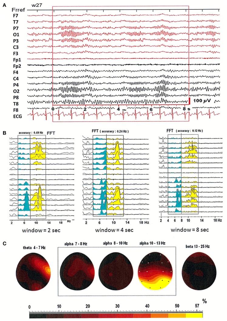
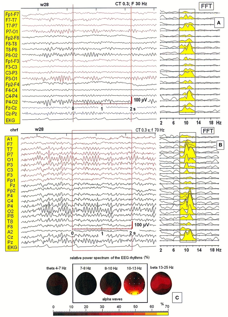
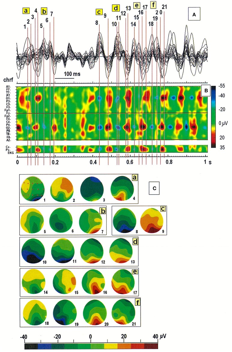
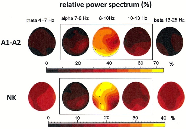
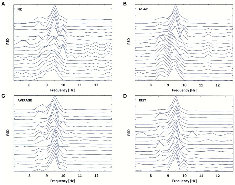
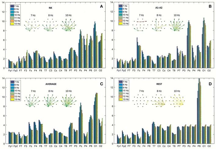
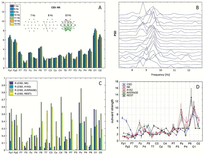
Electroencephalographic (EEG) patterns were analyzed in a group of ambulatory patients who ranged in age and sex using spectral analysis as well as Directed Transfer Function, a method used to evaluate functional brain connectivity. We tested the impact of window size and choice of reference electrode on the identification of two or more peaks with close frequencies in the spectral power distribution, so called "split alpha." Together with the connectivity analysis, examination of spatiotemporal maps showing the distribution of amplitudes of EEG patterns allowed for better explanation of the mechanisms underlying the generation of split alpha peaks. It was demonstrated that the split alpha spectrum can be generated by two or more independent and interconnected alpha wave generators located in different regions of the cerebral cortex, but not necessarily in the occipital cortex. We also demonstrated the importance of appropriate reference electrode choice during signal recording. In addition, results obtained using the original data were compared with results obtained using re-referenced data, using average reference electrode and reference electrode standardization techniques.
Keywords: average reference; directed transfer function; functional brain connectivity; reference electrode standardization technique (REST); spectral analysis; split EEG alpha peaks.
Figures
Similar articles
-
Is So Called "Split Alpha" in EEG Spectral Analysis a Result of Methodological and Interpretation Errors?Front Neurosci. 2020 Nov 26;14:608453. doi: 10.3389/fnins.2020.608453. eCollection 2020. Front Neurosci. 2020. PMID: 33324157 Free PMC article.
-
Hemifield-dependent N1 and event-related theta/delta oscillations: An unbiased comparison of surface Laplacian and common EEG reference choices.Int J Psychophysiol. 2015 Sep;97(3):258-70. doi: 10.1016/j.ijpsycho.2014.12.011. Epub 2015 Jan 3. Int J Psychophysiol. 2015. PMID: 25562833 Free PMC article.
-
How Different EEG References Influence Sensor Level Functional Connectivity Graphs.Front Neurosci. 2017 Jul 5;11:368. doi: 10.3389/fnins.2017.00368. eCollection 2017. Front Neurosci. 2017. PMID: 28725175 Free PMC article.
-
A method to standardize a reference of scalp EEG recordings to a point at infinity.Physiol Meas. 2001 Nov;22(4):693-711. doi: 10.1088/0967-3334/22/4/305. Physiol Meas. 2001. PMID: 11761077 Review.
-
Brain neural synchronization and functional coupling in Alzheimer's disease as revealed by resting state EEG rhythms.Int J Psychophysiol. 2016 May;103:88-102. doi: 10.1016/j.ijpsycho.2015.02.008. Epub 2015 Feb 7. Int J Psychophysiol. 2016. PMID: 25660305 Review.
Cited by
-
Complexity Analysis of EEG Data in Persons With Depression Subjected to Transcranial Magnetic Stimulation.Front Physiol. 2018 Sep 28;9:1385. doi: 10.3389/fphys.2018.01385. eCollection 2018. Front Physiol. 2018. PMID: 30323771 Free PMC article.
-
Is So Called "Split Alpha" in EEG Spectral Analysis a Result of Methodological and Interpretation Errors?Front Neurosci. 2020 Nov 26;14:608453. doi: 10.3389/fnins.2020.608453. eCollection 2020. Front Neurosci. 2020. PMID: 33324157 Free PMC article.
-
EEG Phase Synchronization in Persons With Depression Subjected to Transcranial Magnetic Stimulation.Front Neurosci. 2019 Jan 14;12:1037. doi: 10.3389/fnins.2018.01037. eCollection 2018. Front Neurosci. 2019. PMID: 30692906 Free PMC article.
-
Commentary: Is So-Called "Split Alpha" in EEG Spectral Analysis a Result of Methodological and Interpretation Errors?Front Neurosci. 2021 Sep 24;15:726912. doi: 10.3389/fnins.2021.726912. eCollection 2021. Front Neurosci. 2021. PMID: 34630016 Free PMC article. No abstract available.
-
Relaxing Effects of Breathing Pseudotsuga menziesii and Lavandula angustifolia Essential Oils on Psychophysiological Status in Older Adults.Int J Environ Res Public Health. 2022 Nov 18;19(22):15251. doi: 10.3390/ijerph192215251. Int J Environ Res Public Health. 2022. PMID: 36429972 Free PMC article.
References
-
- Blinowska K. J., Kaminski M. J. (2006). Multivariate signal analysis by parametric models, in Handbook of time Series Analysis. Recent Theoretical Developments and Applications, eds Schelter B., Winterhalder M., Timmer J. (Wernheim: Wiley; – VCH), 373–411. 10.1002/9783527609970.ch15 - DOI
LinkOut - more resources
Full Text Sources
Other Literature Sources