

Hydrogen Sulfide Facilitates the Impaired Sensitivity of Carotid Sinus Baroreflex in Rats with Vascular Calcification
- PMID: 28955233
- PMCID: PMC5601057
- DOI: 10.3389/fphar.2017.00629
Hydrogen Sulfide Facilitates the Impaired Sensitivity of Carotid Sinus Baroreflex in Rats with Vascular Calcification
Abstract
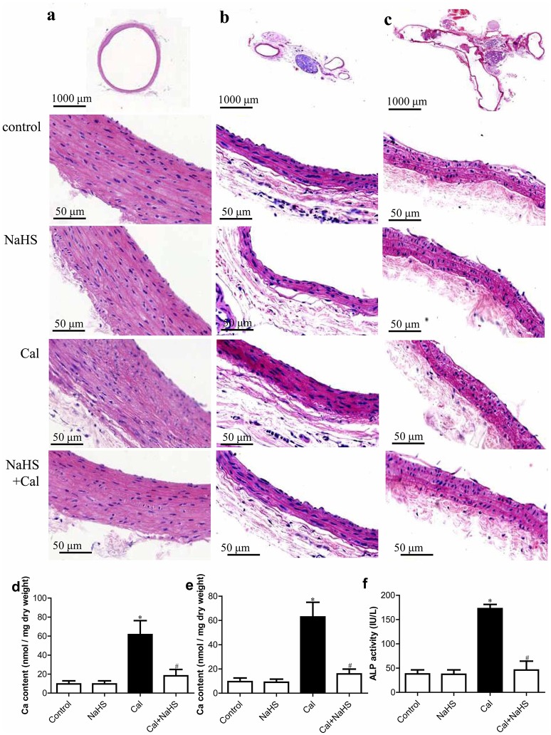
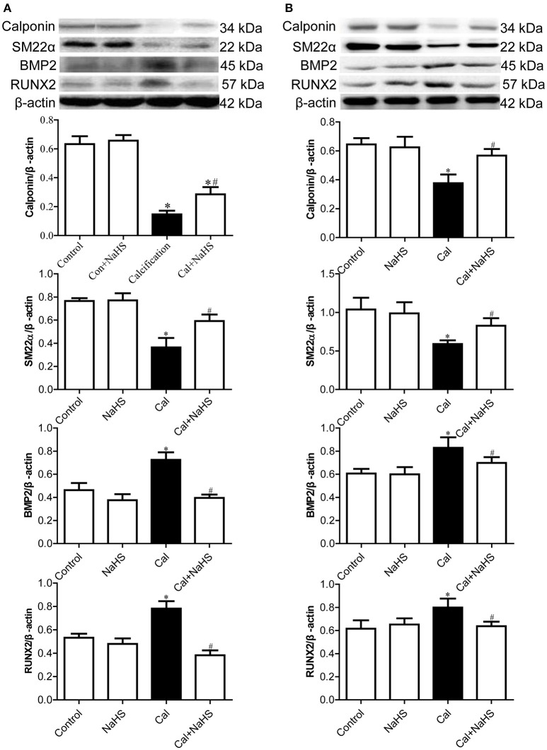
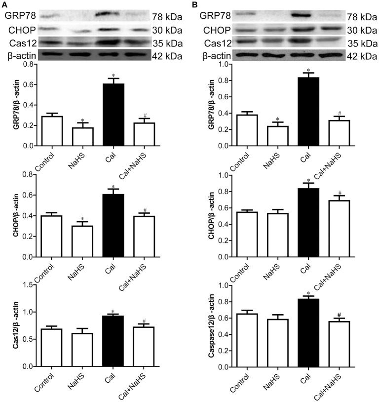
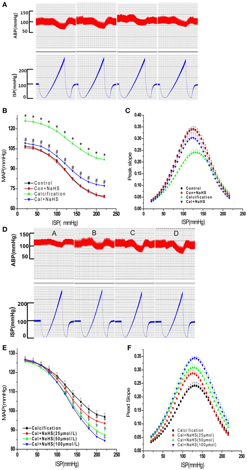
Arterial baroreflex is a general mechanism maintaining cardiovascular homeostasis; its sensitivity is reduced in vascular calcification (VC). Hydrogen sulfide (H2S) treatment facilitates baroreflexive sensitivity in normal and hypertensive rats. Here, we aimed to detect the effect of H2S on baroreflexive sensitivity in rats with VC. The rat VC model was induced by vitamin D3 plus nicotine for 4 weeks. The sensitivity of baroreflex was detected by perfusing the isolated carotid sinus. VC was assessed by hematoxylin and eosin (H&E) staining, Ca2+ content and alkaline phosphatase (ALP) activity. Protein levels were detected by western blot analysis. Vitamin D3 plus nicotine induced structural disorder and elevated Ca2+ content in the aortic and carotid arterial wall and increased plasma ALP activity. In the calcified aorta and carotid artery, protein levels of contractile phenotype markers of vascular smooth muscle cells (VSMCs) were downregulated and that of osteoblast-like phenotype markers and endoplasmic reticulum stress (ERS) markers were upregulated. NaHS treatment ameliorated the histologic disorder and Ca2+ content in the calcified aorta and carotid artery, inhibited the elevated plasma ALP activity, and prevented the transformation of the VSMC phenotype and activation of ERS in rats with VC. Chronic NaHS treatment prevented the impairment of the baroreflex sensitivity and acute NaHS treatment dose-dependently improved the sensitivity in rats with VC. Our results suggested that H2S could directly facilitate the impairment of baroreflex in rats with VC and ameliorate VC, which might provide new target and strategy for regulation of the baroreflex and therapy of VC.
Keywords: baroreflex; endoplasmic reticulum stress; hydrogen sulfide; perfusion of isolated carotid sinus; vascular calcification.
Figures
Similar articles
-
Stellate ganglion block ameliorates vascular calcification by inhibiting endoplasmic reticulum stress.Life Sci. 2018 Jan 15;193:1-8. doi: 10.1016/j.lfs.2017.12.002. Epub 2017 Dec 5. Life Sci. 2018. PMID: 29208463
-
Hydrogen Sulfide Improves Vascular Calcification in Rats by Inhibiting Endoplasmic Reticulum Stress.Oxid Med Cell Longev. 2016;2016:9095242. doi: 10.1155/2016/9095242. Epub 2016 Feb 28. Oxid Med Cell Longev. 2016. PMID: 27022436 Free PMC article.
-
Inhibition of endoplasmic reticulum stress mediates the ameliorative effect of apelin on vascular calcification.J Mol Cell Cardiol. 2021 Mar;152:17-28. doi: 10.1016/j.yjmcc.2020.11.017. Epub 2020 Dec 3. J Mol Cell Cardiol. 2021. PMID: 33279504
-
Hydrogen sulfide ameliorates vascular calcification induced by vitamin D3 plus nicotine in rats.Acta Pharmacol Sin. 2006 Mar;27(3):299-306. doi: 10.1111/j.1745-7254.2006.00283.x. Acta Pharmacol Sin. 2006. PMID: 16490165
-
The Preventive Effects of Xanthohumol on Vascular Calcification Induced by Vitamin D3 Plus Nicotine.Antioxidants (Basel). 2020 Oct 6;9(10):956. doi: 10.3390/antiox9100956. Antioxidants (Basel). 2020. PMID: 33036258 Free PMC article.
Cited by
-
Fibroblast Growth Factor 21 Attenuates Vascular Calcification by Alleviating Endoplasmic Reticulum Stress Mediated Apoptosis in Rats.Int J Biol Sci. 2019 Jan 6;15(1):138-147. doi: 10.7150/ijbs.28873. eCollection 2019. Int J Biol Sci. 2019. PMID: 30662354 Free PMC article.
-
Hydrogen Sulfide (H2S)-Releasing Compounds: Therapeutic Potential in Cardiovascular Diseases.Front Pharmacol. 2018 Sep 21;9:1066. doi: 10.3389/fphar.2018.01066. eCollection 2018. Front Pharmacol. 2018. PMID: 30298008 Free PMC article. Review.
-
Mechanism of homocysteine-mediated endothelial injury and its consequences for atherosclerosis.Front Cardiovasc Med. 2023 Jan 16;9:1109445. doi: 10.3389/fcvm.2022.1109445. eCollection 2022. Front Cardiovasc Med. 2023. PMID: 36727029 Free PMC article. Review.
-
Salusin-β Promotes Vascular Calcification via Nicotinamide Adenine Dinucleotide Phosphate/Reactive Oxygen Species-Mediated Klotho Downregulation.Antioxid Redox Signal. 2019 Dec 20;31(18):1352-1370. doi: 10.1089/ars.2019.7723. Antioxid Redox Signal. 2019. PMID: 31578871 Free PMC article.
-
Endogenous hydrogen sulfide improves vascular remodeling through PPARδ/SOCS3 signaling.J Adv Res. 2020 Jun 20;27:115-125. doi: 10.1016/j.jare.2020.06.005. eCollection 2021 Jan. J Adv Res. 2020. PMID: 33318871 Free PMC article. Review.
References
LinkOut - more resources
Full Text Sources
Other Literature Sources
Miscellaneous