

Kaempferia parviflora Extract Exhibits Anti-cancer Activity against HeLa Cervical Cancer Cells
- PMID: 28955234
- PMCID: PMC5600991
- DOI: 10.3389/fphar.2017.00630
Kaempferia parviflora Extract Exhibits Anti-cancer Activity against HeLa Cervical Cancer Cells
Abstract
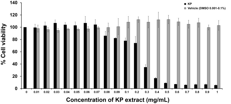
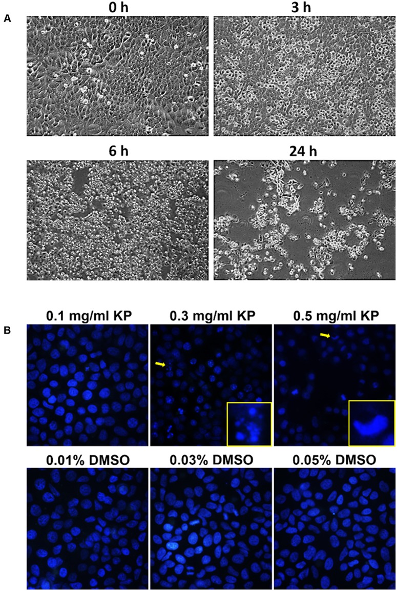
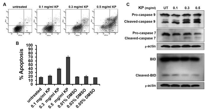
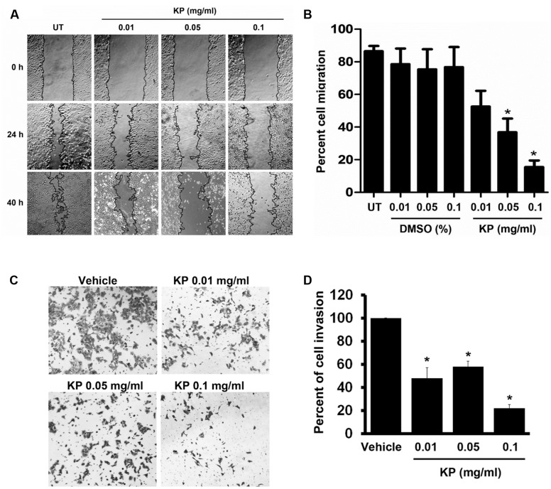
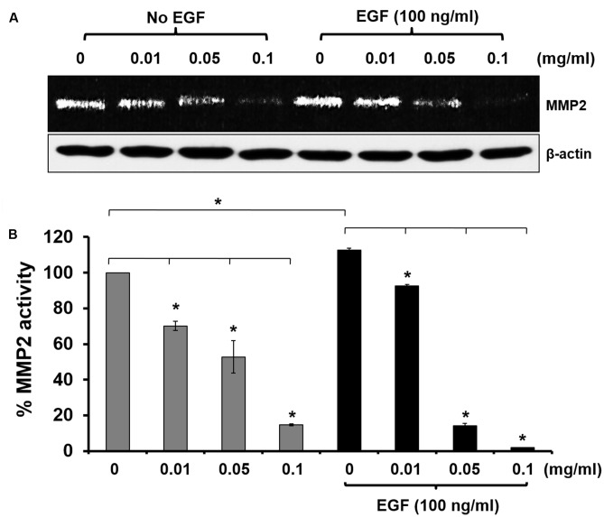
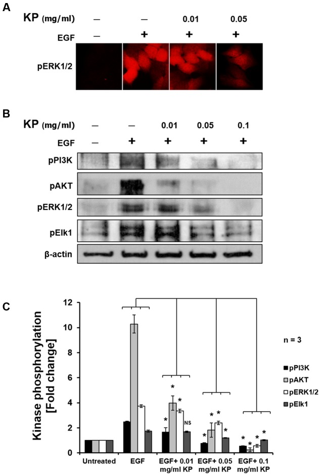
Kaempferia parviflora (KP) has been traditionally used as a folk remedy to treat several diseases including cancer, and several studies have reported cytotoxic activities of extracts of KP against a number of different cancer cell lines. However, many aspects of the molecular mechanism of action of KP remain unclear. In particular, the ability of KP to regulate cancer cell growth and survival signaling is still largely unexplored. The current study aimed to investigate the effects of KP on cell viability, cell migration, cell invasion, cell apoptosis, and on signaling pathways related to growth and survival of cervical cancer cells, HeLa. We discovered that KP reduced HeLa cell viability in a concentration-dependent manner. The potent cytotoxicity of KP against HeLa cells was associated with a dose-dependent induction of apoptotic cell death as determined by flow cytometry and observation of nuclear fragmentation. Moreover, KP-induced cell apoptosis was likely to be mediated through the intrinsic apoptosis pathway since caspase 9 and caspase 7, but not BID, were shown to be activated after KP exposure. Based on the observation that KP induced apoptosis in HeLa cell, we further investigated the effects of KP at non-cytotoxic concentrations on suppressing signal transduction pathways relevant to cell growth and survival. We found that KP suppressed the MAPK and PI3K/AKT signaling pathways in cells activated with EGF, as observed by a significant decrease in phosphorylation of ERK1/2, Elk1, PI3K, and AKT. The data suggest that KP interferes with the growth and survival of HeLa cells. Consistent with the inhibitory effect on EGF-stimulated signaling, KP potently suppressed the migration of HeLa cells. Concomitantly, KP was demonstrated to markedly inhibit HeLa cell invasion. The ability of KP in suppressing the migration and invasion of HeLa cells was associated with the suppression of matrix metalloproteinase-2 production. These data strongly suggest that KP may slow tumor progression and metastasis in patients with cervical cancer. Taken together, the present report provides accumulated evidence revealing the potent anti-cancer activities of Kaempferia parviflora against cervical cancer HeLa cells, and suggests its potential use as an alternative way for cervical cancer prevention and therapy.
Keywords: Kaempferia parviflora; MAPK pathway; PI3K/AKT pathway; anti-cancer; apoptosis; cervical cancer; invasion; migration.
Figures
Similar articles
-
Kaempferia parviflora Extract Inhibits STAT3 Activation and Interleukin-6 Production in HeLa Cervical Cancer Cells.Int J Mol Sci. 2019 Aug 29;20(17):4226. doi: 10.3390/ijms20174226. Int J Mol Sci. 2019. PMID: 31470515 Free PMC article.
-
Anti-cancer effects of Kaempferia parviflora on ovarian cancer SKOV3 cells.BMC Complement Altern Med. 2018 Jun 11;18(1):178. doi: 10.1186/s12906-018-2241-6. BMC Complement Altern Med. 2018. PMID: 29891015 Free PMC article.
-
Ethanolic extract of Kaempferia parviflora interrupts the mechanisms-associated rheumatoid arthritis in SW982 culture model via p38/STAT1 and STAT3 pathways.Phytomedicine. 2019 Jun;59:152755. doi: 10.1016/j.phymed.2018.11.015. Epub 2018 Nov 14. Phytomedicine. 2019. PMID: 31005814
-
Kaempferia parviflora and Its Methoxyflavones: Chemistry and Biological Activities.Evid Based Complement Alternat Med. 2018 Dec 16;2018:4057456. doi: 10.1155/2018/4057456. eCollection 2018. Evid Based Complement Alternat Med. 2018. PMID: 30643531 Free PMC article. Review.
-
A systematic review and meta-analysis of animal and human studies demonstrates the beneficial effects of Kaempferia parviflora on metabolic syndrome and erectile dysfunction.Nutr Res. 2024 Feb;122:80-91. doi: 10.1016/j.nutres.2023.12.001. Epub 2023 Dec 5. Nutr Res. 2024. PMID: 38194854
Cited by
-
Evaluation of Therapeutic Potential of Eugenol-A Natural Derivative of Syzygium aromaticum on Cervical Cancer.Asian Pac J Cancer Prev. 2018 Jul 27;19(7):1977-1985. doi: 10.22034/APJCP.2018.19.7.1977. Asian Pac J Cancer Prev. 2018. PMID: 30051686 Free PMC article.
-
INHBA is a Prognostic Biomarker and Correlated With Immune Cell Infiltration in Cervical Cancer.Front Genet. 2022 Jan 4;12:705512. doi: 10.3389/fgene.2021.705512. eCollection 2021. Front Genet. 2022. PMID: 35058963 Free PMC article.
-
Kaempferia parviflora Extract Inhibits STAT3 Activation and Interleukin-6 Production in HeLa Cervical Cancer Cells.Int J Mol Sci. 2019 Aug 29;20(17):4226. doi: 10.3390/ijms20174226. Int J Mol Sci. 2019. PMID: 31470515 Free PMC article.
-
The industrially important genus Kaempferia: An ethnopharmacological review.Front Pharmacol. 2023 Feb 27;14:1099523. doi: 10.3389/fphar.2023.1099523. eCollection 2023. Front Pharmacol. 2023. PMID: 36923360 Free PMC article. Review.
-
Effect of Ducrosia flabellifolia and Savignya parviflora Extracts on Inhibition of Human Colon and Prostate Cancer Cell Lines.Curr Issues Mol Biol. 2021 Oct 10;43(3):1518-1528. doi: 10.3390/cimb43030107. Curr Issues Mol Biol. 2021. PMID: 34698080 Free PMC article.
References
-
- Banjerdpongchai R., Chanwikruy Y., Rattanapanone V., Sripanidkulchai B. (2009). Induction of apoptosis in the human Leukemic U937 cell line by Kaempferia parviflora Wall.ex.Baker extract and effects of paclitaxel and camptothecin. Asian Pac. J. Cancer Prev. 10 1137–1140. - PubMed
-
- Banjerdpongchai R., Suwannachot K., Rattanapanone V., Sripanidkulchai B. (2008). Ethanolic rhizome extract from Kaempferia parviflora Wall. ex. Baker induces apoptosis in HL-60 cells. Asian Pac. J. Cancer Prev. 9 595–600. - PubMed
-
- Berube M., Deschambeault A., Boucher M., Germain L., Petitclerc E., Guerin S. L. (2005). MMP-2 expression in uveal melanoma: differential activation status dictated by the cellular environment. Mol. Vis. 11 1101–1111. - PubMed
-
- Canavan T. P., Doshi N. R. (2000). Cervical cancer. Am. Fam. Physician 61 1369–1376. - PubMed
LinkOut - more resources
Full Text Sources
Other Literature Sources
Miscellaneous