

A Preliminary Study on the Pattern, the Physiological Bases and the Molecular Mechanism of the Adductor Muscle Scar Pigmentation in Pacific Oyster Crassostrea gigas
- PMID: 28955252
- PMCID: PMC5600958
- DOI: 10.3389/fphys.2017.00699
A Preliminary Study on the Pattern, the Physiological Bases and the Molecular Mechanism of the Adductor Muscle Scar Pigmentation in Pacific Oyster Crassostrea gigas
Abstract
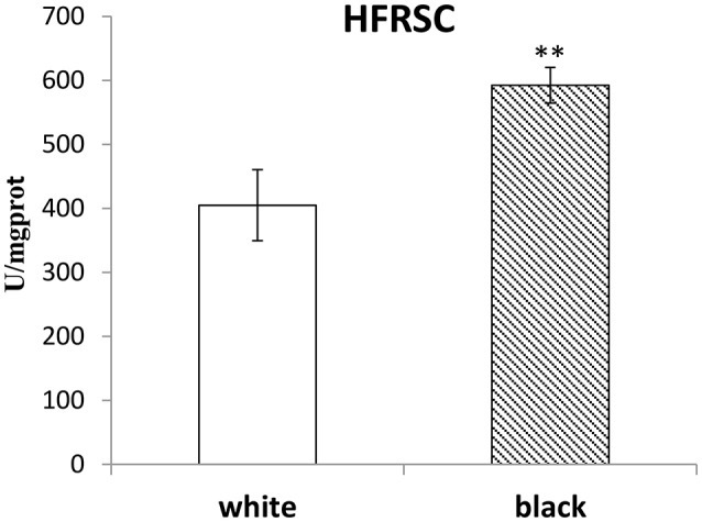
The melanin pigmentation of the adductor muscle scar and the outer surface of the shell are among attractive features and their pigmentation patterns and mechanism still remains unknown in the Pacific oyster Crassostrea gigas. To study these pigmentation patterns, the colors of the adductor muscle scar vs. the outer surface of the shell on the same side were compared. No relevance was found between the colors of the adductor muscle scars and the corresponding outer surface of the shells, suggesting that their pigmentation processes were independent. Interestingly, a relationship between the color of the adductor muscle scars and the dried soft-body weight of Pacific oysters was found, which could be explained by the high hydroxyl free radical scavenging capacity of the muscle attached to the black adductor muscle scar. After the transcriptomes of pigmented and unpigmented adductor muscles and mantles were studied by RNAseq and compared, it was found that the retinol metabolism pathway were likely to be involved in melanin deposition on the adductor muscle scar and the outer surface of the shell, and that the different members of the tyrosinase or Cytochrome P450 gene families could play a role in the independent pigmentation of different organs.
Keywords: Pacific oyster; adductor muscle scar; dried soft-body weight; melanin; outer surface of shell; pigmentation.
Figures


Similar articles
-
Shell Biosynthesis and Pigmentation as Revealed by the Expression of Tyrosinase and Tyrosinase-like Protein Genes in Pacific Oyster (Crassostrea gigas) with Different Shell Colors.Mar Biotechnol (NY). 2021 Oct;23(5):777-789. doi: 10.1007/s10126-021-10063-2. Epub 2021 Sep 6. Mar Biotechnol (NY). 2021. PMID: 34490547
-
Transcriptomic and Physiological Analysis Reveal Melanin Synthesis-Related Genes and Pathways in Pacific Oysters (Crassostrea gigas).Mar Biotechnol (NY). 2024 Apr;26(2):364-379. doi: 10.1007/s10126-024-10302-2. Epub 2024 Mar 14. Mar Biotechnol (NY). 2024. PMID: 38483671
-
Extraction and Identification of the Pigment in the Adductor Muscle Scar of Pacific Oyster Crassostrea gigas.PLoS One. 2015 Nov 10;10(11):e0142439. doi: 10.1371/journal.pone.0142439. eCollection 2015. PLoS One. 2015. PMID: 26555720 Free PMC article.
-
Expression of tyrosinase-like protein genes and their functional analysis in melanin synthesis of Pacific oyster (Crassostrea gigas).Gene. 2022 Oct 5;840:146742. doi: 10.1016/j.gene.2022.146742. Epub 2022 Jul 19. Gene. 2022. PMID: 35868415
-
Mitf involved in shell pigmentation by activating tyrosinase-mediated melanin synthesis in Pacific oyster (Crassostrea gigas).Gene. 2024 Mar 1;897:148086. doi: 10.1016/j.gene.2023.148086. Epub 2023 Dec 15. Gene. 2024. PMID: 38104952
Cited by
-
A Novel Tyrosinase Gene Plays a Potential Role in Modification the Shell Organic Matrix of the Triangle Mussel Hyriopsis cumingii.Front Physiol. 2020 Feb 19;11:100. doi: 10.3389/fphys.2020.00100. eCollection 2020. Front Physiol. 2020. PMID: 32153421 Free PMC article.
-
Transcriptomic and proteomic analyses of genetic factors influencing adductor muscle coloration in QN Orange scallops.BMC Genomics. 2019 May 9;20(1):363. doi: 10.1186/s12864-019-5717-y. BMC Genomics. 2019. PMID: 31072381 Free PMC article.
-
Comparative transcriptomic and proteomic analysis of yellow shell and black shell pearl oysters, Pinctada fucata martensii.BMC Genomics. 2019 Jun 8;20(1):469. doi: 10.1186/s12864-019-5807-x. BMC Genomics. 2019. PMID: 31176356 Free PMC article.
-
Integrated Analysis of Coding Genes and Non-coding RNAs Associated with Shell Color in the Pacific Oyster (Crassostrea gigas).Mar Biotechnol (NY). 2021 Jun;23(3):417-429. doi: 10.1007/s10126-021-10034-7. Epub 2021 Apr 30. Mar Biotechnol (NY). 2021. PMID: 33929611
References
-
- Batista F. M., Ben-Hamadou R., Fonseca V. G., Taris N., Ruano F., Reis-Henriques M. A., et al. (2008). Comparative study of shell shape and muscle scar pigmentation in the closely related cupped oysters Crassostrea angulata, C. gigas and their reciprocal hybrids. Aquat. Living Resour. 21, 31–38. 10.1051/alr:2008019 - DOI
-
- Brake J., Evans F., Langdon C. (2004). Evidence for genetic control of pigmentation of shell and mantle edge in selected families of Pacific oysters, Crassostrea gigas. Aquaculture 229, 89–98. 10.1016/S0044-8486(03)00325-9 - DOI
LinkOut - more resources
Full Text Sources
Other Literature Sources