

Inattention Predicts Increased Thickness of Left Occipital Cortex in Men with Attention-Deficit/Hyperactivity Disorder
- PMID: 28955255
- PMCID: PMC5601484
- DOI: 10.3389/fpsyt.2017.00170
Inattention Predicts Increased Thickness of Left Occipital Cortex in Men with Attention-Deficit/Hyperactivity Disorder
Abstract
Background: Attention-deficit/hyperactivity disorder (ADHD) in adulthood is a serious and frequent psychiatric disorder with the core symptoms inattention, impulsivity, and hyperactivity. The principal aim of this study was to investigate associations between brain morphology, i.e., cortical thickness and volumes of subcortical gray matter, and individual symptom severity in adult ADHD.
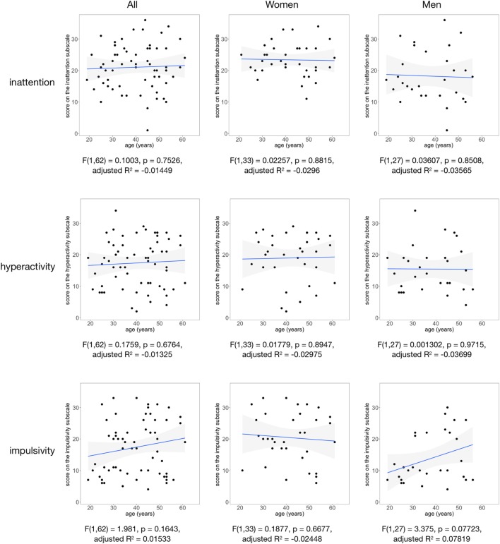
Methods: Surface-based brain morphometry was performed in 35 women and 29 men with ADHD using FreeSurfer. Linear regressions were calculated between cortical thickness and the volumes of subcortical gray matter and the inattention, hyperactivity, and impulsivity subscales of the Conners Adult ADHD Rating Scales (CAARS). Two separate analyses were performed. For the first analysis, age was included as additional regressor. For the second analysis, both age and severity of depression were included as additional regressors. Study participants were recruited between June 2012 and January 2014.
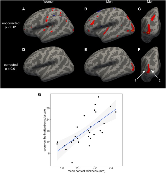
Results: Linear regression identified an area in the left occipital cortex of men, covering parts of the middle occipital sulcus and gyrus, in which the score on the CAARS inattention subscale predicted increased mean cortical thickness [F(1,27) = 26.27, p < 0.001, adjusted R2 = 0.4744]. No significant associations were found between cortical thickness and the scores on CAARS subscales in women. No significant associations were found between the volumes of subcortical gray matter and the scores on CAARS subscales, neither in men nor in women. These results remained stable when severity of depression was included as additional regressor, together with age.
Conclusion: Increased cortical thickness in the left occipital cortex may represent a mechanism to compensate for dysfunctional attentional networks in male adult ADHD patients.
Keywords: FreeSurfer; attention; attention-deficit/hyperactivity disorder; cortical thickness; gray matter; morphometry; occipital cortex.
Figures
Similar articles
-
Hyperactivity/restlessness is associated with increased functional connectivity in adults with ADHD: a dimensional analysis of resting state fMRI.BMC Psychiatry. 2019 Jan 25;19(1):43. doi: 10.1186/s12888-019-2031-9. BMC Psychiatry. 2019. PMID: 30683074 Free PMC article. Clinical Trial.
-
Inferior Frontal Gyrus Volume Loss Distinguishes Between Autism and (Comorbid) Attention-Deficit/Hyperactivity Disorder-A FreeSurfer Analysis in Children.Front Psychiatry. 2018 Oct 23;9:521. doi: 10.3389/fpsyt.2018.00521. eCollection 2018. Front Psychiatry. 2018. PMID: 30405459 Free PMC article.
-
Characterization of cortical and subcortical abnormalities in drug-naive boys with attention-deficit/hyperactivity disorder.J Affect Disord. 2019 May 1;250:397-403. doi: 10.1016/j.jad.2019.03.048. Epub 2019 Mar 8. J Affect Disord. 2019. PMID: 30877863
-
Gray Matter Network Associated With Attention in Children With Attention Deficit Hyperactivity Disorder.Front Psychiatry. 2022 Jul 4;13:922720. doi: 10.3389/fpsyt.2022.922720. eCollection 2022. Front Psychiatry. 2022. PMID: 35859604 Free PMC article.
-
Gray matter volume associations in youth with ADHD features of inattention and hyperactivity/impulsivity.Hum Brain Mapp. 2024 Apr;45(5):e26589. doi: 10.1002/hbm.26589. Hum Brain Mapp. 2024. PMID: 38530121 Free PMC article.
Cited by
-
Neural Encoding of the Reliability of Directional Information During the Preparation of Targeted Movements.Front Neurosci. 2021 Aug 24;15:679408. doi: 10.3389/fnins.2021.679408. eCollection 2021. Front Neurosci. 2021. PMID: 34504412 Free PMC article.
-
Reduced fronto-striatal volume in attention-deficit/hyperactivity disorder in two cohorts across the lifespan.Neuroimage Clin. 2020;28:102403. doi: 10.1016/j.nicl.2020.102403. Epub 2020 Aug 28. Neuroimage Clin. 2020. PMID: 32949876 Free PMC article.
-
Sex Differences in Cortical Structural Alterations in Major Depressive Disorder With Suicidal Ideation.Depress Anxiety. 2025 Jul 2;2025:1706750. doi: 10.1155/da/1706750. eCollection 2025. Depress Anxiety. 2025. PMID: 40636274 Free PMC article.
-
Hyperactivity/restlessness is associated with increased functional connectivity in adults with ADHD: a dimensional analysis of resting state fMRI.BMC Psychiatry. 2019 Jan 25;19(1):43. doi: 10.1186/s12888-019-2031-9. BMC Psychiatry. 2019. PMID: 30683074 Free PMC article. Clinical Trial.
-
Acute Effects of Aerobic Exercise on Executive Function and Attention in Adult Patients With ADHD.Front Psychiatry. 2019 Mar 26;10:132. doi: 10.3389/fpsyt.2019.00132. eCollection 2019. Front Psychiatry. 2019. PMID: 30971959 Free PMC article.
References
LinkOut - more resources
Full Text Sources
Other Literature Sources