. 2017 Sep 12:8:1567.
doi: 10.3389/fpls.2017.01567. eCollection 2017.

Purification and In Vitro Activity of Mitochondria Targeted Nitrogenase Cofactor Maturase NifB

Affiliations

Purification and In Vitro Activity of Mitochondria Targeted Nitrogenase Cofactor Maturase NifB

Stefan Burén et al. Front Plant Sci. .

Abstract

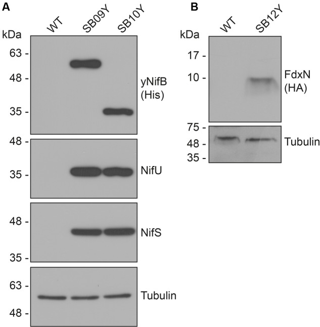
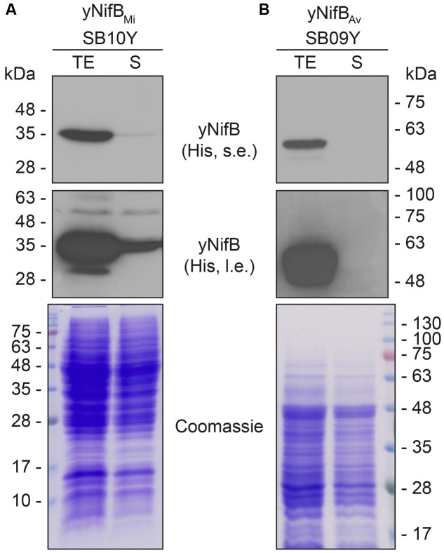
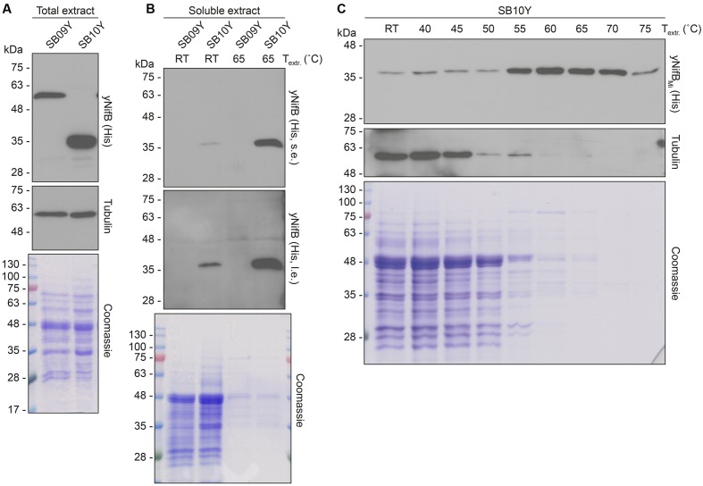
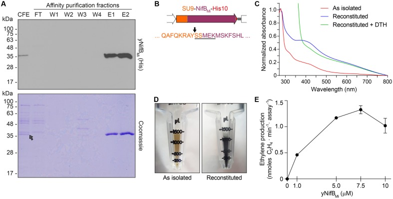
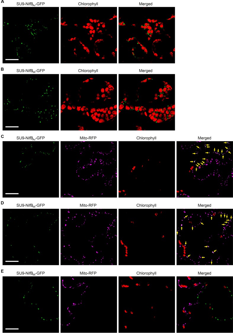
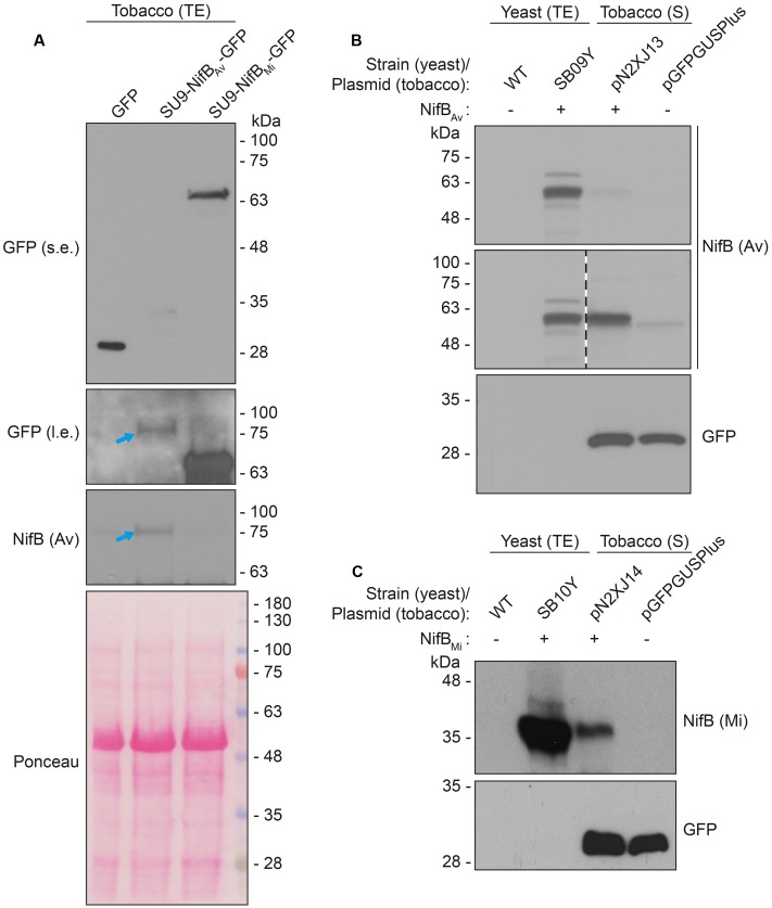
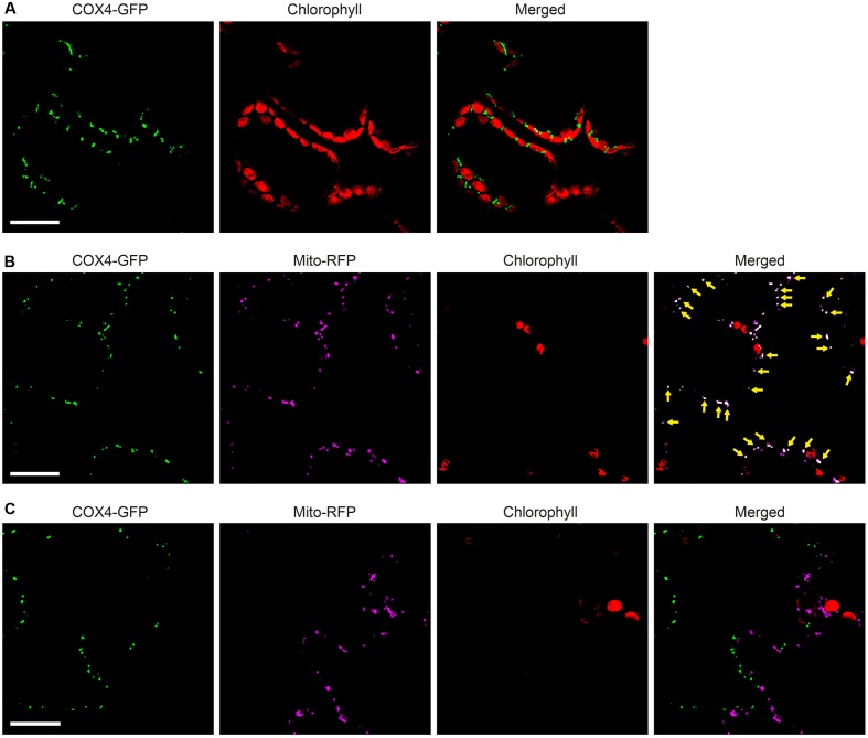
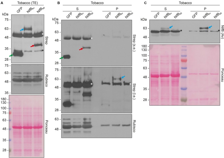
Active NifB is a milestone in the process of engineering nitrogen fixing plants. NifB is an extremely O2-sensitive S-adenosyl methionine (SAM)-radical enzyme that provides the key metal cluster intermediate (NifB-co) for the biosyntheses of the active-site cofactors of all three types of nitrogenases. NifB and NifB-co are unique to diazotrophic organisms. In this work, we have expressed synthetic codon-optimized versions of NifB from the γ-proteobacterium Azotobacter vinelandii and the thermophilic methanogen Methanocaldococcus infernus in Saccharomyces cerevisiae and in Nicotiana benthamiana. NifB proteins were targeted to the mitochondria, where O2 consumption is high and bacterial-like [Fe-S] cluster assembly operates. In yeast, NifB proteins were co-expressed with NifU, NifS, and FdxN proteins that are involved in NifB [Fe-S] cluster assembly and activity. The synthetic version of thermophilic NifB accumulated in soluble form within the yeast cell, while the A. vinelandii version appeared to form aggregates. Similarly, NifB from M. infernus was expressed at higher levels in leaves of Nicotiana benthamiana and accumulated as a soluble protein while A. vinelandii NifB was mainly associated with the non-soluble cell fraction. Soluble M. infernus NifB was purified from aerobically grown yeast and biochemically characterized. The purified protein was functional in the in vitro FeMo-co synthesis assay. This work presents the first active NifB protein purified from a eukaryotic cell, and highlights the importance of screening nif genes from different organisms in order to sort the best candidates to assemble a functional plant nitrogenase.

Keywords: SAM-radical; iron-molybdenum cofactor; mitochondria; nitrogen fixing plants; yeast.


Figures

FIGURE 1
FIGURE 1
Expression of NifB, NifU, NifS and FdxN proteins in S. cerevisiae. (A) Western blot analysis of yNifBAv and yNifBMi, as well as NifU and NifS, in total protein extracts from strains SB09Y and SB10Y. (B) Western blot analysis of C-terminally HA-tagged FdxN in total protein extract from SB12Y strain. Extracts in (A) and (B) were prepared from aerobically grown cells following galactose induction, and proteins in the extract separated by SDS-PAGE before transferred to membranes. Antibodies recognizing NifUAv, NifSAv, His epitope, HA epitope, and tubulin were used. Tubulin immunoblot signal intensity is used as loading control. See Table 1 for details about recombinant yeast strains.
FIGURE 2
FIGURE 2
Solubility of A. vinelandii and M. infernus NifB proteins expressed in S. cerevisiae. (A,B) Western blot analysis of yNifBMi (A) and yNifBAv (B) present in total protein extracts (TE) and the soluble fraction (S) of yeast strains SB10Y and SB09Y. Conditions for strain growth, induction of protein expression, total extract preparation, and separation by SDS-PAGE are as in Figure 1. Antibodies recognizing the His epitope were used. Short (s.e.) and long (l.e.) film exposures are shown. Coomassie stained SDS gels (below) of the protein extracts are included as loading controls.
FIGURE 3
FIGURE 3
Levels of soluble yNifBAv and yNifBMi in heat-treated yeast extracts. (A,B) Total NifB levels (A) and levels of soluble NifB upon 65°C heat-treatment (B) of protein extracts from yeast expressing yNifBAv (SB09Y) or yNifBMi (SB10Y). Antibodies recognizing the His epitope were used. Short (s.e.) and long (l.e.) film exposures are shown. RT means room temperature. Tubulin and/or Coomassie stained SDS gels of the same protein extracts are included as loading controls. (C) Western blot analysis of soluble yNifBMi in SB10Y protein extracts upon heat-treatment at increasing temperatures. Heat-induced precipitation of yeast proteins in the extract at the different temperatures is shown using antibodies recognizing tubulin, as well as by Coomassie staining of proteins from the extract resolved by SDS-PAGE.
FIGURE 4
FIGURE 4
Purification and biochemical properties of yNifBMi. (A) SDS-PAGE and Western blot analysis of yNifBMi purification. CFE, 65°C heated SB10Y cell-free extract; FT, affinity chromatography flow through; W1-W4 and E1-E2, affinity chromatography wash and elution fractions containing increasing concentrations of imidazole (see Materials and Methods for details). Grey arrow in the Coomassie stained panel points to the position of yNifBMi in the gel. (B) SU9 processing site (black arrow) of yNifBMi. Underlined sequence indicates the N-terminal amino acids of yNifBMi identified by Edman degradation. (C) UV-visible spectra of as isolated, reconstituted, and dithionite (DTH)-reduced reconstituted yNifBMi. (D) Typical color of as isolated and reconstituted yNifBMi purified preparations. (E) Titration of FeMo-co synthesis and nitrogenase reconstitution assay with yNifBMi. The indicated concentrations of yNifBMi monomer were used. NifB activity was determined by acetylene reduction assay of reconstituted NifDK from ΔnifB A. vinelandii UW140 cell-free extracts. Data represent mean ± standard deviation (n = 2) at each yNifBMi concentration.
FIGURE 5
FIGURE 5
Expression of mitochondria targeted (SU9) NifBAv and NifBMi GFP fusions in N. benthamiana leaves. (A,B) Mesophyll cells expressing SU9-NifBAv-GFP (A) or SU9-NifBMi-GFP (B). GFP (green) and chlorophyll autofluorescence (red) of chloroplasts is shown. (C–E) Epidermal cells expressing SU9-NifBAv-GFP (C) and SU9-NifBMi-GFP (D,E), together with a fluorescent mitochondria marker (Mito-RFP). GFP (green), Mito-RFP (magenta) and chlorophyll autofluorescence (red) of chloroplasts is shown. Co-localization of SU9-NifBAv-GFP or SU9-NifBMi-GFP constructs with Mito-RFP labeled structures is shown as white in the merged images, and highlighted with yellow arrows. Adjacent cells expressing SU9-NifBMi-GFP or Mito-RFP are shown as control to verify the specificity of the signal recorded in each channel (E). Scale bars show 30 μm. Confocal Microscopy conditions are specified in Materials and Methods.
FIGURE 6
FIGURE 6
Expression and solubility of mitochondria targeted (SU9) NifBAv and NifBMi in N. benthamiana leaves. (A) Western blot analysis of total protein extracts (TE) prepared from infiltrated N. benthamiana leaves expressing GFP, SU9-NifBAv-GFP or SU9-NifBMi-GFP. Blue arrows indicate the polypeptide recognized both by GFP and NifBAv specific antibodies. Short (s.e.) and long (l.e.) film exposures of the GFP antibody probed membrane are shown. (B) Migration of SU9-NifBAv-His10 when expressed in S. cerevisiae and N. benthamiana. Migration in SDS-PAGE was determined after Western blot analysis using NifBAv specific antibodies. Total protein extracts (TE) from W303-1a S. cerevisiae cells (WT) or cells expressing SU9-NifBAv-His10 (SB09Y) were prepared. Soluble protein extracts (S) from N. benthamiana leaf cells infiltrated with A. tumefaciens containing control vector (pGFPGUSPlus) or vector for expression of SU9-NifBAv-His10 (pN2XJ13). Dotted line indicate different exposures of the right part of the membrane. See Supplementary Figure S9 for entire gel of the cropped exposure. (C) Migration of SU9-NifBMi-His10 when expressed in S. cerevisiae and N. benthamiana. Migration in SDS-PAGE was determined after Western blot analysis using NifBMi specific antibodies. Total protein extracts (TE) from W303-1a S. cerevisiae cells (WT) or cells expressing SU9-NifBMi-His10 (SB10Y) were prepared. Soluble protein extracts (S) from N. benthamiana leaf cells infiltrated with A. tumefaciens containing control vector (pGFPGUSPlus) or vector for expression of SU9-NifBMi-His10 (pN2XJ14). As control of N. benthamiana leaf infiltration, GFP expressed from the pGFPGUSPlus vector backbone was detected (B,C).
FIGURE 7
FIGURE 7
Functionality of COX4 leader sequence for mitochondria targeting of GFP in N. benthamiana leaves. (A) Mesophyll cells expressing COX4-twinStrep-GFP. GFP (green) and chlorophyll autofluorescence (red) of chloroplasts is shown. (B,C) Epidermal cells expressing COX4-twinStrep-GFP together with a fluorescent mitochondria marker (Mito-RFP). GFP (green), Mito-RFP (magenta) and chlorophyll autofluorescence (red) of chloroplasts is shown. Co-localization of COX4-twinStrep-GFP with Mito-RFP labeled structures (B) is shown as white in the merged image, and highlighted with yellow arrows. Adjacent cells expressing COX4-twinStrep-GFP or Mito-RFP (C) are shown as control to verify the specificity of the signal recorded in each channel. Scale bars show 30 μm. Confocal Microscopy conditions are specified in Materials and Methods.
FIGURE 8
FIGURE 8
Expression and solubility of mitochondria targeted (COX4) NifBAv and NifBMi in N. benthamiana leaves. (A) Western blot analysis of total protein extracts (TE) prepared from infiltrated N. benthamiana leaves expressing COX4-twinStrep-GFP (GFP), COX4-twinStrep-NifBAv (NifBAv) or COX4-twinStrep-NifBMi (NifBMi) and separated by SDS-PAGE. The COX4-twinStrep-GFP (green arrow), COX4-twinStrep-NifBAv (blue arrow), COX4-twinStrep-NifBMi (red arrow) proteins are highlighted. A pronounced non-specific polypeptide detected using the Strep-tag antibodies (white star) co-migrated with the large subunit of Rubisco. The membrane probed with antibodies against Rubisco was also stained with Ponceau and is included as loading control. (B,C) Western blot analysis of the soluble (S) and non-soluble pellet (P) fractions of N. benthamiana leaf total extracts used in (A), using Strep-tag antibodies (B) or NifBAv antibodies (C). The COX4-twinStrep-GFP (green arrow), COX4-twinStrep-NifBAv (blue arrow), COX4-twinStrep-NifBMi (red arrow) proteins are highlighted. Non-specific bands detected using the Strep-tag antibodies (white stars) co-migrated with Rubisco (B). Non-specific bands detected with NifBAv antibodies (black stars) are also indicated (C). Short (s.e.) and long (l.e.) film exposures of the Strep-tag antibody probed membrane are shown (B). Ponceau staining of the NifBAv antibody probed membrane is shown as loading control (C).

Similar articles

Cited by

References

    1. Allen R. S., Tilbrook K., Warden A. C., Campbell P. C., Rolland V., Singh S. P., et al. (2017). Expression of 16 nitrogenase proteins within the plant mitochondrial matrix. Front. Plant Sci. 8:287 10.3389/fpls.2017.00287 - DOI - PMC - PubMed
    1. Balk J., Pilon M. (2011). Ancient and essential: the assembly of iron-sulfur clusters in plants. Trends Plant Sci. 16 218–226. 10.1016/j.tplants.2010.12.006 - DOI - PubMed
    1. Beinert H. (1983). Semi-micro methods for analysis of labile sulfide and of labile sulfide plus sulfane sulfur in unusually stable iron-sulfur proteins. Anal. Biochem. 131 373–378. 10.1016/0003-2697(83)90186-0 - DOI - PubMed
    1. Bishop P. E., Joerger R. D. (1990). Genetics and molecular biology of alternative nitrogen fixation systems. Annu. Rev. Plant Physiol. Plant Mol. Biol. 41 109–125. 10.1146/annurev.pp.41.060190.000545 - DOI
    1. Borlaug N. E. (2002). Feeding a world of 10 billion people: the miracle ahead. Vitr. Cell. Dev. Biol. Plant 38 221–228. 10.1079/IVP2001279 - DOI