

Unusual case of splenic sarcoidosis without morphological lesions detected by PET-CT in a patient with breast cancer: functional imaging between pitfalls and therapeutic guide
- PMID: 28955402
- PMCID: PMC5606293
- DOI: 10.3332/ecancer.2017.766
Unusual case of splenic sarcoidosis without morphological lesions detected by PET-CT in a patient with breast cancer: functional imaging between pitfalls and therapeutic guide
Abstract
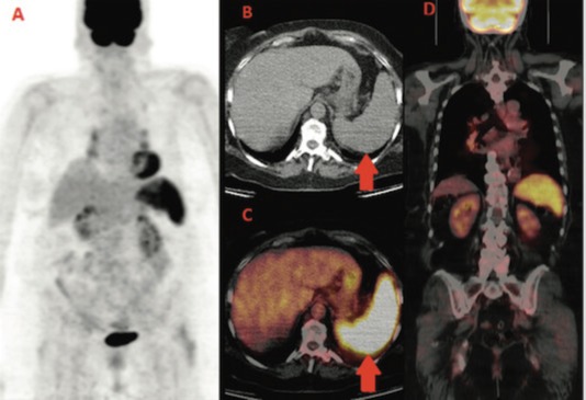
A 60-year-old woman under treatment with letrozole for metastatic breast cancer underwent 18F-FDG PET-CT for restaging. A new widespread intense splenic FDG uptake without nodular lesions and multiple FDG-avid mediastinal and abdominal nodes were observed. Based on these findings, a nodal and transbronchial lung biopsy was performed. Histological results were compatible with sarcoidosis. The patient began steroid treatment and 6 weeks after a PET-CT showed normalisation of both splenic and nodal uptake. In our case, 18F-FDG PET-CT has been useful in detecting a rare case of splenic sarcoidosis without typical nodular lesions on CT images, impacting the patient's treatment and prognosis.
Keywords: 18F-FDG; breast cancer; positron emission tomography; spleen sarcoidosis.
Figures


Similar articles
-
F-18 fluorodeoxyglucose and F-18 fluorothymidine positron emission tomography/computed tomography imaging in a case of neurosarcoidosis.Clin Nucl Med. 2010 Feb;35(2):67-70. doi: 10.1097/RLU.0b013e3181c7c149. Clin Nucl Med. 2010. PMID: 20090446
-
More advantages in detecting bone and soft tissue metastases from prostate cancer using 18F-PSMA PET/CT.Hell J Nucl Med. 2019 Jan-Apr;22(1):6-9. doi: 10.1967/s002449910952. Epub 2019 Mar 7. Hell J Nucl Med. 2019. PMID: 30843003
-
(18)F-FDG and (18)F-FLT PET/CT imaging in the characterization of mediastinal lymph nodes.Ann Nucl Med. 2016 Apr;30(3):207-16. doi: 10.1007/s12149-015-1047-6. Epub 2015 Dec 11. Ann Nucl Med. 2016. PMID: 26661845 Clinical Trial.
-
Sarcoidosis with bone involvement mimicking metastatic disease at (18)F-FDG PET/CT: problem solving by diffusion whole-body MRI.Ecancermedicalscience. 2015 May 7;9:537. doi: 10.3332/ecancer.2015.537. eCollection 2015. Ecancermedicalscience. 2015. PMID: 26015806 Free PMC article. Review.
-
Atypical sarcoidosis: case reports and review of the literature.Eur Rev Med Pharmacol Sci. 2009 Mar;13 Suppl 1:37-44. Eur Rev Med Pharmacol Sci. 2009. PMID: 19530510 Review.
References
-
- Baughman RP, Teirstein AS, Judson MA, et al. on behalf of Case Control Etiologic Study of Sarcoidosis (ACCESS) research group Clinical characteristics of patients in a case control study of sarcoidosis. Am J Respir Crit Care Med. 2001;164:1885–1889. doi: 10.1164/ajrccm.164.10.2104046. - DOI - PubMed
-
- Rizzato G, Palmieri G, Agrati AM, et al. The organ-specific extrapulmonary presentation of sarcoidosis: a frequent occurrence but a challenge to an early diagnosis A 3-year-long prospective observational study. Sarcoidosis Vasc Diffuse Lung Dis. 2004;21:119–126. - PubMed
Publication types
LinkOut - more resources
Full Text Sources
Other Literature Sources