. 2017 Jul 13;3(1):e000436.
doi: 10.1136/rmdopen-2017-000436. eCollection 2017.

Nardilysin is involved in autoimmune arthritis via the regulation of tumour necrosis factor alpha secretion

Affiliations

Nardilysin is involved in autoimmune arthritis via the regulation of tumour necrosis factor alpha secretion

Takayuki Fujii et al. RMD Open. .

Abstract

Objective: Tumour necrosis factor alpha (TNF-α) plays an important role in rheumatoid arthritis (RA). TNF-α is synthesised as a membrane-anchored precursor and is fully activated by a disintegrin and metalloproteinase 17 (ADAM17)-mediated ectodomain shedding. Nardilysin (NRDC) facilitates ectodomain shedding via activation of ADAM17. This study was undertaken to elucidate the role of NRDC in RA.

Methods: NRDC-deficient (Nrdc-/- ) mice and macrophage-specific NRDC-deficient (NrdcdelM ) mice were examined in murine RA models, collagen antibody-induced arthritis (CAIA) and K/BxN serum transfer arthritis (K/BxN STA). We evaluated the effect of gene deletion or silencing of Nrdc on ectodomain shedding of TNF-α in macrophages or monocytes. NRDC concentration in synovial fluid from patients with RA and osteoarthritis (OA) were measured. We also examined whether local gene silencing of Nrdc ameliorated CAIA.

Results: CAIA and K/BxN STA were significantly attenuated in Nrdc-/- mice and NrdcdelM mice. Gene deletion or silencing of Nrdc in macrophages or THP-1 cells resulted in the reduction of TNF-α shedding. The level of NRDC is higher in synovial fluid from RA patients compared with that from OA patients. Intra-articular injection of anti-Nrdcsmall interfering RNA ameliorated CAIA.

Conclusion: These data indicate that NRDC plays crucial roles in the pathogenesis of autoimmune arthritis and could be a new therapeutic target for RA treatment.

Keywords: Autoantibodies; Rheumatoid arthritis; Synovial fluid; TNF-alpha.


Conflict of interest statement

Competing interests: None declared.

Figures

Figure 1
Figure 1
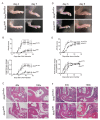
CAIA is attenuated in Nrdc/ mice. (A) Photographs of the hind paws 4 days and 7 days after the induction of CAIA. Upper panels, wild-type (WT); lower panels, Nrdc/ mice. (B) Arthritis score (upper) and ankle thickness (lower) were measured daily after the induction of CAIA. n=5 per genotype (mean ± SEM). *p<0.05; **p<0.01, Welch’s t-test. (C) H&E staining of the ankle joints 14 days after the induction of CAIA in WT (upper panels) and Nrdc/ (lower panels) mice. Scale bars: 500 µm in the 40× magnification and 200 µm in the 100× magnification. Symbols in the 100x magnification images: arrow, bone erosion; arrow head, cartilage erosion; *, hypertrophic synovium.
Figure 2
Figure 2
Macrophage-specific Nrdc-deficient (NrdcdelM) mice are resistant to CAIA and K/BxN STA. (A) Photographs of the hind paws 4 days and 7 days after the induction of CAIA. Upper panels, Nrdcflox/flox; lower panels, NrdcdelM mice. (B) Arthritis score (upper) and ankle thickness (lower) were measured daily after the induction of CAIA. n=5 per genotype (mean ± SEM). *p<0.05; **p<0.01, Welch’s t-test. (C) H&E staining of the ankle joints 14 days after the induction of CAIA in Nrdcflox/flox (upper panels) and NrdcdelM (lower panels) mice. Scale bars: 500 µm in the 40× magnification and 200 µm in the 100× magnification. Symbols in the 100x magnification images: arrow, bone erosion; *, synovial hypertrophy. (D) Photographs of the hind paws 3 days and 5 days after the induction of K/BxN STA. Upper panels, Nrdcflox/flox; lower panels, NrdcdelM mice. (E) Arthritis score (upper) and ankle thickness (lower) were measured daily after the induction of K/BxN STA. n=4 per genotype (mean ± SEM). *p<0.05; **p<0.01, Welch’s t-test. (F) H&E staining of the ankle joints 14 days after the induction of K/BxN STA in Nrdcflox/flox (upper panels) and NrdcdelM (lower panels) mice. Scale bars: 500 µm in the 40× magnification and 200 µm in the 100× magnification. Symbols in the 100× magnification images: arrow, bone erosion; arrow head, cartilage erosion; *, synovial hypertrophy.
Figure 3
Figure 3
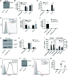
Secretion of TNF-α from macrophages and monocytic cells were reduced by the deletion of Nrdc. (A) Western blot analysis of NRDC in BMDM from Nrdcflox/flox (left) and NrdcdelM mice (right). β-Actin was used as a loading control. (B) TNF-α mRNA levels in unstimulated or LPS-stimulated BMDMs were analysed by reverse transcription and quantitative PCR (qRT-PCR). β-Actin mRNA levels were used as internal control. n=6 (mean ± SEM). (C) TNF-α secreted from BMDMs in conditioned medium was measured by ELISA. n=6 (mean ± SEM). (D) Membrane-bound TNF-α in BMDMs was stained 3 hours after LPS stimulation and analysed by flow cytometry. Filled histogram, unstimulated control; blue open histogram, Nrdcflox/flox; red open histogram, NrdcdelM. Representative histograms out of four independent experiments are shown. (E) ADAM17 mRNA levels in unstimulated or LPS-stimulated BMDMs were analysed by qRT-PCR. β-Actin mRNA levels were used as internal control. n=6 (mean ± SEM). (F) NRDC mRNA levels in THP-1 cells, transfected with lentiviral vectors for negative control RNA interference (RNAi) (control microRNA (miR)) or RNAi targeting Nrdc (anti-NRDC miR), were analysed using qRT-PCR. β-Actin mRNA was used as an internal control. n=6 (mean±SEM). (G) NRDC and ADAM17 protein expressions in THP-1 cells transfected with lentiviral vectors for control miR (left) or anti-NRDC miR 1 (right) were analysed by Western blot. β-Actin was used as a loading control. (H) TNF-α mRNA levels in THP-1 cells, transfected with lentiviral vectors as described in (G), were analysed using qRT-PCR. n=8 (mean ± SEM). (I) Secreted TNF-α (conditioned medium) and cell-contained TNF-α (cell extracts) in THP-1 cells described above were measured by ELISA. n=8 (mean±SEM). (J) F4/80 expression in peritoneal macrophage (PMs). Filled histogram, isotype control; open histogram, F4/80. Representative histograms out of three independent experiments are shown. (K) NRDC expression in PMs, isolated from Nrdcflox/flox (left) and NrdcdelM mice (right) after the reverse passive Arthus reaction, was examined by Western blot. β-Actin was used as a loading control. (L) Membrane-bound TNF-α was stained and analysed by flow cytometry. Filled histogram, unstimulated control; blue open histogram, Nrdcflox/flox; red open histogram, NrdcdelM. Representative histograms out of three independent experiments are shown. (M) Measurement of secreted TNF-α concentration in the peritoneal lavage of Nrdcflox/flox (n=5), NrdcdelM (n=5) and control (n=4) mice. *p<0.05; **p<0.01, Welch’s t-test.
Figure 4
Figure 4
NRDC and TNF-α increase in synovial fluid from patients with RA. (A) NRDC concentration in synovial fluid from OA (n=17) and RA (n=20) patients. **p<0.01, Welch’s t-test. (B) TNF-α concentration in synovial fluid from OA and RA patients. **p<0.01, Welch’s t-test. (C) NRDC in synovial fluid has a tendency of positive correlation with TNF-α in RA patients. Solid line, approximate line; broken line, 95% CI. r=0.44, p=0.054, Spearman’s rank correlation test. (D) NRDC in synovial fluid is significantly correlated with CRP in RA patients. Solid line, approximate line; broken line, 95% CI. r=0.50, p<0.05, Spearman’s rank correlation test.
Figure 5
Figure 5
Intra-articular Nrdc gene silencing with siRNA attenuates CAIA. (A) Screening of siRNAs against NRDC in RAW264.7, WEHI 13 VAR and mouse embryonic fibroblasts (MEF). (B) Schedule of the induction of CAIA and intra-articular injection of siRNA. (C) Photographs of the hind paws 4 days after the induction of CAIA. Anti-NRDC siRNA was injected in the left ankle, while control siRNA was injected in the right ankle joints of BALB/c WT mice. (D) Ankle thickness was measured daily after the induction of CAIA (n=5 per treatment). *p<0.05, Welch’s t-test.

Similar articles

Cited by

References

    1. Terao C, Ikari K, Nakayamada S, et al. . A twin study of rheumatoid arthritis in the Japanese population. Mod Rheumatol 2016;26:685–9.10.3109/14397595.2015.1135856 - DOI - PubMed
    1. Keffer J, Probert L, Cazlaris H, et al. . Transgenic mice expressing human tumour necrosis factor: a predictive genetic model of arthritis. Embo J 1991;10:4025–31. - PMC - PubMed
    1. Müssener A, Litton MJ, Lindroos E, et al. . Cytokine production in synovial tissue of mice with collagen-induced arthritis (CIA). Clin Exp Immunol 1997;107:485–93.10.1046/j.1365-2249.1997.3181214.x - DOI - PMC - PubMed
    1. Ji H, Pettit A, Ohmura K, et al. . Critical roles for interleukin 1 and tumor necrosis factor alpha in antibody-induced arthritis. J Exp Med 2002;196:77–85.10.1084/jem.20020439 - DOI - PMC - PubMed
    1. Williams RO, Feldmann M, Maini RN. Anti-tumor necrosis factor ameliorates joint disease in murine collagen-induced arthritis. Proc Natl Acad Sci U S A 1992;89:9784–8.10.1073/pnas.89.20.9784 - DOI - PMC - PubMed