. 2017 Sep 25:3:17056.
doi: 10.1038/cddiscovery.2017.56. eCollection 2017.

Necroptotic debris including damaged mitochondria elicits sepsis-like syndrome during late-phase tularemia

Affiliations

Necroptotic debris including damaged mitochondria elicits sepsis-like syndrome during late-phase tularemia

Anju Singh et al. Cell Death Discov. .

Abstract

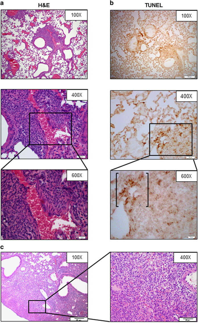
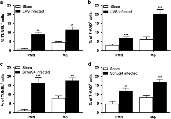
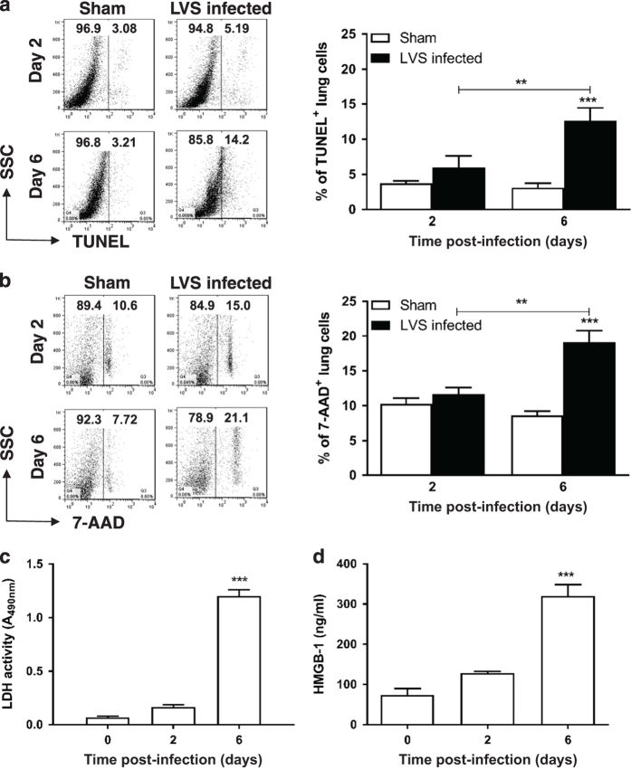
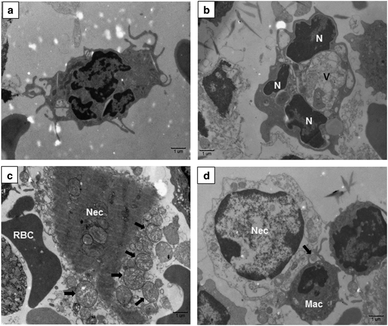
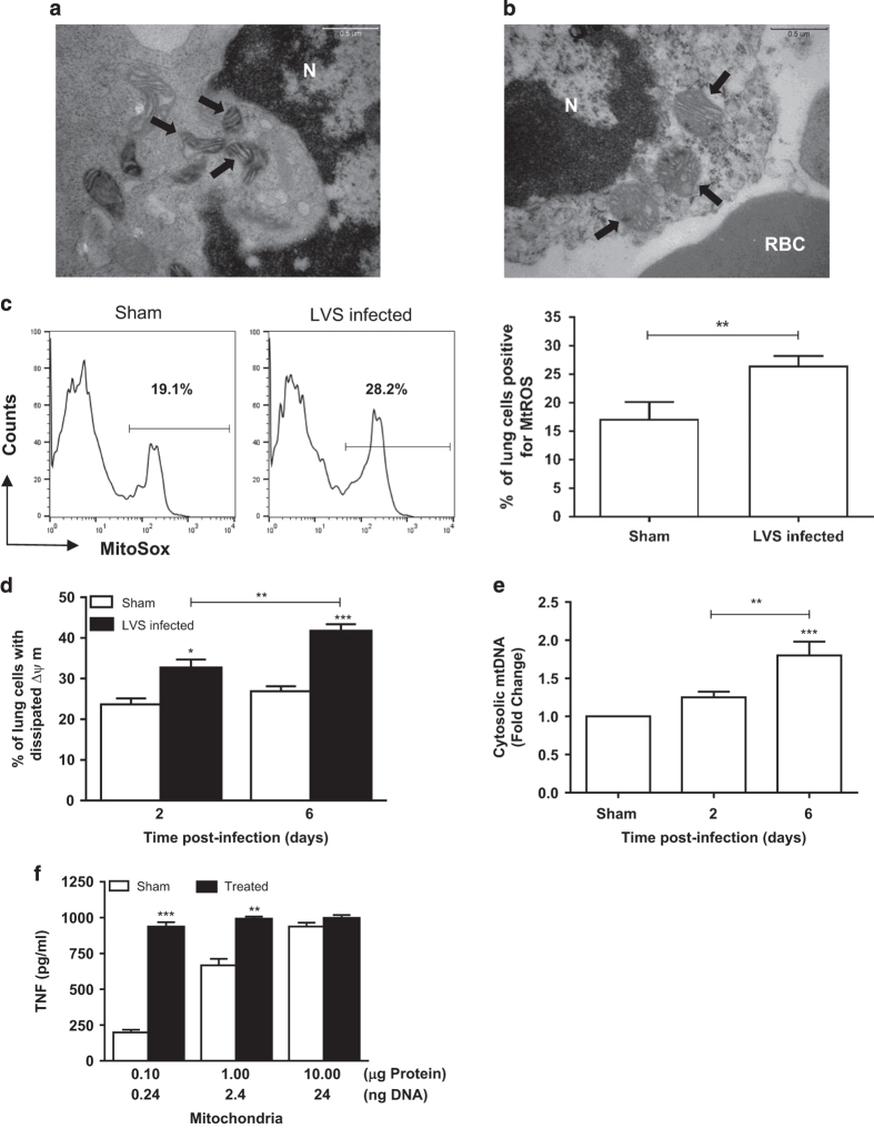
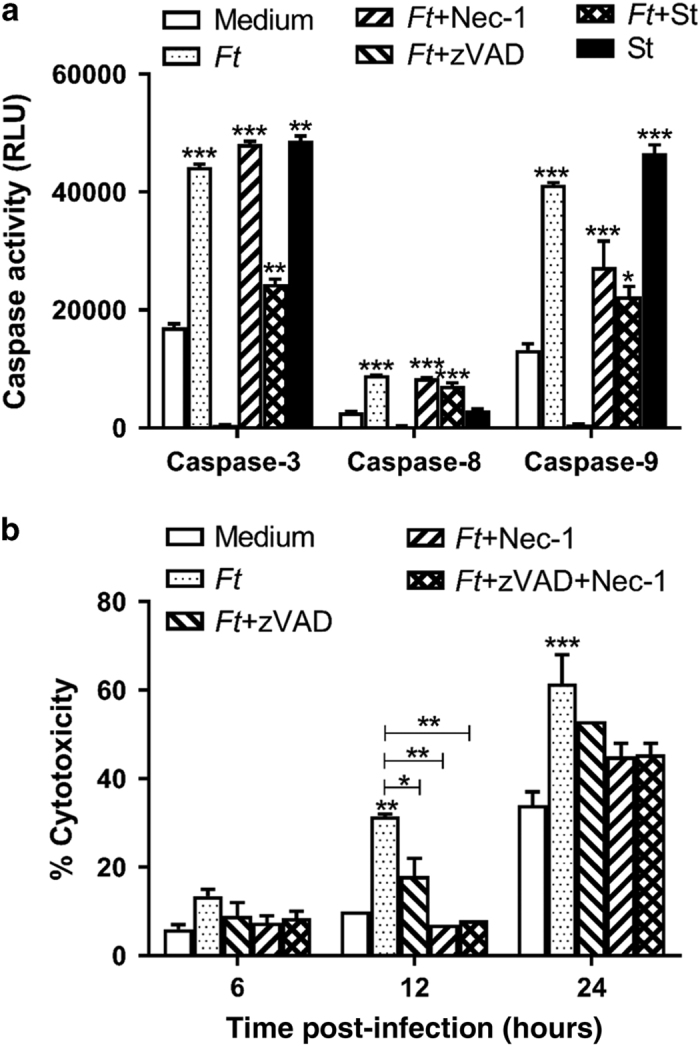
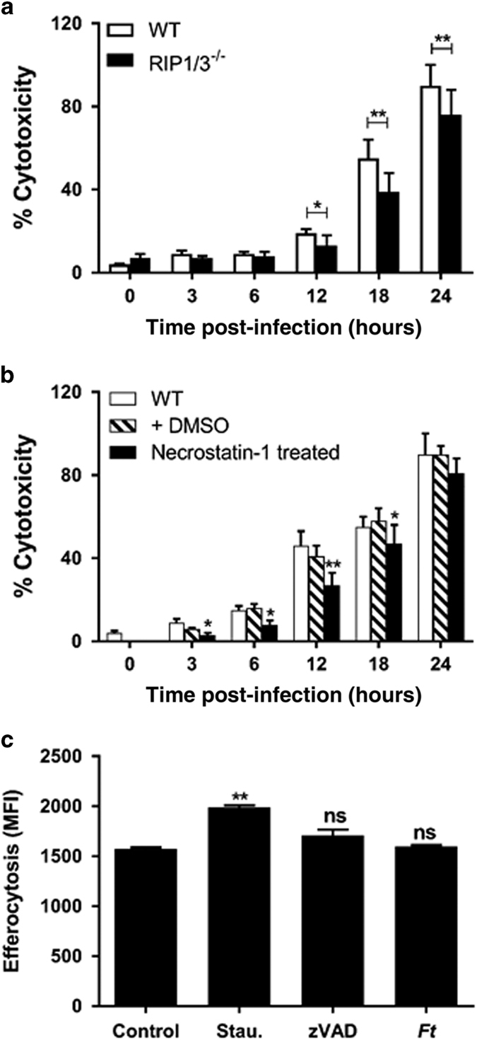
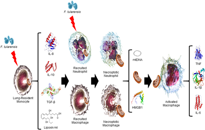
Infection with Francisella tularensis ssp. tularensis (Ft) strain SchuS4 causes an often lethal disease known as tularemia in rodents, non-human primates, and humans. Ft subverts host cell death programs to facilitate their exponential replication within macrophages and other cell types during early respiratory infection (⩽72 h). The mechanism(s) by which cell death is triggered remains incompletely defined, as does the impact of Ft on mitochondria, the host cell's organellar 'canary in a coal mine'. Herein, we reveal that Ft infection of host cells, particularly macrophages and polymorphonuclear leukocytes, drives necroptosis via a receptor-interacting protein kinase 1/3-mediated mechanism. During necroptosis mitochondria and other organelles become damaged. Ft-induced mitochondrial damage is characterized by: (i) a decrease in membrane potential and consequent mitochondrial oncosis or swelling, (ii) increased generation of superoxide radicals, and (iii) release of intact or damaged mitochondria into the lung parenchyma. Host cell recognition of and response to released mitochondria and other damage-associated molecular patterns engenders a sepsis-like syndrome typified by production of TNF, IL-1β, IL-6, IL-12p70, and IFN-γ during late-phase tularemia (⩾72 h), but are absent early during infection.


Conflict of interest statement

The authors declare no conflict of interest.

Figures

Figure 1
Figure 1
Respiratory infection with Ft LVS induces apoptosis and secondary necrosis. Perfused lungs from day 3 Ft-infected (103 CFU) C57BL/6 mice were recovered and processed for histological evaluation. Paraformaldehyde-fixed, paraffin-embedded sections were stained with hematoxylin–eosin (H&E) (a) or TUNEL (b). Evidence of apoptosis and necrotic cell debris was mixed with the ground substance in the lung parenchyma. The ×600 panels represent magnifications of the boxed areas seen in the ×400 panels. Bracketed area (b, ×600) represents the TUNEL-positive fragmented nucleic acid released from the dead/damaged cells. Results are representative of six individual mice. (c) By day 6 p.i., necrotizing inflammation is a hallmark pathology of pulmonary tularemia. Note the accumulation of mixed cellular infiltrates (including PMNs, macrophages, and lymphocytes) in alveolar lumen/interstitium and necrotic areas in the lung parenchyma (×100). Inset picture: necrotic (Nec) lung parenchyma and dead/dying cells (arrow) in the vicinity of necrotic areas (×400).
Figure 2
Figure 2
Myeloid cells undergo apoptosis/necrosis during the course of infection with Ft. Total lung cells were isolated from C57BL/6 mice infected with 103 CFU of Ft LVS (a and b) or 20 CFU of SchuS4 (c and d) at day 3 p.i. The cells were stained with TUNEL (a and c) or 7-aminoactinomycin D (7-AAD) (b and d) to quantify the percentage of myeloid cells undergoing apoptosis and necrosis. The lungs of Ft SchuS4-infected mice were not perfused before isolation of cells as to minimize potential liquid dispersal of select agent. Data are presented as the mean±S.E.M. from three independent experiments (n=6 mice per group or 18 mice total). **P<0.01, and ***P<0.001.
Figure 3
Figure 3
Apoptotic/necrotic cells accumulate during the course of infection with Ft LVS. Total lung cells were isolated from Ft-infected (103 CFU) C57BL/6 mice at different time points and were stained with TUNEL (a) or 7-aminoactinomycin D (7-AAD) (b) to quantify the percentage of apoptotic and necrotic cells, respectively. **P<0.01 and ***P<0.001. Lactate dehydrogenase (LDH) (c) and HMGB1 (d) levels were determined in the bronchoalveolar lavage fluid (BALF) samples isolated from the Ft-infected mice at various time points p.i. Data are presented as the mean±S.E.M. from two independent experiments (n=6 mice per group or 12 mice total). ***P<0.001. All results shown were subjected to one-way analysis of variance (ANOVA) with Bonferroni’s post-test.
Figure 4
Figure 4
Electron microscopic analysis of leukocytes in normal and Ft LVS-infected lung. (a) Normal PMN isolated from sham-inoculated lung showing multilobed nucleus with normal distribution of chromatin structures. (b) Activated PMN exhibiting signs of apoptosis with condensed/marginated nuclear chromatin (N) and cytoplasmic vacuoles (v). (c) Necrotic cell (Nec) showing grossly swollen mitochondria being released into the extracellular space (arrows). Red blood cell, RBC. (d) Necrotic cell (Nec) being recognized by adjacent macrophage (Mac). Free mitochondrion (arrow) outside the cell appears to be phagocytized by a macrophage.
Figure 5
Figure 5
Damaged mitochondria in Ft-infected lungs represent a source of DAMPs that elicit TH1-type proinflammatory cytokines. (a) Electron micrograph of mitochondria (arrows) within a white blood cell isolated from sham-inoculated lung. Mitochondria have predominantly lamellar cristae. N, nucleus. (b) Electron micrograph of white blood cell isolated from infected lung tissue showing mitochondria (arrows) with abnormal, round/dilated cristae, some forming protrusions at the mitochondrial surface, and swollen size. RBC, red blood cell. (c) Superoxide levels in the mitochondria within lung cells from uninfected (Sham) and day 6 infected mice were determined by staining isolated cells with MitoSOX Red and were quantified by flow cytometry (left panel) as % cells positive for mitochondrial ROS (MtROS) (right panel). (d) The percentage of lung cells from uninfected (Sham) and Ft-infected mice whose mitochondria exhibit dissipated membrane potentials at various time points p.i. were quantified by flow cytometry using JC-1 staining. (e) Quantitative PCR analysis for the presence of mtDNA in the cytosol of lung cells from uninfected (Sham) and Ft-infected mice was performed as described in Materials and methods. (f) Mitochondria isolated from uninfected (Sham) and Ft-infected lungs at day 6 p.i. were evaluated for their proinflammatory capacity. Wild-type C57BL/6 BMDMs (2.5×10⁵ cells/well) were incubated with various amounts of mitochondria for 24 h. Supernatants were collected and assayed for the presence of TNF by ELISA (see also Supplementary Figure S2). Data are presented as the mean±S.E.M. from two to three independent experiments (n=6 mice per group or 12–18 mice total). *P<0.05, **P<0.01, and ***P<0.001. All results shown were subjected to one-way analysis of variance (ANOVA) with Bonferroni’s post-test.
Figure 6
Figure 6
Ft infection induces cell death in a caspase-dependent and -independent manner. (a) C57BL/6 BMDMs were infected with Ft LVS at a multiplicity of infection (MOI) of 100 in the absence or presence of zVAD-fmk (20 μM), necrostatin-1 (20 μM), and staurosporine (1 μM). Levels of active caspase-8, -9, and -3 were measured at 24 h p.i. (b) BMDMs were infected with Ft LVS at an MOI of 100 in the absence or presence of zVAD-fmk, necrostatin-1, or both. Lactate dehydrogenase (LDH) release was measured at the indicated time points. Data are presented as the mean±S.E.M. from three independent experiments. *P<0.05, **P<0.01, and ***P<0.001. All results shown were subjected to one-way analysis of variance (ANOVA) with Bonferroni’s post-test.
Figure 7
Figure 7
Ft induces necroptotic cell death in macrophages. (a) Wild-type (WT) and RIP1/3−/− BMDMs were infected with Ft LVS at a multiplicity of infection (MOI) of 100 and lactate dehydrogenase (LDH) release was measured at the indicated time points. (b) WT BMDMs either were left untreated or were treated with dimethyl sulfoxide (DMSO) alone or necrostatin-1 (20 μM) and LDH release was measured at the indicated time points. (c) WT BMDMs were treated for 24 h with medium alone or medium containing 5 μM staurosporine (to induce apoptosis), or LPS+zVAD (to induce necroptosis) or with Ft alone. Dead cells were collected, counted, and resuspended in fresh medium and were applied to naïve BMDMs at a 2 : 1 ratio. After 2 h, cells were washed with cold phosphate-buffered saline (PBS) and the fluorescence intensity was quantified using a plate reader. Data are presented as the mean±S.E.M. from three independent experiments. *P<0.05, **P<0.01. All results shown were subjected to one-way analysis of variance (ANOVA) with Bonferroni’s post-test.
Figure 8
Figure 8
Schematic representation of F. tularensis-induced generation of mitochondrial DAMPs and sepsis-like syndrome. Initial infection of lung-resident cells by F. tularensis results in a principally anti-inflammatory response, typified by production of interleukin-10 (IL-10), tumor growth factor-β (TGFβ), and lipoxin A4 (LXA4). However, simultaneous release of potent chemokines (for example, keratinocyte-derived chemoattractant (KC)/IL-8 and monocyte chemotactic protein-1 (MCP-1)), whose production is augmented by LXA4, drives the recruitment of PMNs and macrophages that support exponential replication of bacteria following their infection. F. tularensis-induced delay in apoptosis of these infected cells gives way to caspase-3-mediated, RIP1/3-dependent necroptosis and subsequent release of mitochondria and other DAMPs, of both mitochondrial and non-mitochondrial origin. A target of such DAMPs are activated macrophages that secrete the proinflammatory cytokines that can result in end-organ failure and death.

Similar articles

Cited by

References

    1. Parmely MJ, Fischer JL, Pinson DM. Programmed cell death and the pathogenesis of tissue injury induced by type A Francisella tularensis. FEMS Microbiol Lett 2009; 301: 1–11. - PMC - PubMed
    1. Cui Z, Song E, Hu DN, Chen M, Rosen R, McCormick SA. Butein-induces apoptosis in human uveal melanoma cells through mitochondrial apoptosis pathway. Curr Eye Res 2012; 37: 730–739. - PubMed
    1. Wang C, Youle RJ. The role of mitochondria in apoptosis(). Annu Rev Genet 2009; 43: 95–118. - PMC - PubMed
    1. Karin M, Lin A. NF-[kappa]B at the crossroads of life and death. Nat Immunol 2002; 3: 221–227. - PubMed
    1. Taylor RC, Cullen SP, Martin SJ. Apoptosis: controlled demolition at the cellular level. Nat Rev Mol Cell Biol 2008; 9: 231–241. - PubMed

LinkOut - more resources