

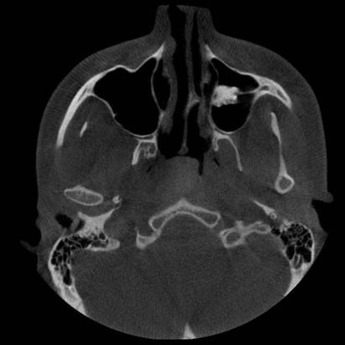
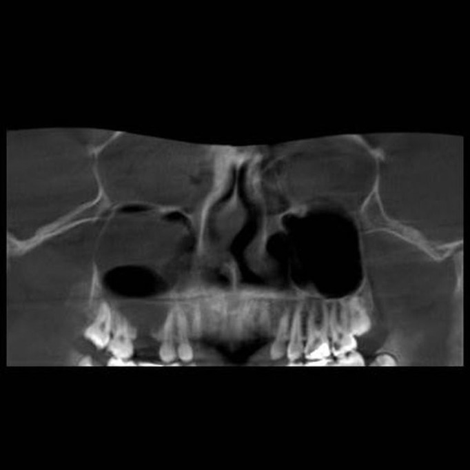
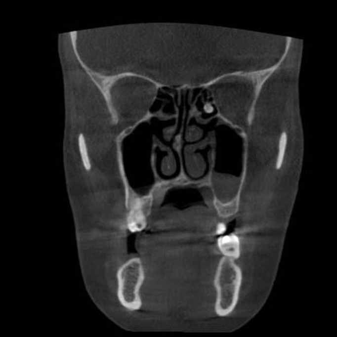
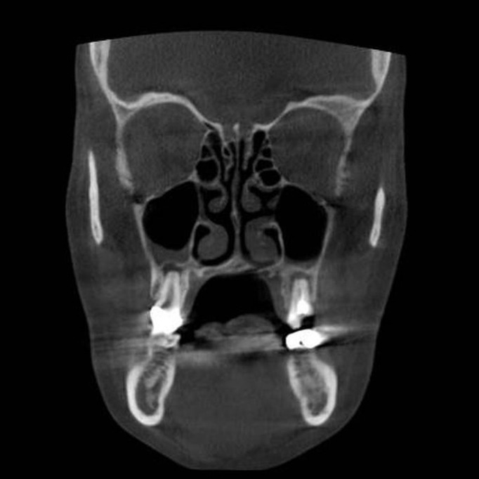
Paranasal sinus pathoses on cone beam computed tomography
- PMID: 28955552
- PMCID: PMC5573450
- DOI: 10.17096/jiufd.47796
Paranasal sinus pathoses on cone beam computed tomography
Abstract
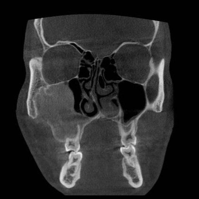
Purpose: The aim of this study was to investigate paranasal sinus pathoses detected on cone-beam computed tomography (CBCT) in an adult population.
Patients and methods: Three observers retrospectively inspected 353 consecutive CBCT scans obtained in a dentomaxillofacial radiology department for paranasal sinus pathoses. Descriptive statistics and chi-square tests were used to determine the prevalence of categorical parameters.
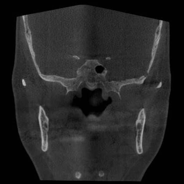
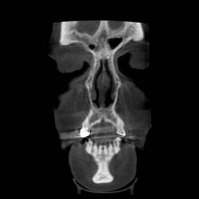
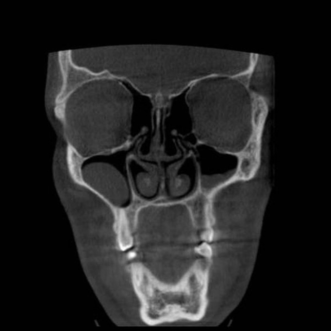
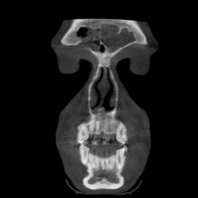
Results: The age of the patients ranged from 18 to 85 years (mean 41.27±16.76). There were 172 (48.7%) females and 181 (51.3%) males. There was a significant difference between the genders (p=0.02), with males (53.5%) having more sinus pathoses than females (46.5%). When the left and right sinuses were considered together, pathoses were most commonly seen in the maxillary sinuses (57.1%), followed by the ethmoid (53.7 %), frontal (22.6%), and sphenoid sinuses (15.8%). Mucosal thickening was the most frequently observed abnormality (51.7%), followed by hypoplasia (17.5%) and sinusitis (17.3%).
Conclusion: CBCT is a preferable imaging method for evaluation of paranasal sinuses. Dentomaxillofacial radiologists should examine the whole volume of CBCT images to ensure they do not overlook paranasal sinus pathoses.
Keywords: Cone-beam computed tomography; maxillofacial region; paranasal sinuses; sinusitis.
Conflict of interest statement
Conflict of interest: None declared.
Figures
Similar articles
-
Exploring the age and gender-based distribution of paranasal sinus osteomas using cone beam computed tomography: A retrospective cross-sectional study.Heliyon. 2024 Jul 31;10(15):e35222. doi: 10.1016/j.heliyon.2024.e35222. eCollection 2024 Aug 15. Heliyon. 2024. PMID: 39170231 Free PMC article.
-
Occurrence of maxillary sinus abnormalities detected by cone beam CT in asymptomatic patients.BMC Oral Health. 2012 Aug 10;12:30. doi: 10.1186/1472-6831-12-30. BMC Oral Health. 2012. PMID: 22883529 Free PMC article.
-
Assessment of paranasal sinus parameters according to ancient skulls' gender and age by using cone-beam computed tomography.Folia Morphol (Warsz). 2019;78(2):344-350. doi: 10.5603/FM.a2018.0089. Epub 2018 Oct 3. Folia Morphol (Warsz). 2019. PMID: 30280374
-
Prevalence of sinus membrane thickening and association with unhealthy teeth: a retrospective review of 831 consecutive patients with 1,662 cone-beam scans.J Oral Maxillofac Surg. 2014 Dec;72(12):2454-60. doi: 10.1016/j.joms.2014.06.442. Epub 2014 Jun 27. J Oral Maxillofac Surg. 2014. PMID: 25236817 Review.
-
Cone beam computed tomography for the nasal cavity and paranasal sinuses.Dent Clin North Am. 2014 Jul;58(3):627-51. doi: 10.1016/j.cden.2014.04.003. Dent Clin North Am. 2014. PMID: 24993926 Review.
Cited by
-
Is There a Relationship Between Maxillary Sinus's Inferior Pneumatisation and Sinonasal Variations? A Retrospective CBCT Study.J Oral Maxillofac Res. 2023 Sep 30;14(3):e3. doi: 10.5037/jomr.2023.14303. eCollection 2023 Jul-Sep. J Oral Maxillofac Res. 2023. PMID: 37969952 Free PMC article.
-
Anatomical variations and accessory structures in the maxilla in relation to implantological procedures: an observational retrospective study of 212 cases using cone-bean computed tomography.Int J Implant Dent. 2022 Nov 28;8(1):59. doi: 10.1186/s40729-022-00459-7. Int J Implant Dent. 2022. PMID: 36441355 Free PMC article.
-
Insight in to the Awareness of CBCT as an Imaging Modality in the Diagnosis and Management of ENT Disorders: A Cross Sectional Study.Indian J Otolaryngol Head Neck Surg. 2022 Dec;74(Suppl 3):5283-5293. doi: 10.1007/s12070-020-02209-w. Epub 2020 Oct 14. Indian J Otolaryngol Head Neck Surg. 2022. PMID: 36742614 Free PMC article.
-
Convolutional neural network for automatic maxillary sinus segmentation on cone-beam computed tomographic images.Sci Rep. 2022 May 7;12(1):7523. doi: 10.1038/s41598-022-11483-3. Sci Rep. 2022. PMID: 35525857 Free PMC article.
-
Management of Schneiderian membrane perforations during maxillary sinus floor augmentation with lateral approach in relation to subsequent implant survival rates: a systematic review and meta-analysis.Int J Implant Dent. 2021 Jul 12;7(1):91. doi: 10.1186/s40729-021-00346-7. Int J Implant Dent. 2021. PMID: 34250560 Free PMC article.
References
-
- Porter RS, Kaplan JL. The Merck Manual of Diagnosis and Therapy. 19th ed. Rahway, NJ, USA: Merck Publishing Group Merck and Co., Inc.; 2011.
-
- White SC, Pharoah MJ. Oral radiology: principles and interpretation. 6th ed. New Delhi, India: Mosby; 2009.
-
- Ingle JI, Bakland LK. Endodontics. 5th ed. Canada: BC Decker Inc; 2002.
-
- Donizeth-Rodrigues C, Fonseca-Da Silveira M, Gonçalves-De Alencar AH, Garcia-Santos-Silva MA, Francisco-De-Mendonça E, Estrela C. Three-dimensional images contribute to the diagnosis of mucous retention cyst in maxillary sinus. Med Oral Patol Oral Cir Bucal. 2013 Jan 1;18(1) doi: 10.4317/medoral.18141. - DOI - PMC - PubMed
LinkOut - more resources
Full Text Sources
Other Literature Sources