

Preparation and characterization of cockle shell aragonite nanocomposite porous 3D scaffolds for bone repair
- PMID: 28955752
- PMCID: PMC5614679
- DOI: 10.1016/j.bbrep.2017.04.008
Preparation and characterization of cockle shell aragonite nanocomposite porous 3D scaffolds for bone repair
Abstract
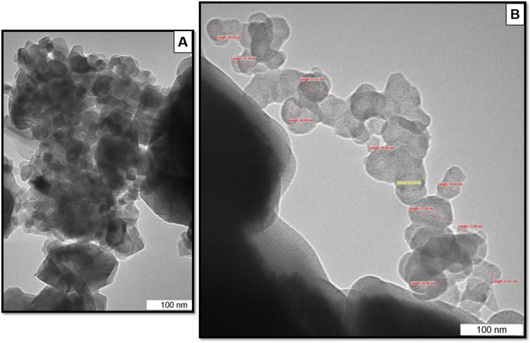
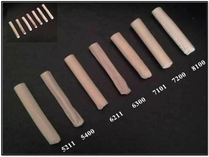
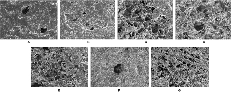
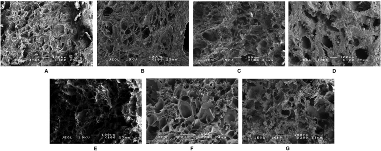
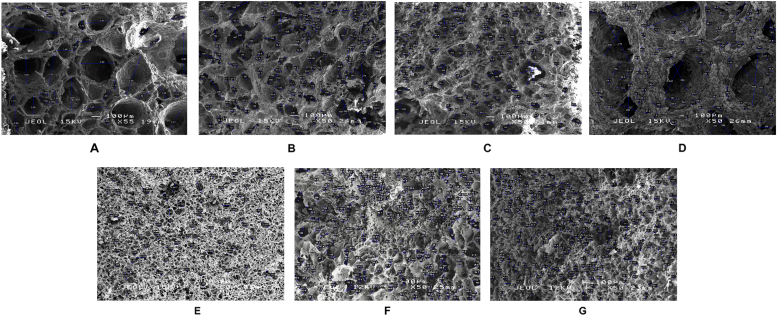
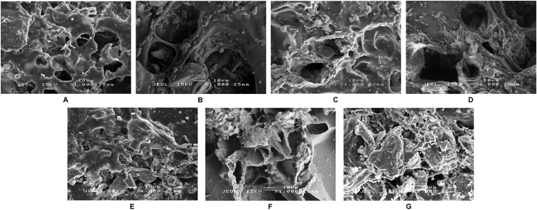
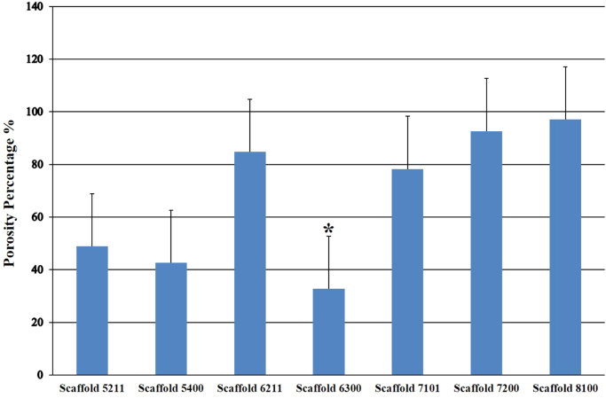
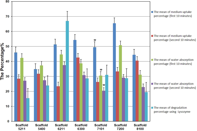
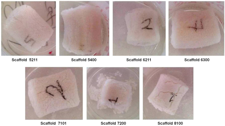
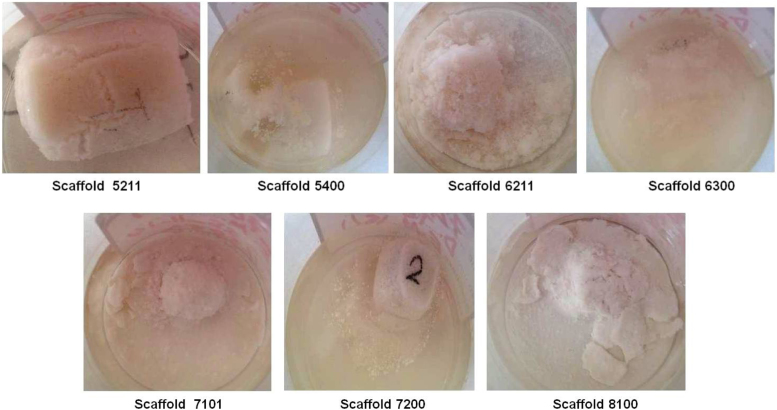
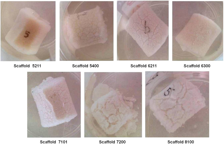
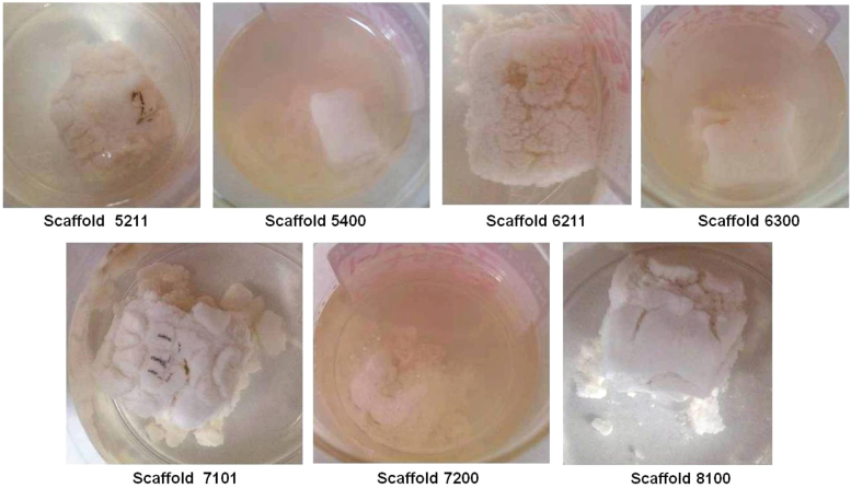
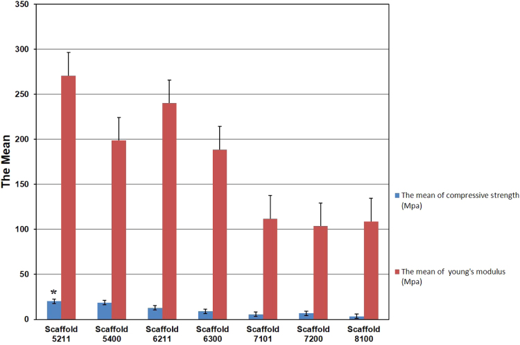
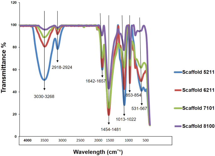
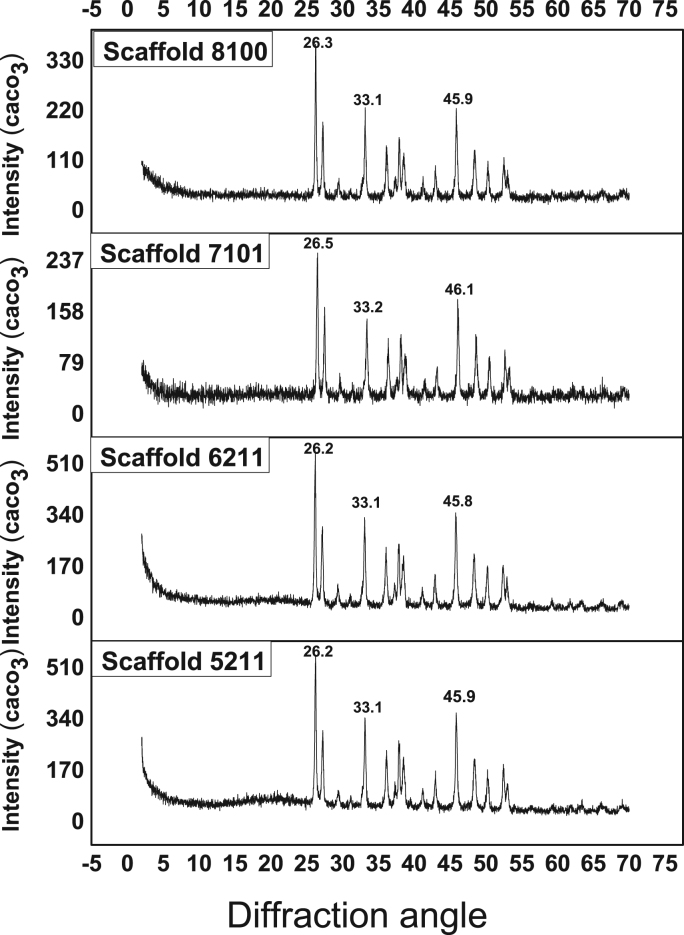
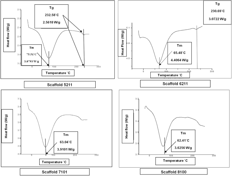
The demands for applicable tissue-engineered scaffolds that can be used to repair load-bearing segmental bone defects (SBDs) is vital and in increasing demand. In this study, seven different combinations of 3 dimensional (3D) novel nanocomposite porous structured scaffolds were fabricated to rebuild SBDs using an extraordinary blend of cockle shells (CaCo3) nanoparticles (CCN), gelatin, dextran and dextrin to structure an ideal bone scaffold with adequate degradation rate using the Freeze Drying Method (FDM) and labeled as 5211, 5400, 6211, 6300, 7101, 7200 and 8100. The micron sized cockle shells powder obtained (75 µm) was made into nanoparticles using mechano-chemical, top-down method of nanoparticles synthesis with the presence of the surfactant BS-12 (dodecyl dimethyl bataine). The phase purity and crystallographic structures, the chemical functionality and the thermal characterization of the scaffolds' powder were recognized using X-Ray Diffractometer (XRD), Fourier transform infrared (FTIR) spectrophotometer and Differential Scanning Calorimetry (DSC) respectively. Characterizations of the scaffolds were assessed by Scanning Electron Microscopy (SEM), Degradation Manner, Water Absorption Test, Swelling Test, Mechanical Test and Porosity Test. Top-down method produced cockle shell nanoparticles having averagely range 37.8±3-55.2±9 nm in size, which were determined using Transmission Electron Microscope (TEM). A mainly aragonite form of calcium carbonate was identified in both XRD and FTIR for all scaffolds, while the melting (Tm) and transition (Tg) temperatures were identified using DSC with the range of Tm 62.4-75.5 °C and of Tg 230.6-232.5 °C. The newly prepared scaffolds were with the following characteristics: (i) good biocompatibility and biodegradability, (ii) appropriate surface chemistry and (iii) highly porous, with interconnected pore network. Engineering analyses showed that scaffold 5211 possessed 3D interconnected homogenous porous structure with a porosity of about 49%, pore sizes ranging from 8.97 to 337 µm, mechanical strength 20.3 MPa, Young's Modulus 271±63 MPa and enzymatic degradation rate 22.7 within 14 days.
Keywords: %, Percentage; 3D porous nanocomposite scaffold; 3D, 3 Dimensional; 5211, cockle shells nanoparticles 50%, gelatin 25%, dextran 10%, and dextrin 15%; 5400, cockle shells nanoparticles 50%, gelatin 40%, dextran 5%, and dextrin 5%.; 6211, cockle shells nanoparticles 60%, gelatin 20%, dextran 10%, and dextrin 10%; 6300, cockle shells nanoparticles 60%, gelatin 30%, dextran 5%, and dextrin 5%; 7101, cockle shells nanoparticles 70%, gelatin 15%, dextran 5%, and dextrin 10%; 7200, cockle shells nanoparticles 70%, gelatin 20%, dextran 5%, and dextrin 5%; 8100, cockle shells nanoparticles 80%, gelatin 10%, dextran 5%, and dextrin 5%; ACN, Aragonite Calcium Carbonate Nanoparticles; ANOVA, One-Way Analysis of Variance; Aragonite; BS-12, dodecyl dimethyl bataine; Bone; C-H, Carbon-Hydrogen group; C-O, Carbon-Oxygen group; CCN, Calcium Carbonate Nanoparticles; Ca10PO46OH2, Chemical structure of Hydroxyapatite; CaCO3, Calcium carbonate; Characterization; Cockle shells; DSC, Differential Scanning Calorimetry; DW, Deionized Water; ECM, Extracellular Matrix; FDM, Freeze Drying Method; FTIR, Fourier Transform Infrared; HA, Hydroxyapatite; Hf, Heat of fusion; JCPDS, Joint Committee of Powder Diffraction Society; MPa, Megapascals (MPa or N/mm2) pascal (Pa) unit=one Newton per square meter; NC, Natural coral; PBS, Phosphate Buffer Solution; Pet, Density of Ethanol; R, Radius; S.E., Standard Error; SBD, Segmental Bone Defects; SEM, Scanning Electron Microscopy; T, Thickness; TEM, Transmission Electron Microscopy; Tg, Glass transition Temperature; Tm, Melting Temperature; U/mL, Unit per milliliter; W0, Dry Weight (Initial Weight); W1, Dry Weight; W2, Wet Weight; Wd, Dry Weight; Ww, Wet Weight; XRD, X-Ray Diffraction; cm, Centimeter; mL, Milliliter; min, Minutes; nm, Nanometer; °C, Degree Celsius; µm, Micrometer.
Figures
Similar articles
-
Surface-functionalized cockle shell-based calcium carbonate aragonite polymorph as a drug nanocarrier.Nanotechnol Sci Appl. 2017 May 16;10:79-94. doi: 10.2147/NSA.S120868. eCollection 2017. Nanotechnol Sci Appl. 2017. PMID: 28572724 Free PMC article.
-
Evaluation of a novel nanocrystalline hydroxyapatite powder and a solid hydroxyapatite/Chitosan-Gelatin bioceramic for scaffold preparation used as a bone substitute material.Turk J Chem. 2020 Aug 18;44(4):884-900. doi: 10.3906/kim-1912-40. eCollection 2020. Turk J Chem. 2020. PMID: 33488200 Free PMC article.
-
Generation of graphene oxide and nano-bioglass based scaffold for bone tissue regeneration.Biomed Mater. 2022 Sep 30;17(6). doi: 10.1088/1748-605X/ac92b4. Biomed Mater. 2022. PMID: 36113451
-
Preparation of novel bioactive nano-calcium phosphate-hydrogel composites.Sci Technol Adv Mater. 2010 Feb 22;11(1):014103. doi: 10.1088/1468-6996/11/1/014103. eCollection 2010 Feb. Sci Technol Adv Mater. 2010. PMID: 27877318 Free PMC article. Review.
-
Advances in Biodegradable 3D Printed Scaffolds with Carbon-Based Nanomaterials for Bone Regeneration.Materials (Basel). 2020 Nov 11;13(22):5083. doi: 10.3390/ma13225083. Materials (Basel). 2020. PMID: 33187218 Free PMC article. Review.
Cited by
-
Green Ca-source of cockle shells converted to calcium acetate for environmental sustainability.Heliyon. 2024 May 31;10(11):e32153. doi: 10.1016/j.heliyon.2024.e32153. eCollection 2024 Jun 15. Heliyon. 2024. PMID: 38868018 Free PMC article.
-
Development of 3D Printable Calcium Phosphate Cement Scaffolds with Cockle Shell Powders.Materials (Basel). 2023 Sep 10;16(18):6154. doi: 10.3390/ma16186154. Materials (Basel). 2023. PMID: 37763431 Free PMC article.
-
Coaxial Electrospun Polycaprolactone/Gelatin Nanofiber Membrane Loaded with Salidroside and Cryptotanshinone Synergistically Promotes Vascularization and Osteogenesis.Int J Nanomedicine. 2024 Jun 27;19:6519-6546. doi: 10.2147/IJN.S461141. eCollection 2024. Int J Nanomedicine. 2024. PMID: 38957181 Free PMC article.
-
Progress in Modern Marine Biomaterials Research.Mar Drugs. 2020 Nov 25;18(12):589. doi: 10.3390/md18120589. Mar Drugs. 2020. PMID: 33255647 Free PMC article. Review.
-
In vivo evaluation of a novel nanocomposite porous 3D scaffold in a rabbit model: histological analysis.Int J Nanomedicine. 2017 Dec 1;12:8587-8598. doi: 10.2147/IJN.S145663. eCollection 2017. Int J Nanomedicine. 2017. PMID: 29238193 Free PMC article.
References
-
- Oest M.E., Dupont K.M., Kong H.J., Mooney D.J., Guldberg R.E. Quantitative assessment of scaffold and growth factor-mediated repair of critically sized bone defects. J. Orthop. Res. 2007:941–950. - PubMed
-
- Blackwood K.A., Bock N., Dargaville T.R., Woodruff M.A. Scaffolds for growth factor delivery as applied to bone tissue engineering: review article. Int. J. Polym. Sci. 2012;2012:1–25.
-
- Blom A.W., Cunningham J.L., Hughes G., Lawes T.J., Smith N., Blunn G., Learmonth I.D., Goodship A.E. The compatibility of ceramic bone graft substitutes as allograft extenders for use in impaction grafting of the femur. J. Bone Jt. Surg. 2005;87-B(3):421–425. - PubMed
LinkOut - more resources
Full Text Sources
Other Literature Sources
Research Materials
Miscellaneous
