

Transcriptional modulation of pattern recognition receptors in chronic colitis in mice is accompanied with Th1 and Th17 response
- PMID: 28955789
- PMCID: PMC5613238
- DOI: 10.1016/j.bbrep.2017.08.009
Transcriptional modulation of pattern recognition receptors in chronic colitis in mice is accompanied with Th1 and Th17 response
Erratum in
-
Corrigendum "Transcriptional modulation of pattern recognition receptors in chronic colitis in mice is accompanied with Th1 and Th17 response" [Biochem. Biophys. Rep. 12 (2017) 29-39].Biochem Biophys Rep. 2018 Feb 15;13:147-148. doi: 10.1016/j.bbrep.2018.02.002. eCollection 2018 Mar. Biochem Biophys Rep. 2018. PMID: 29988850 Free PMC article.
Abstract
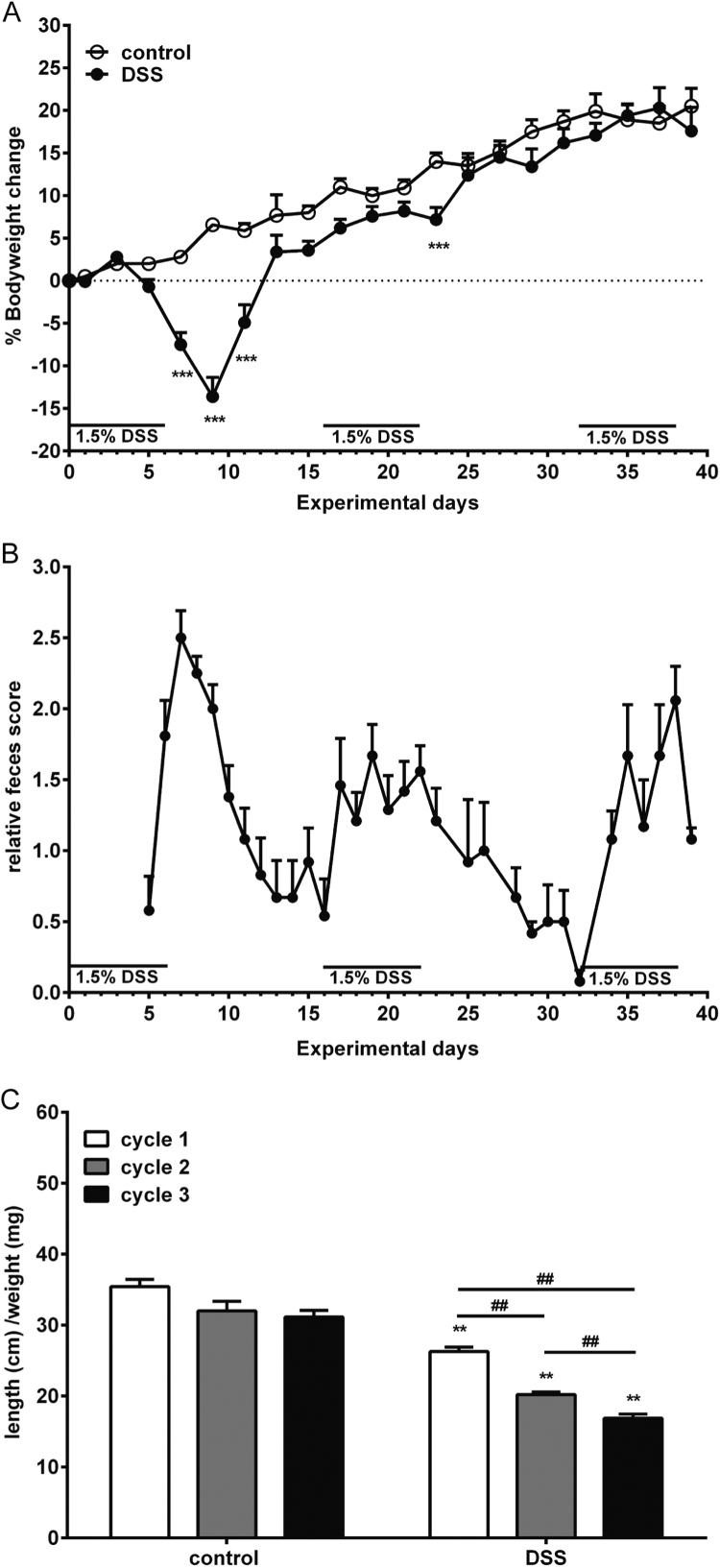
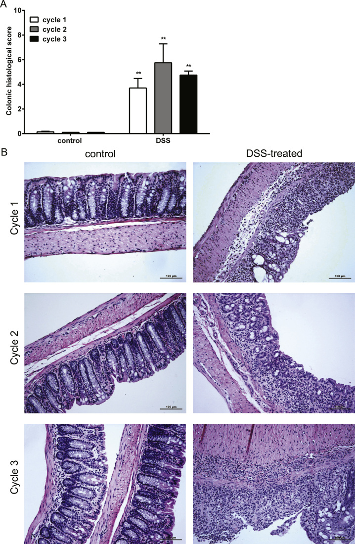
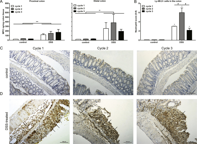
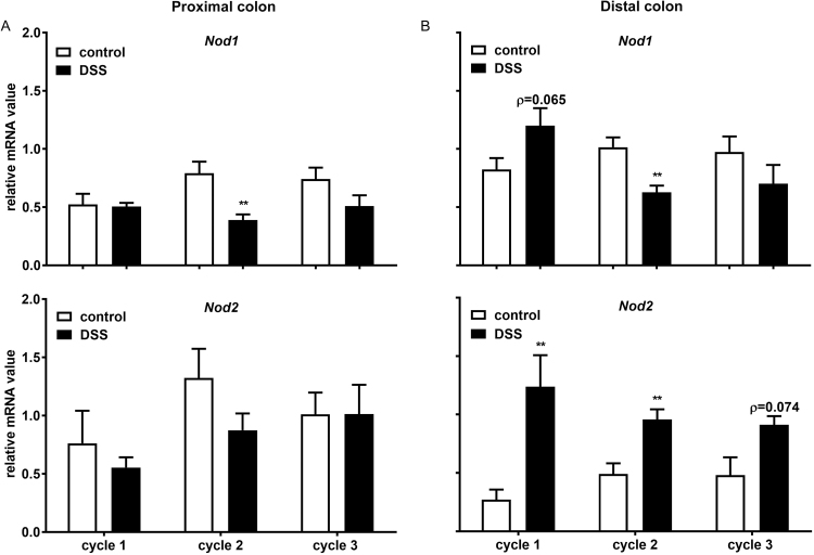
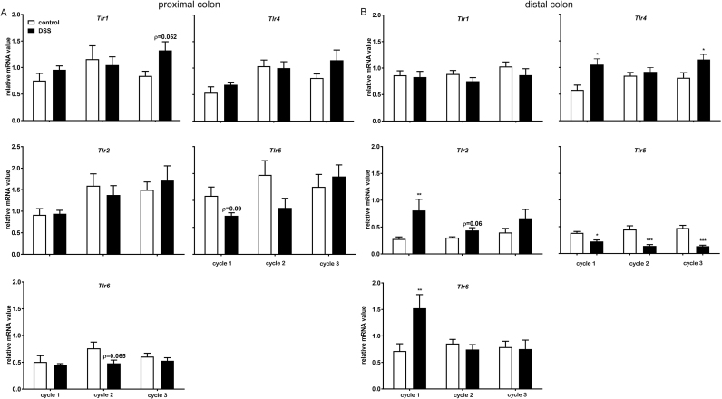
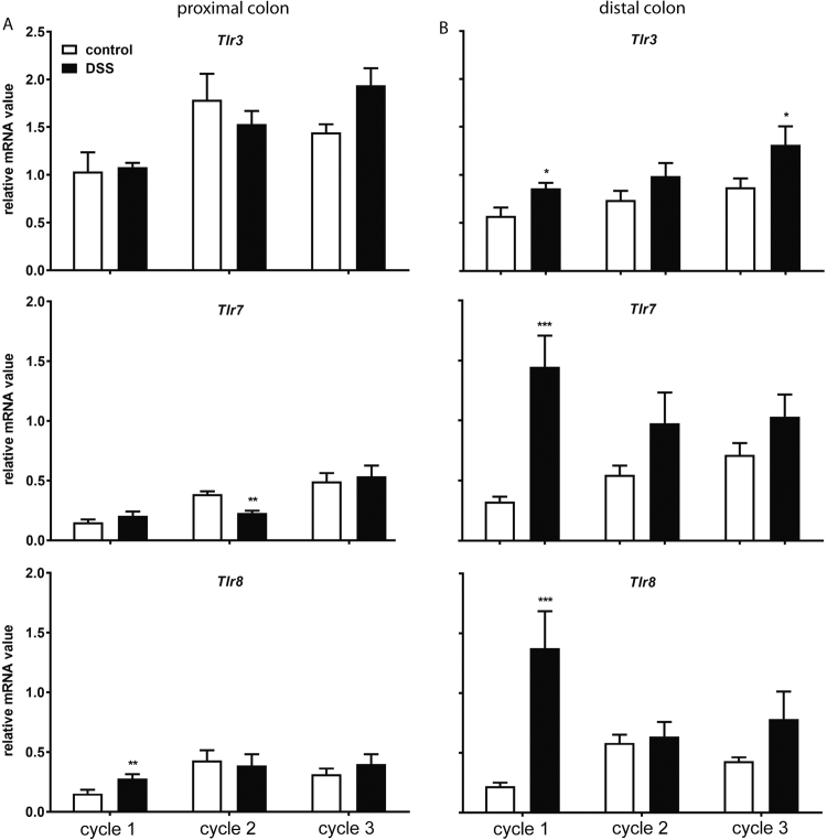
Pattern recognition receptors (PRRs) may contribute to inflammatory bowel diseases (IBD) development due to their microbial-sensing ability and the unique microenvironment in the inflamed gut. In this study, the PRR mRNA expression profile together with T cell-associated factors in the colon was examined using a chronic colitis mice model. 8-12 week old C57BL/6 mice were exposed to multiple dextran sodium sulfate (DSS) treatments interspersed with a rest period to mimic the course of chronic colitis. The clinical features and histological data were collected. The mRNA expressions of colonic PRRs, T cell-associated components were measured. Finally, the colons were scored for Foxp3+ cells. During chronic colitis, the histological data, but not the clinical manifestations demonstrated characteristic inflammatory symptoms in the distal colon. In contrast to acute colitis, the expression of all Toll-like receptors (Tlrs), except Tlr5 and Tlr9, was unaffected after repeated DSS treatments. The expression of Nod1 was decreased, while Nod2 increased. After third DSS treatment, only the expressions of Tlr3 and Tlr4 were significantly enhanced. Unlike other PRRs, decreased Tlr5 and increased Tlr9 mRNA expression persisted during the chronic colitis period. As the colitis progress, only the mRNA expression of Ifnγ and Il17 staid increased during chronic colitis, while the acute colitis-associated increase of Il23, and Il10 and Il12 was abolished. Finally, increased histological score of Foxp3+ cell in colon was found during the chronic colitis period. This study provides an expression pattern of PRRs during chronic colitis that is accompanied by a Th1- and Th17 cell-mediated immune response.
Keywords: Inflammatory bowel disease; Th cell responses; Toll like receptors.
Figures

Similar articles
-
Transcriptional modulation of pattern recognition receptors in acute colitis in mice.Biochim Biophys Acta. 2013 Dec;1832(12):2162-72. doi: 10.1016/j.bbadis.2013.07.004. Epub 2013 Jul 12. Biochim Biophys Acta. 2013. PMID: 23851050
-
Epithelial toll-like receptor 5 is constitutively localized in the mouse cecum and exhibits distinctive down-regulation during experimental colitis.Clin Vaccine Immunol. 2006 Jan;13(1):132-8. doi: 10.1128/CVI.13.1.132-138.2006. Clin Vaccine Immunol. 2006. PMID: 16426010 Free PMC article.
-
IL-33 alleviates DSS-induced chronic colitis in C57BL/6 mice colon lamina propria by suppressing Th17 cell response as well as Th1 cell response.Int Immunopharmacol. 2015 Dec;29(2):846-853. doi: 10.1016/j.intimp.2015.08.032. Epub 2015 Sep 8. Int Immunopharmacol. 2015. PMID: 26359542
-
Anti-tumor necrosis factor α therapy associates to type 17 helper T lymphocytes immunological shift and significant microbial changes in dextran sodium sulphate colitis.World J Gastroenterol. 2019 Mar 28;25(12):1465-1477. doi: 10.3748/wjg.v25.i12.1465. World J Gastroenterol. 2019. PMID: 30948910 Free PMC article.
-
Berberine ameliorates chronic relapsing dextran sulfate sodium-induced colitis in C57BL/6 mice by suppressing Th17 responses.Pharmacol Res. 2016 Aug;110:227-239. doi: 10.1016/j.phrs.2016.02.010. Epub 2016 Mar 9. Pharmacol Res. 2016. PMID: 26969793
Cited by
-
Cross Talk between Gut Microbiota and Intestinal Mucosal Immunity in the Development of Ulcerative Colitis.Infect Immun. 2021 Aug 16;89(9):e0001421. doi: 10.1128/IAI.00014-21. Epub 2021 Aug 16. Infect Immun. 2021. PMID: 33526559 Free PMC article. Review.
-
The Prophylactic Use of Bovine Colostrum in a Murine Model of TNBS-Induced Colitis.Animals (Basel). 2020 Mar 15;10(3):492. doi: 10.3390/ani10030492. Animals (Basel). 2020. PMID: 32183497 Free PMC article.
-
Intestinal inflammation induced by dextran sodium sulphate causes liver inflammation and lipid metabolism disfunction in laying hens.Poult Sci. 2020 Mar;99(3):1663-1677. doi: 10.1016/j.psj.2019.11.028. Epub 2020 Jan 22. Poult Sci. 2020. PMID: 32111331 Free PMC article.
-
Low-protein diets supplemented with casein hydrolysate favor the microbiota and enhance the mucosal humoral immunity in the colon of pigs.J Anim Sci Biotechnol. 2019 Oct 10;10:79. doi: 10.1186/s40104-019-0387-9. eCollection 2019. J Anim Sci Biotechnol. 2019. PMID: 31624591 Free PMC article.
-
Inflammation, Autoinflammation and Autoimmunity in Inflammatory Bowel Diseases.Curr Issues Mol Biol. 2023 Jun 30;45(7):5534-5557. doi: 10.3390/cimb45070350. Curr Issues Mol Biol. 2023. PMID: 37504266 Free PMC article. Review.
References
-
- Baumgart D.C., Sandborn W.J. Inflammatory bowel disease: clinical aspects and established and evolving therapies. Lancet. 2007;369(9573):1641–1657. - PubMed
-
- Ananthakrishnan A.N. Epidemiology and risk factors for IBD. Nat. Rev. Gastroenterol. Hepatol. 2015 - PubMed
-
- Brown S.J., Mayer L. The immune response in inflammatory bowel disease. Am. J. Gastroenterol. 2007;102(9):2058–2069. - PubMed
-
- Walsh D. Pattern recognition receptors-Molecular orchestrators of inflammation in inflammatory bowel disease. Cytokine Growth Factor Rev. 2012 - PubMed
-
- Maloy K.J., Powrie F. Intestinal homeostasis and its breakdown in inflammatory bowel disease. Nature. 2011;474(7351):298–306. - PubMed
LinkOut - more resources
Full Text Sources
Other Literature Sources
Research Materials
