

Increased level of phosphorylated desmin and its degradation products in heart failure
- PMID: 28955862
- PMCID: PMC5600436
- DOI: 10.1016/j.bbrep.2016.02.014
Increased level of phosphorylated desmin and its degradation products in heart failure
Abstract
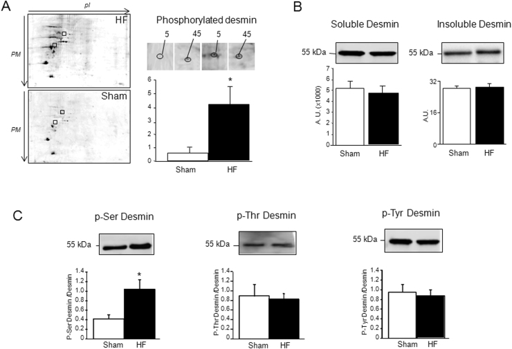
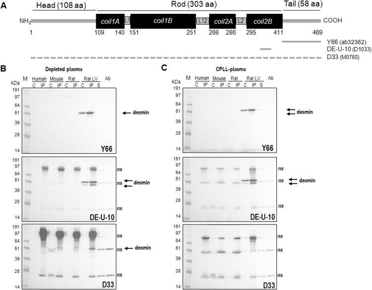
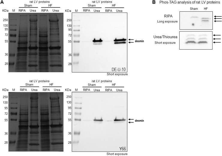
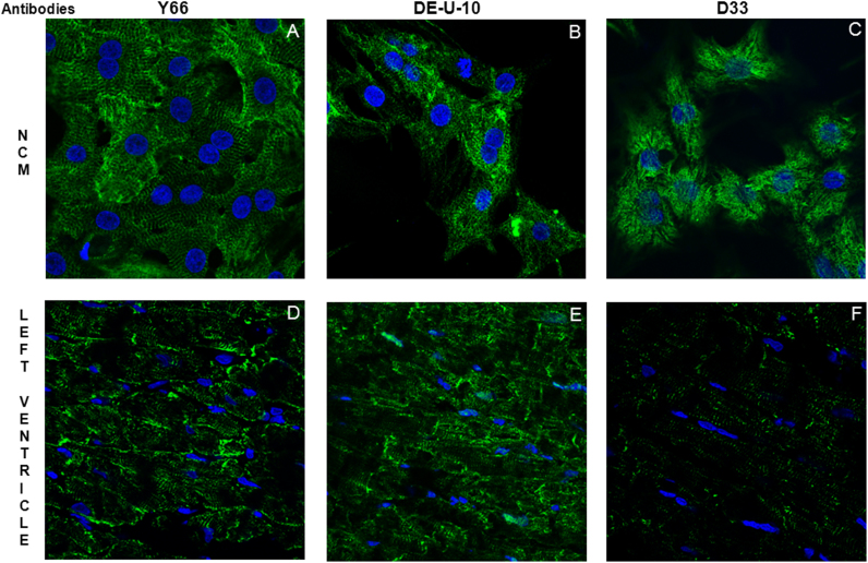
Although several risk factors such as infarct size have been identified, the progression/severity of heart failure (HF) remains difficult to predict in clinical practice. Using an experimental rat model of ischemic HF and phosphoproteomic technology, we found an increased level of phosphorylated desmin in the left ventricle (LV) of HF-rats. The purpose of the present work is to assess whether desmin is a circulating or only a tissue biomarker of HF. We used several antibodies in order to detect desmin, its proteolytic fragments and its phosphorylated form in LV and plasma by western blot, phosphate affinity electrophoresis, mass spectrometry and immunofluorescence. Plasma was treated with combinatorial peptide ligand library or depleted for albumin and immunoglobulins to increase the sensitivity of detection. We found a 2-fold increased serine-desmin phosphorylation in the LV of HF-rats, mainly in the insoluble fraction, suggesting the formation of desmin aggregates. Desmin cleavage products were also detected in the LV of HF rats, indicating that the increased phosphorylation of desmin results in more susceptibility to proteolytic activity, likely mediated by calpain activity. The native desmin and its degradation products were undetectable in the plasma of rat, mouse or human. These data suggest the potential of serine-phosphorylated form of desmin and its degradation products, but not of desmin itself, as tissue but not circulating biomarkers of HF.
Keywords: Desmin; Heart failure; Mass spectrometry; Phosphorylation; Plasma; Western blot.
Figures
Similar articles
-
Interplay Between Phosphorylation and O-GlcNAcylation of Sarcomeric Proteins in Ischemic Heart Failure.Front Endocrinol (Lausanne). 2018 Oct 5;9:598. doi: 10.3389/fendo.2018.00598. eCollection 2018. Front Endocrinol (Lausanne). 2018. PMID: 30344511 Free PMC article.
-
Circulating plasma serine208-phosphorylated troponin T levels are indicator of cardiac dysfunction.J Cell Mol Med. 2013 Oct;17(10):1335-44. doi: 10.1111/jcmm.12112. Epub 2013 Aug 2. J Cell Mol Med. 2013. PMID: 23905701 Free PMC article.
-
Desmin Phosphorylation Triggers Preamyloid Oligomers Formation and Myocyte Dysfunction in Acquired Heart Failure.Circ Res. 2018 May 11;122(10):e75-e83. doi: 10.1161/CIRCRESAHA.117.312082. Epub 2018 Feb 26. Circ Res. 2018. PMID: 29483093 Free PMC article.
-
Desmin modifications associate with amyloid-like oligomers deposition in heart failure.Cardiovasc Res. 2014 Apr 1;102(1):24-34. doi: 10.1093/cvr/cvu003. Epub 2014 Jan 9. Cardiovasc Res. 2014. PMID: 24413773 Free PMC article.
-
Desmin aggrephagy in rat and human ischemic heart failure through PKCζ and GSK3β as upstream signaling pathways.Cell Death Discov. 2021 Jun 26;7(1):153. doi: 10.1038/s41420-021-00549-2. Cell Death Discov. 2021. PMID: 34226534 Free PMC article.
Cited by
-
Interplay Between Phosphorylation and O-GlcNAcylation of Sarcomeric Proteins in Ischemic Heart Failure.Front Endocrinol (Lausanne). 2018 Oct 5;9:598. doi: 10.3389/fendo.2018.00598. eCollection 2018. Front Endocrinol (Lausanne). 2018. PMID: 30344511 Free PMC article.
-
Mitochondrial-Targeted Therapies Require Mitophagy to Prevent Oxidative Stress Induced by SOD2 Inactivation in Hypertrophied Cardiomyocytes.Antioxidants (Basel). 2022 Apr 6;11(4):723. doi: 10.3390/antiox11040723. Antioxidants (Basel). 2022. PMID: 35453408 Free PMC article.
-
A Risk Classification System With Five-Gene for Survival Prediction of Glioblastoma Patients.Front Neurol. 2019 Jul 16;10:745. doi: 10.3389/fneur.2019.00745. eCollection 2019. Front Neurol. 2019. PMID: 31379707 Free PMC article.
-
Woven bone formation and mineralization by rat mesenchymal stromal cells imply increased expression of the intermediate filament desmin.Front Endocrinol (Lausanne). 2023 Sep 4;14:1234569. doi: 10.3389/fendo.2023.1234569. eCollection 2023. Front Endocrinol (Lausanne). 2023. PMID: 37732119 Free PMC article.
-
Costameres, dense plaques and podosomes: the cell matrix adhesions in cardiovascular mechanosensing.J Muscle Res Cell Motil. 2019 Jun;40(2):197-209. doi: 10.1007/s10974-019-09529-7. Epub 2019 Jun 18. J Muscle Res Cell Motil. 2019. PMID: 31214894 Free PMC article. Review.
References
-
- Go A.S., Mozaffarian D., Roger V.L., Benjamin E.J., Berry J.D., Blaha M.J., Dai S., Ford E.S., Fox C.S., Franco S., Fullerton H.J., Gillespie C., Hailpern S.M., Heit J.A., Howard V.J., Huffman M.D., Judd S.E., Kissela B.M., Kittner S.J., Lackland D.T., Lichtman J.H., Lisabeth L.D., Mackey R.H., Magid D.J., Marcus G.M., Marelli A., Matchar D.B., McGuire D.K., Mohler E.R., Moy 3rd, C.S., Mussolino M.E., Neumar R.W., Nichol G., Pandey D.K., Paynter N.P., Reeves M.J., Sorlie P.D., Stein J., Towfighi A., Turan T.N., Virani S.S., Wong N.D., Woo D., Turnerl M.B. American heart association statistics committee and stroke statistics subcommittee. Heart disease and stroke statistics-2014 update: a report from the American heart association. Circulation. 2014;129:e28–e292. - PMC - PubMed
-
- Lindsey M.L., Mayr M., Gomes A.V., Delles C., Arrell D.K., Murphy A.M., Lange R.A., Costello C.E., Jin Y.F., Laskowitz D.T., Sam F., Terzic A., Van Eyk J., Srinivas P.R. Transformative impact of Proteomics on cardiovascular health and disease: a scientific statement from the American heart association. Circulation. 2015;132:852–872. - PubMed
-
- Cieniewski-Bernard C., Mulder P., Henry J.P., Drobecq H., Dubois E., Pottiez G., Thuillez C., Amouyel P., Richard V., Pinet F. Proteomic analysis of left ventricular remodeling in an experimental model of heart failure. J. Proteome Res. 2008;7:5004–5016. - PubMed
-
- Dubois E., Richard V., Mulder P., Lamblin N., Drobecq H., Henry J.P., Amouyel P., Thuillez C., Bauters C., Pinet F. Decreased serine207 phosphorylation of troponin T as a biomarker for left ventricular remodelling after myocardial infarction. Eur. Heart J. 2011;32:115–123. - PubMed
LinkOut - more resources
Full Text Sources
Other Literature Sources
Research Materials
Miscellaneous
