

CD44 induced enhancement of phosphatase activity and calcium influx: Modifications of EGR-1 expression and cell proliferation
- PMID: 28955875
- PMCID: PMC5600419
- DOI: 10.1016/j.bbrep.2016.03.016
CD44 induced enhancement of phosphatase activity and calcium influx: Modifications of EGR-1 expression and cell proliferation
Abstract
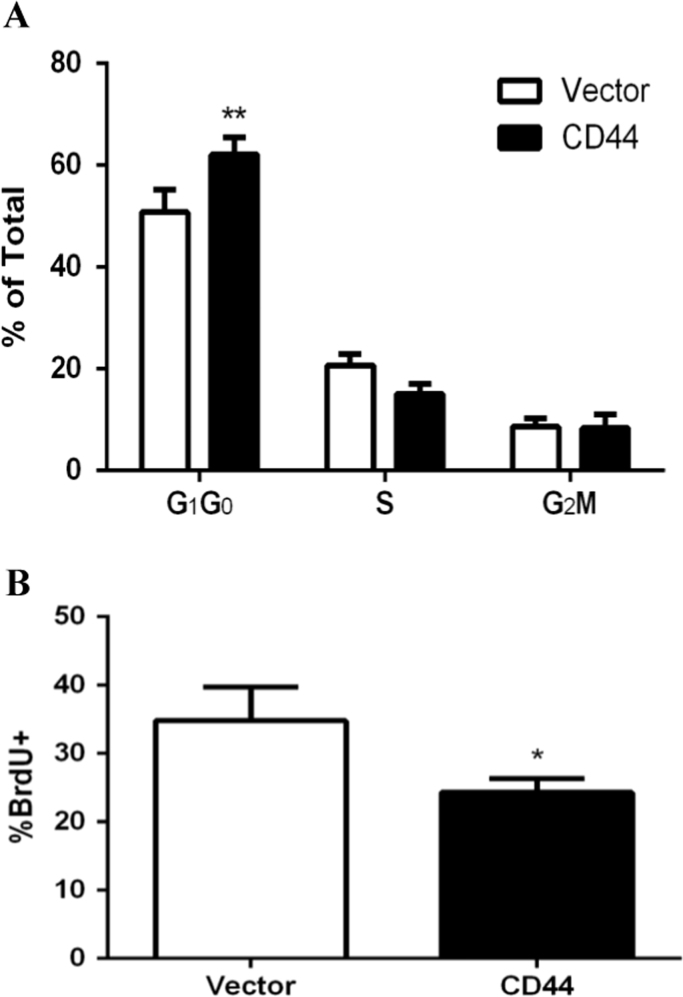
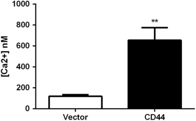
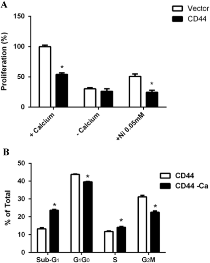
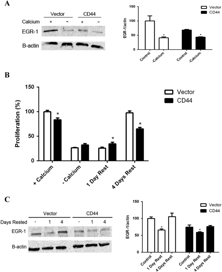
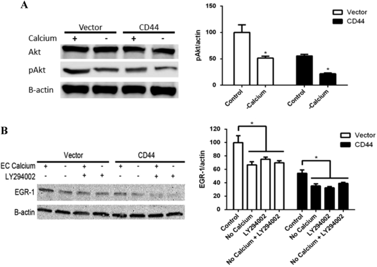
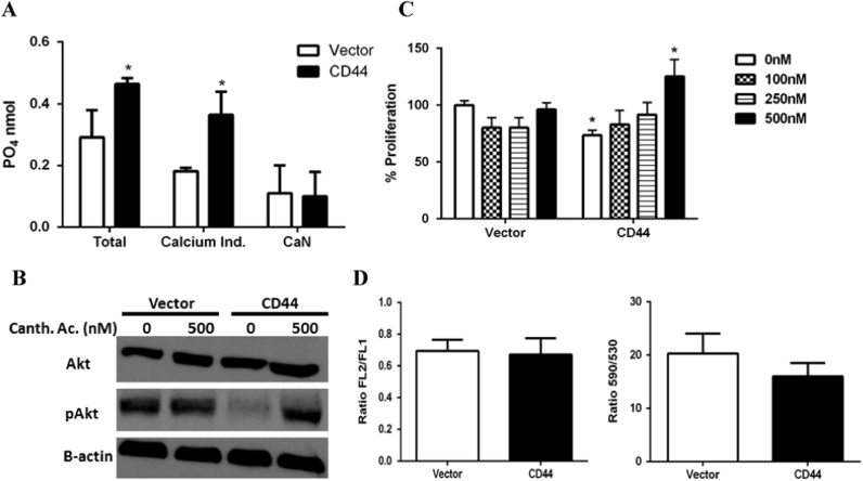
The purpose of this study was to investigate how CD44 impaired Akt phosphorylation, EGR-1 expression and cell proliferation. E6.1 Jurkat cells, which lack endogenous CD44 expression, were engineered to express CD44. Previously we showed that Akt is hypophosphorylated, EGR-1 expression is reduced and proliferation is impaired in CD44 expressing E6.1 Jurkat cells. The cell cycle was studied using flow cytometry and the role of calcium (Ca2+) in Akt phosphorylation and EGR-1 expression was investigated using Western blotting. Phosphatase activity was assessed using a commercially available kit. CD44 expressing cells showed disruption at the G1 to S transition. Chelation of Ca2+ from the culture media impaired Akt phosphorylation and EGR-1 expression in both CD44 expressing cells and the open vector control. Moreover, Ni2+ disrupted cell proliferation in both cell types suggesting Ca2+ import through calcium release activated calcium channels (CRAC). Staining of cells with fura-2 AM showed significantly higher Ca2+ in CD44 expressing cells as compared with the vehicle control. Finally, non-calcium mediated phosphatase activity was significantly greater in CD44 expressing cells. We propose that the enhanced phosphatase activity in the CD44 cells increased the dephosphorylation rate of Akt; at the same time, the increased intracellular concentration of Ca2+ in the CD44 cells ensured that the phosphorylation of Akt remains intact albeit at lower concentrations as compared with the vector control. Reduced Akt phosphorylation resulted in lowered expression of EGR-1 and hence, reduced the cell proliferation rate.
Keywords: Acute Lymphoblastic Leukemia; CD44; Calcium; Jurkat; Proliferation.
Figures
Similar articles
-
Inhibition of cell proliferation by CD44: Akt is inactivated and EGR-1 is down-regulated.Cell Prolif. 2010 Aug;43(4):385-95. doi: 10.1111/j.1365-2184.2010.00689.x. Cell Prolif. 2010. PMID: 20590664 Free PMC article.
-
Loss of Egr-1 sensitizes pancreatic β-cells to palmitate-induced ER stress and apoptosis.J Mol Med (Berl). 2015 Jul;93(7):807-18. doi: 10.1007/s00109-015-1272-4. Epub 2015 Mar 5. J Mol Med (Berl). 2015. PMID: 25737480
-
P2X(7) receptor stimulation upregulates Egr-1 biosynthesis involving a cytosolic Ca(2+) rise, transactivation of the EGF receptor and phosphorylation of ERK and Elk-1.J Cell Physiol. 2007 Oct;213(1):36-44. doi: 10.1002/jcp.21085. J Cell Physiol. 2007. PMID: 17474086
-
N-4-tert-butyl benzyl haloperidol chloride suppresses Ca2+-dependent Egr-1 expression and subsequently inhibits vascular smooth muscle cell proliferation induced by angiotensin II.Cell Physiol Biochem. 2009;23(4-6):295-304. doi: 10.1159/000218176. Epub 2009 May 6. Cell Physiol Biochem. 2009. PMID: 19471097
-
Antisense-mediated loss of calcium homoeostasis endoplasmic reticulum protein (CHERP; ERPROT213-21) impairs Ca2+ mobilization, nuclear factor of activated T-cells (NFAT) activation and cell proliferation in Jurkat T-lymphocytes.Biochem J. 2003 Jul 1;373(Pt 1):133-43. doi: 10.1042/BJ20030013. Biochem J. 2003. PMID: 12656674 Free PMC article.
Cited by
-
CD44 Intracellular Domain: A Long Tale of a Short Tail.Cancers (Basel). 2023 Oct 18;15(20):5041. doi: 10.3390/cancers15205041. Cancers (Basel). 2023. PMID: 37894408 Free PMC article. Review.
-
Role of CD44 in Regulating TLR2 Activation of Human Macrophages and Downstream Expression of Proinflammatory Cytokines.J Immunol. 2018 Jan 15;200(2):758-767. doi: 10.4049/jimmunol.1700713. Epub 2017 Dec 1. J Immunol. 2018. PMID: 29196459 Free PMC article.
-
CD44 Receptor Mediates Urate Crystal Phagocytosis by Macrophages and Regulates Inflammation in A Murine Peritoneal Model of Acute Gout.Sci Rep. 2020 Apr 1;10(1):5748. doi: 10.1038/s41598-020-62727-z. Sci Rep. 2020. PMID: 32238827 Free PMC article.
-
G-protein-coupled estrogen receptor 30 regulation of signaling downstream of protein kinase Cε mediates sex dimorphism in hyaluronan-induced antihyperalgesia.Pain. 2025 Mar 1;166(3):539-556. doi: 10.1097/j.pain.0000000000003419. Epub 2024 Oct 10. Pain. 2025. PMID: 39787533
-
miR-let-7c-3p targeting on Egr-1 contributes to the committed differentiation of leukemia cells into monocyte/macrophages.Oncol Lett. 2022 Jun 22;24(2):273. doi: 10.3892/ol.2022.13393. eCollection 2022 Aug. Oncol Lett. 2022. PMID: 35782903 Free PMC article.
References
-
- DeGrendele H.C., Estess P., Siegelman M.H. Requirement for CD44 in activated T cell extravasation into an inflammatory site. Science. 1997;278:672–675. - PubMed
-
- Ponta H., Sherman L., Herrlich P.A. CD44: from adhesion molecules to signalling regulators. Nat. Rev. Mol. Cell Biol. 2003;4:33–45. - PubMed
-
- Singleton P.A., Bourguignon L.Y. CD44 interaction with ankyrin and IP3 receptor in lipid rafts promotes hyaluronan-mediated Ca2+ signaling leading to nitric oxide production and endothelial cell adhesion and proliferation. Exp. Cell Res. 2004;295:102–118. - PubMed
LinkOut - more resources
Full Text Sources
Other Literature Sources
Miscellaneous
