. 2016 May 18:7:56-62.
doi: 10.1016/j.bbrep.2016.05.013. eCollection 2016 Sep.

Actin exposure upon tissue injury is a targetable wound site-specific protein marker

Affiliations

Actin exposure upon tissue injury is a targetable wound site-specific protein marker

Erik D Pendleton et al. Biochem Biophys Rep. .

Abstract

Background: Identification of wound-specific markers would represent an important step toward damaged tissue detection and targeted delivery of biologically important materials to injured sites. Such delivery could minimize the amount of therapeutic materials that must be administered and limit potential collateral damage on nearby normal tissues. Yet, biological markers that are specific for injured tissue sites remain elusive.

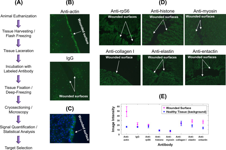
Methods: In this study, we have developed an immunohistological approach for identification of protein epitopes specifically exposed in wounded tissue sites.

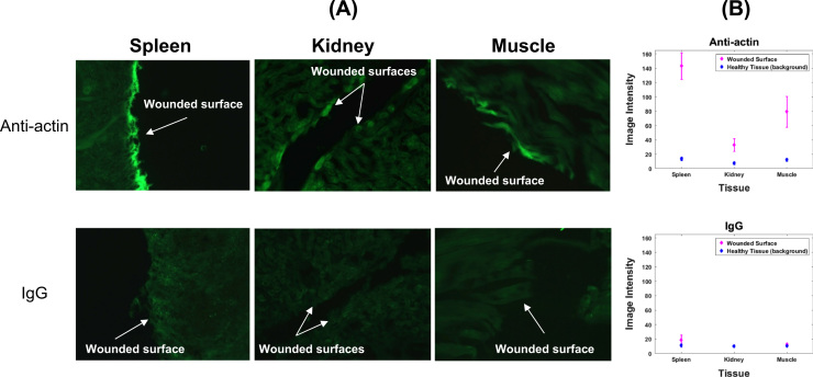
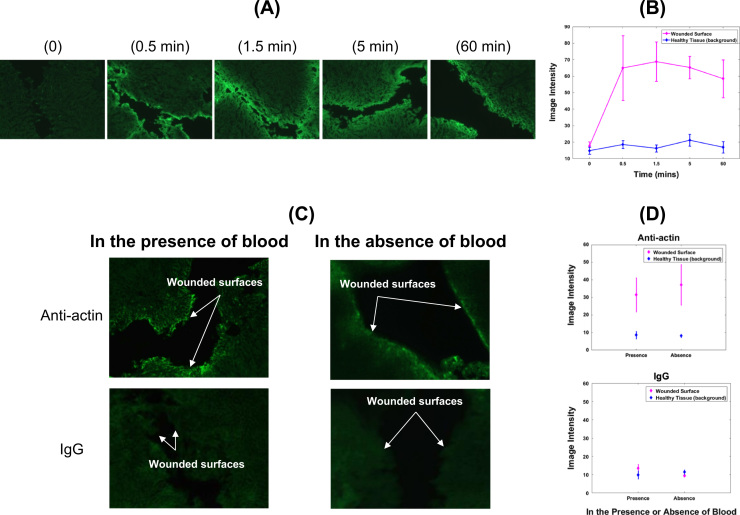
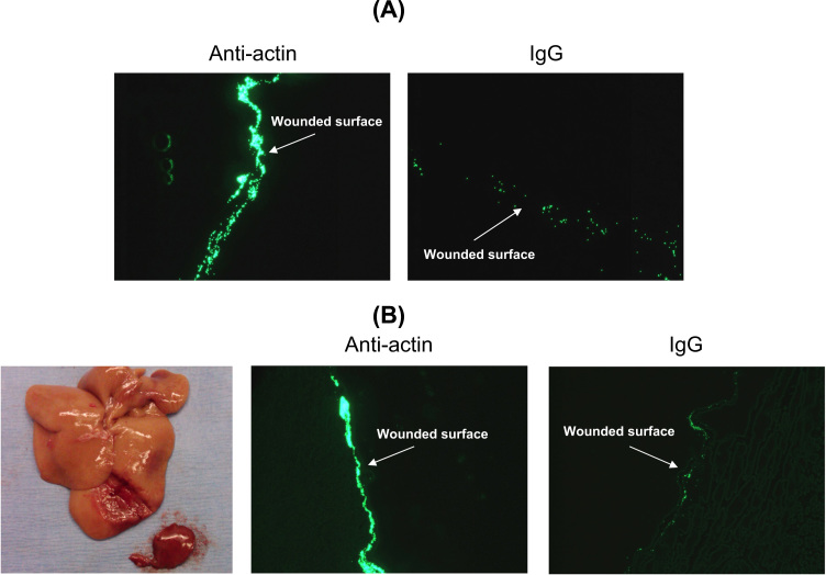
Results: Using ex-vivo tissue samples in combination with fluorescently-labeled antibodies we show that actin, an intracellular cytoskeletal protein, is specifically exposed upon injury. The targetability of actin in injured sites has been demonstrated in vivo through the specific delivery of anti-actin conjugated particles to the wounded tissue in a lethal rat model of grade IV liver injury.

Conclusions: These results illustrate that identification of injury-specific protein markers and their targetability for specific delivery is feasible.

General significance: Identification of wound-specific targets has important medical applications as it could enable specific delivery of various products, such as expression vectors, therapeutic drugs, hemostatic materials, tissue healing, or scar prevention agents, to internal sites of penetrating or surgical wounds regardless of origin, geometry or location.

Keywords: Actin; Bleeding; Hemorrhage; Injury; Protein marker; Wound.


Figures

Fig. 1.
Fig. 1
Identification of actin as a wound-specific marker. (A). Outline of the experimental procedure for identification of protein targets exposed in injured tissue sites. Tissue samples are removed from the animal upon euthanization and lacerated under controlled conditions. Tissue samples are incubated with labeled (fluorescein) antibodies against the candidate target epitopes or with labeled non-specific antibodies (IgG) as negative controls. Tissue samples are then fixed, subjected to fast freezing and cryosectioning, and examined using fluorescence imaging microscopy. Microscopy images are captured and analyzed to assess whether the target epitopes were exposed in injured tissue areas as a means of their specific interaction with their cognate labeled antibodies. (B). Traumatized rat liver tissue treated with fluoresceinated (FITC-labeled) antibody against actin (top panel) or IgG (bottom panel). Images are representative of a series of horizontal slices across a vertical injury. Pictures were taken using inverted microscopy at 10× magnification. (C). Fluorescence image of tissue sample treated with FITC-labeled anti-actin (green) and fluorescent nuclear counterstain, DAPI (blue). (D). Screening against intracellular (top panels) or extracellular matrix (bottom panels) proteins for specific wound exposure. (E). Quantitative analysis (mean and standard deviation) of image intensities at the wounded surface versus the neighboring healthy tissue areas for the wound targets tested.
Fig. 2.
Fig. 2
Actin recognition in injured sites of various tissues. (A). Traumatized rat spleen (left), kidney (middle) or muscle (right) tissues were treated with fluoresceinated (FITC-labeled) antibody against actin (top panels) or IgG as negative control (bottom panels). (B). Quantitative analysis (mean and standard deviation) of image intensities at the wounded surface versus the neighboring healthy areas for spleen, kidney and muscle tissues treated with anti-actin (top panel) or IgG (bottom panel) antibodies.
Fig. 3.
Fig. 3
Availability of actin epitope in wounded tissue. (A). Fluorescence images from experiments assessing wound-specific binding of FITC-labeled anti-actin antibodies upon incubation with injured rat liver ex vivo for various time periods. (B). Quantitative analysis (mean and standard deviation) of image intensities at the wounded surface versus the neighboring healthy tissue areas upon incubation of injured tissue samples with anti-actin antibody for various incubation times. (C). Targeting of actin in wounded rat liver tissue immersed in blood (left panels) or in the absence of blood (right panels). Excised wounded liver tissue in each case was treated with either FITC-labeled antibody against actin (top panels) or FITC-labeled IgG antibody as negative control (bottom panels). (D). Quantitative analysis of image intensities at the wounded surface versus the neighboring healthy tissue areas for tissue samples treated with anti-actin (top panel) or IgG (bottom panel) antibodies in the presence or absence of blood.
Fig. 4.
Fig. 4
Anti-actin-conjugated beads binding to wounded liver tissue ex vivo and in vivo. (A). Anti-actin-conjugated beads binding to injured liver tissue ex vivo. Excised liver tissue was incubated with labeled polystyrene beads conjugated to either anti-actin antibodies (left panel) or IgG as negative control (right panel). (B). Assessment of actin as a wound-specific biomarker in vivo. Transport and wound-specific binding was tested in a grade IV anesthetized rat lethal liver injury model. Lacerated liver tissue was incubated with labeled polystyrene beads conjugated to either anti-actin antibody or IgG (negative control). Following treatment, the anesthetized animals were euthanized, and the livers harvested, washed, fixed and sectioned for fluorescence microscopy. Left panel: Typical liver injury generated in this procedure. Middle panel: Beads tagged with anti-actin antibodies. Right panel: Beads tagged with IgG.

Similar articles

References

    1. Agnihotri S.A., Mallikarjuna N.N., Aminabhavi T.M. Recent advances on chitosan-based micro- and nanoparticles in drug delivery. J. Control. Release. 2004;100:5–28. - PubMed
    1. Ahrens S., Zelenay S., Sancho D., Hanč P., Kjær S. F-actin is an evolutionarily conserved damage-associated molecular pattern recognized by DNGR-1, a receptor for dead cells. Immunity. 2012;36:635–645. - PubMed
    1. Emoto K., Nagasaki Y., Iijima M., Kato M., Kataoka K. Preparation of non-fouling surface through the coating with core-polymerized block copolymer micelles having aldehyde-ended PEG shell. Colloids Surf. B: Biointerfaces. 2000;18:337–346. - PubMed
    1. Gasper M.M., Blanco D., Cruz M.E., Alonso M.J. Formulation of L-asparaginase-loaded poly(lactide-co-glycolide) nanoparticles: influence of polymer properties on enzyme loading, activity and in vitro release. J. Control. Release. 1998;52:53–62. - PubMed
    1. Gref R., Minamitake Y., Peracchia M.T., Trubetskoy V., Torchilin V., Langer R. Biodegradable long-circulating polymeric nanospheres. Science. 1994;263:1600–1603. - PubMed

LinkOut - more resources