

Specific titin and myomesin domains stimulate myoblast proliferation
- PMID: 28956009
- PMCID: PMC5614584
- DOI: 10.1016/j.bbrep.2016.12.007
Specific titin and myomesin domains stimulate myoblast proliferation
Abstract
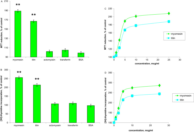
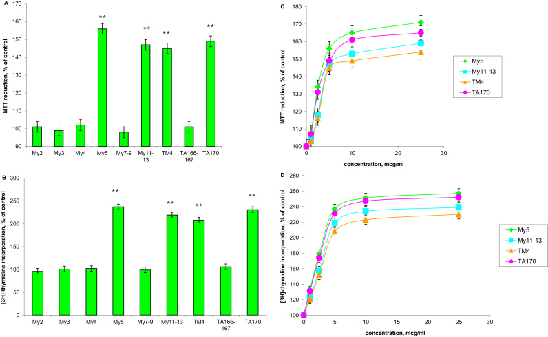
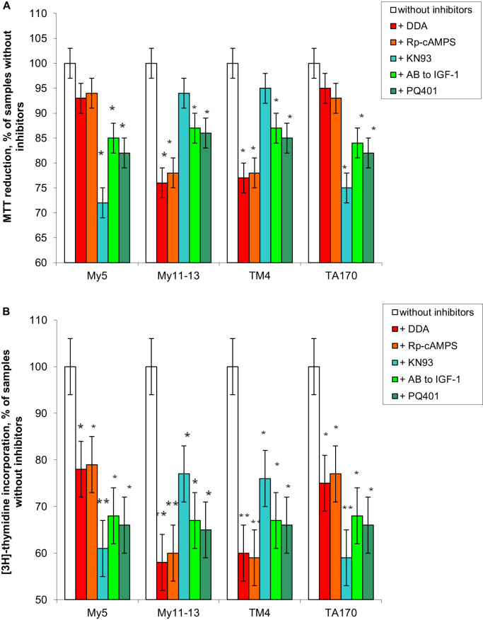
Myofibrillar proteins titin and myomesin stimulated myoblast proliferation as determined by MTT-test and labelled thymidine incorporation in the DNA. Specific Fn type III and Ig-like domains of these proteins were able to exert mitogenic effects as well. Proliferative effect of Fn type III domains was highly sensitive to inhibition of Ca2+/calmodulin dependent protein kinase, whereas the effect of Ig-like domains showed greater sensitivity to the inhibition of adenylyl cyclase - cAMP - PKA pathway. IGF-1 autocrine signalling inhibition partially suppressed mitogenic effects revealed by both domain types.
Keywords: Fn type III domain; IGF-1; Ig-like domain; Myoblast proliferation; Myomesin; Titin.
Figures
Similar articles
-
Induction of insulin-like growth factor 1 splice forms by subfragments of myofibrillar proteins.Mol Cell Endocrinol. 2015 Jan 5;399:69-77. doi: 10.1016/j.mce.2014.08.010. Epub 2014 Aug 22. Mol Cell Endocrinol. 2015. PMID: 25152160
-
Molecular structure of the sarcomeric M band: mapping of titin and myosin binding domains in myomesin and the identification of a potential regulatory phosphorylation site in myomesin.EMBO J. 1997 Jan 15;16(2):211-20. doi: 10.1093/emboj/16.2.211. EMBO J. 1997. PMID: 9029142 Free PMC article.
-
Study of the mechanical properties of myomesin proteins using dynamic force spectroscopy.J Mol Biol. 2005 May 20;348(5):1127-37. doi: 10.1016/j.jmb.2005.03.040. Epub 2005 Apr 7. J Mol Biol. 2005. PMID: 15854649
-
Growth hormone and the insulin-like growth factor system in myogenesis.Endocr Rev. 1996 Oct;17(5):481-517. doi: 10.1210/edrv-17-5-481. Endocr Rev. 1996. PMID: 8897022 Review.
-
Stretching molecular springs: elasticity of titin filaments in vertebrate striated muscle.Histol Histopathol. 2000 Jul;15(3):799-811. doi: 10.14670/HH-15.799. Histol Histopathol. 2000. PMID: 10963124 Review.
Cited by
-
Potassium chloride released from contracting skeletal muscle may stimulate development of its hypertrophy.Biochem Biophys Rep. 2019 Mar 21;18:100627. doi: 10.1016/j.bbrep.2019.100627. eCollection 2019 Jul. Biochem Biophys Rep. 2019. PMID: 30957033 Free PMC article.
-
The Influence of Myofibrils on the Proliferation and Differentiation of Myoblasts Cocultured with Macrophages.Dokl Biochem Biophys. 2018 Mar;479(1):72-76. doi: 10.1134/S1607672918020060. Epub 2018 May 19. Dokl Biochem Biophys. 2018. PMID: 29779100
References
-
- Schiaffino S., Dyar K.A., Ciciliot S., Blaauw B., Sandri M. Mechanisms regulating skeletal muscle growth and atrophy. FEBS J. 2013;280:4294–4314. - PubMed
-
- Clemmons D.R. Role of IGF-I in skeletal muscle mass maintenance. Trends Endocrinol. Metab. 2009;20:349–356. - PubMed
-
- Banerjee A., Guttridge D.C. Mechanisms for maintaining muscle. Curr. Opin. Support Palliat. Care. 2012;6:451–456. - PubMed
-
- Yang S.Y., Goldspink G. Different roles of the IGF-I Ec peptide (MGF) and mature IGF-I in myoblast proliferation and differentiation. FEBS Lett. 2002;522:156–160. - PubMed
-
- Kandalla P.K., Goldspink G., Butler-Browne G., Mouly V. Mechano Growth Factor E peptide (MGF-E), derived from an isoform of IGF-1, activates human muscle progenitor cells and induces an increase in their fusion potential at different ages. Mech. Ageing Dev. 2011;132:154–162. - PubMed
LinkOut - more resources
Full Text Sources
Other Literature Sources
Miscellaneous
