. 2018 Feb;75(4):757-773.
doi: 10.1007/s00018-017-2664-0. Epub 2017 Sep 27.

Amyloid precursor protein products concentrate in a subset of exosomes specifically endocytosed by neurons

Affiliations

Amyloid precursor protein products concentrate in a subset of exosomes specifically endocytosed by neurons

Karine Laulagnier et al. Cell Mol Life Sci. 2018 Feb.

Abstract

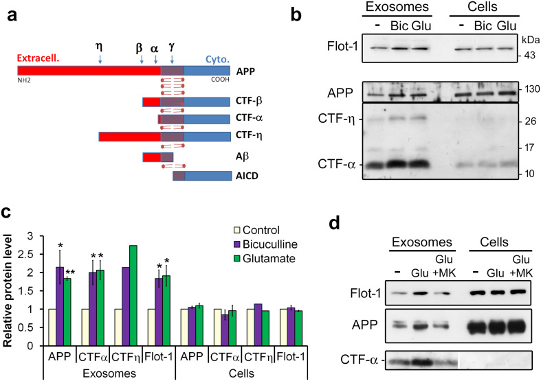
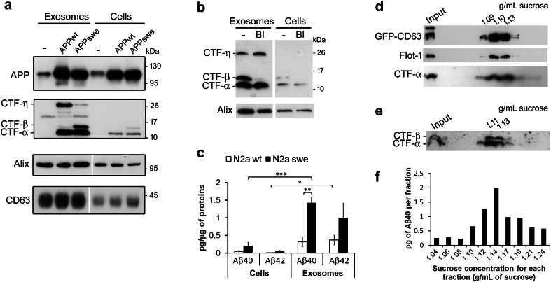
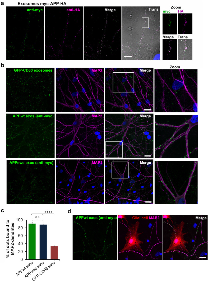
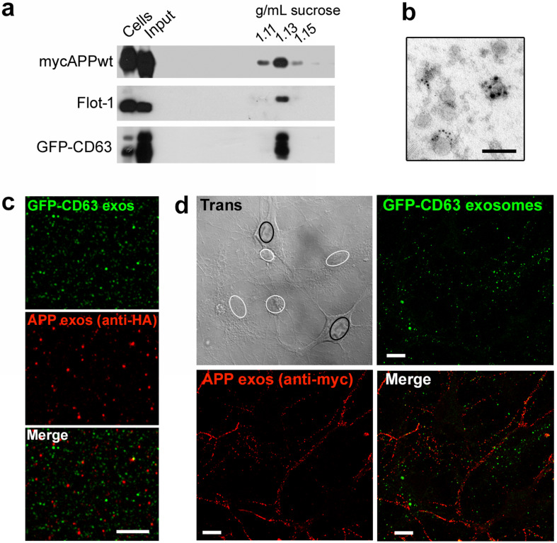
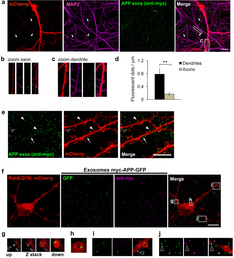
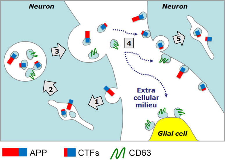
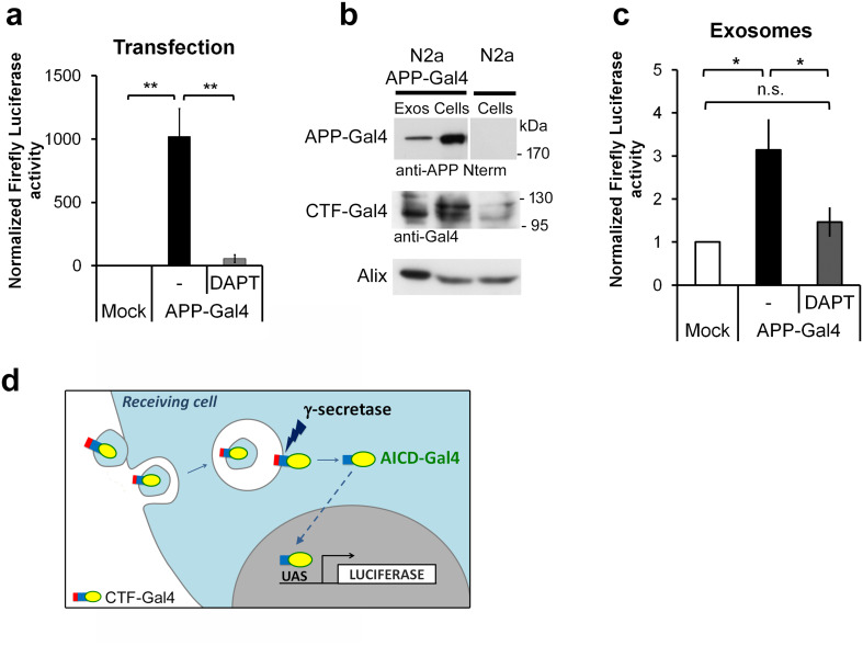
Amyloid beta peptide (Aβ), the main component of senile plaques of Alzheimer's disease brains, is produced by sequential cleavage of amyloid precursor protein (APP) and of its C-terminal fragments (CTFs). An unanswered question is how amyloidogenic peptides spread throughout the brain during the course of the disease. Here, we show that small lipid vesicles called exosomes, secreted in the extracellular milieu by cortical neurons, carry endogenous APP and are strikingly enriched in CTF-α and the newly characterized CTF-η. Exosomes from N2a cells expressing human APP with the autosomal dominant Swedish mutation contain Aβ peptides as well as CTF-α and CTF-η, while those from cells expressing the non-mutated form of APP only contain CTF-α and CTF-η. APP and CTFs are sorted into a subset of exosomes which lack the tetraspanin CD63 and specifically bind to dendrites of neurons, unlike exosomes carrying CD63 which bind to both neurons and glial cells. Thus, neuroblastoma cells secrete distinct populations of exosomes carrying different cargoes and targeting specific cell types. APP-carrying exosomes can be endocytosed by receiving cells, allowing the processing of APP acquired by exosomes to give rise to the APP intracellular domain (AICD). Thus, our results show for the first time that neuronal exosomes may indeed act as vehicles for the intercellular transport of APP and its catabolites.

Keywords: CTF-C83; CTF-C99; Extracellular vesicles; Intercellular communication; Neurodegenerative disorders.


Conflict of interest statement

The authors declare that they have no conflict of interest.

Figures

Fig. 1
Fig. 1
CTFs are enriched in neuronal exosomes. a Schematic drawing of APP and its proteolytic fragments described in the text. α, β, γ, and η refer to cleavage sites by the relevant secretases. b Exosomes released by DIV 15 cortical neurons contain APP and are enriched in CTF-α and a 26 kDa CTF proposed to be CTF-η. Western blot (WB) analysis of Flotillin-1 (Flot-1), full-length APP (APP) and C-terminal fragments (CTF-η and CTF-α) in cells and exosomes. Exosomes were harvested from supernatants of cortical neurons incubated during 20 min in control medium (−), or medium containing 40 μM bicuculline (Bic) or 50 μM glutamate (Glu). c Glutamate receptor activation increases exosomal release of APP-CTF-α and CTF-η. Densitometry of WB shows the glutamate-induced increase of APP, CTF-α, and Flotillin-1 in exosome pellets but not in cells. Means of three experiments ± Standard Error Mean “SEM” (*p < 0.05; unpaired Student t test). In the case of CTF-η, quantification is the mean of two experiments; the values of the relative protein levels are: 2.3 and 1.9 for bicuculline, and 2.9 and 2.5 for glutamate. d Inhibition of NMDA receptors by MK801 blocks glutamate-induced increase in exosomal release of APP and CTF-α. WB of exosomes released by cortical neurons incubated in control medium (−), added of 50 μM glutamate (Glu), or 50 μM glutamate together with 5 μM MK801 (Glu + MK). Note that the film showing CTF-α has been cut to remove irrelevant lanes of the same gel
Fig. 2
Fig. 2
Exosomes released by N2a cells expressing human APPwt and APPswe are enriched in CTFs and Aβ peptides. a Exosomes contain APP and are enriched in CTF-α and two distinct CTFs depending on the APP form expressed. The upper panel is a WB of cells and exosomes from N2a cells (−), or N2a expressing APPwt (APPwt) or APPswe (APPswe) labelled with an antibody against the N-terminal part of APP to reveal full-length APP. CTFs were revealed using antibodies against the C-terminal part of APP. The lower panels are WB of cells and exosomes from N2a cells or APPwt- or APPswe-expressing N2a cells showing that expression of these proteins does not apparently change the amount of two exosome markers, Alix or CD63. Note that for both WB of Alix and CD63, the film have been cut to remove irrelevant lanes of the same gel. b BACE-1 inhibition (BI) blocks the appearance of CTF-β and increases the amount of CTF-η. Alix was used as an exosome marker. c Aβ peptides are enriched in exosomes compared to cells. Aβ42 and Aβ40 were quantified by ELISA. Means of 4–5 experiments ± SEM. *p < 0.05 **p < 0.01 ***p < 0.001. d–f Aβ40, CTF-α, and CTF-β are present in the exosomal fractions containing CD63. Exosomes released by GFP-CD63 N2a cells transfected with APPwt (d) or APPswe (e, f) were separated on sucrose density gradients. Two fractions were collected and analyzed by WB (d, e). ELISA was also used to detect Aβ40 in the fractions (f). Sucrose concentration of exosome-containing fractions is indicated in g/mL
Fig. 3
Fig. 3
APP exosomes bind specifically to neurons. a Exosomes purified from myc-APP-HA-expressing N2a supernatants were incubated during 2 h on DIV 17 hippocampal cells. Double immunofluorescence was used to localize the myc-tagged N-terminal- (green) and the HA-tagged C-terminal (magenta) parts of APP on exosomes. Colocalizations of the tags appear as white dots and demonstrate full-length APP-containing exosomes labelling of neurites. b APP-containing exosomes bind specifically to neurites in contrast to CD63 containing exosomes which bind to all cells. Exosomes purified from supernatants of GFP-CD63 N2a (GFP-CD63 exosomes) or N2a cells expressing myc-APPwt (APPwt exos) or mycAPPswe (APPswe exos) were incubated on hippocampal cultures and APP exosomes localized by immunofluorescence with anti-myc antibody (green). Co-staining with anti-MAP2 (magenta) shows that APP exosomes label MAP2-positive dendrites in contrast to GFP-CD63 exosomes which bind to all cells of the culture. c Percentage of each class of exosomes bound to MAP2-positive neuron cell bodies and dendrites. d Hippocampal cells transfected with mCherry were incubated with APP exosomes which were revealed by myc immunostaining (APPwt exos). Neurons were labelled using anti-MAP2 (Magenta). The picture shows APP exosomes bound to MAP2-positive dendrites but not to the underlying mCherry-expressing, MAP2-negative glial cell. Nuclei were stained with Hoechst (blue in a–c). Bars 10 μm
Fig. 4
Fig. 4
APP and CD63 define exosome subpopulations with different target cells. Exosomes were purified from culture supernatants of N2a cells expressing both GFP-CD63 and myc-APPwt (a, d) or myc-APPwt-HA (b, c). a Exosomes were separated on a linear sucrose gradient, as shown in Fig. 2. Myc-APPwt, Flotillin-1, and GFP-CD63 appear in exosomal fractions containing 1.11–1.15 g/mL of sucrose. b Co-immunogold staining with anti-HA (6 nm gold particles) and anti-CD63 (irregular silver deposits > 10 nm) demonstrates the presence of myc-APP-HA and GFP-CD63, respectively, in different vesicles. Bar  100 nm. c Immunofluorescence staining of purified exosomes with anti-HA confirms that APP and GFP-CD63 are localized on different objects (Pearson’s correlation coefficient between APP staining and GFP calculated on 20,000 objects with JACoP of ImageJ: r = 0.01) Bar  5 μm d APP exosomes [APP exos (anti-myc), red] and CD63 exosomes have different binding specificities towards neural cells. Exosomes were incubated on hippocampal cells during 2 h. On the transmitted light picture, circles delineate nuclei of neurons (black) and glial cells (white). Bar  10 μm
Fig. 5
Fig. 5
APP exosomes mainly bind to dendrites and can be endocytosed by hippocampal neurons. Hippocampal neurons expressing mCherry were incubated for 20 h with APP exosomes. a APP exosomes were revealed with anti-myc [APPexos (anti-myc), green] and dendrites labelled with anti-MAP2 (magenta). Arrowheads point to thin mCherry-filled processes identified as axons because of their MAP2-negativity. b, c Higher magnifications illustrate that APP exosomes bind less to axons (b) than to dendrites (c). Bars  20 μm d quantification of the number of APP exosomes counted per μm of dendrites or axons. Mean of four experiments ± SEM (p** < 0.01, unpaired Student t test). e APP exosomes revealed by anti-myc [APP exos (anti-myc), green] bound to mCherry-expressing dendrites. Photographs illustrate APP exosomes localized on dendritic spine necks (empty arrowhead), dendritic spine heads (white arrowheads), or dendritic shaft (white arrow). Bar  10 μm. f Endocytosis of APP exosomes by hippocampal neurons. Myc-APP-GFP exosomes were incubated for 2 h on Rab5-Q79L-mCherry-expressing neurons (red) and immunostained with anti-myc (magenta). Rab5-Q79L-mCherry delineates enlarged endosomes in which GFP can be detected (green). gj Higher magnifications illustrating individual dilated Rab5-Q79L-mCherry endosomes containing GFP–exosomes. g shows a Z-stack of four slices into an enlarged endosomes. hj are individual Z slices of enlarged endosomes. Note that colocalized myc and GFP spots appearing as white dots are only detected on the cell surface, outside endosomes (white empty arrowheads in g, i, j). Bars  10 μm
Fig. 6
Fig. 6
Schematic illustration of the results obtained. APP is endocytosed (step 1) and processed into CTFs inside endosomes which also contain CD63 (step 2). APP-CTFs are sorted into different populations of intraluminal vesicles accumulating inside late endosomes called multivesicular bodies (MVBs). MVBs can fuse with the plasma membrane thereby releasing the intraluminal vesicles which once outside are referred to as exosomes (step 3). CD63–exosomes indifferently bind to neurons and glial cells whereas APP–exosomes bind specifically to, and can be endocytosed by neurons (steps 4 and 5). Note that for simplification, we have represented a differential sorting of APP and CD63 into ILV of the same MVB, but the possibility remains that APP and CD63 are sorted into different MVB populations. Note that our work cannot exclude that APP fragments are sorted into vesicles budding from the cell surface (not represented here)
Fig. 7
Fig. 7
Exosomal CTFs can be processed in receiving cells. a Luciferase activation in N2a cells co-transfected with APP fused with Gal4 (APP-Gal4) and the UAS-Firefly luciferase reporter demonstrates APP cleavage by γ-secretase into AICD-Gal4. UAS-Firefly luciferase was transfected alone (Mock) or together with APP-Gal4. Renilla luciferase was co-expressed to normalize Firefly activity and luciferase activities measured 48 h after the transfections. γ-secretase inhibitor (DAPT) was added 20 h before measurement. Average of five experiments for APP-Gal4 and three experiments for DAPT conditions are shown ± SEM. (**p < 0.01, unpaired Student t test). b WB analysis of exosomes secreted by N2a cells expressing APP-Gal4 demonstrates the presence of APP-Gal4 and CTF-Gal4 in exosomes. c APP-Gal4 exosomes incubated for 20 h on N2a transfected with the UAS-Firefly luciferase reporter only, activate luciferase. The γ-secretase inhibitor DAPT blocks this activation indicating cleavage of CTF-Gal4 into AICD in the recipient cells. Firefly luciferase activity was measured as in a. Average of four experiments ± SEM. *p < 0.05, unpaired Student t test. d Schematic representation of the way exosomes containing APP-Gal4 would induce luciferase activity in cells transfected with UAS-Firefly luciferase only. Exosomes containing CTF-Gal4 are endocytosed by UAS-luciferase transfected cells. Once inside endosomes, exosomes fuse with the limiting endosomal membrane. CTF-Gal4 is thereby inserted into the membrane and accessible to cleavage by γ-secretase. AICD-Gal4 is released and enters the nucleus to activate transcription of the luciferase gene

References

    1. Gruenberg J, Stenmark H. The biogenesis of multivesicular endosomes. Nat Rev Mol Cell Biol. 2004;5:317–323. doi: 10.1038/nrm1360. - DOI - PubMed
    1. Denzer K, Kleijmeer MJ, Heijnen HF, et al. Exosome: from internal vesicle of the multivesicular body to intercellular signaling device. J Cell Sci. 2000;113:3365–3374. - PubMed
    1. Laulagnier K, Schieber NL, Maritzen T, et al. Role of AP1 and Gadkin in the traffic of secretory endo-lysosomes. Mol Biol Cell. 2011;22:2068–2082. doi: 10.1091/mbc.E11-03-0193. - DOI - PMC - PubMed
    1. Edgar JR, Willén K, Gouras GK, Futter CE. ESCRTs regulate amyloid precursor protein sorting in multivesicular bodies and intracellular amyloid-β accumulation. J Cell Sci. 2015;128:2520–2528. doi: 10.1242/jcs.170233. - DOI - PMC - PubMed
    1. White IJ, Bailey LM, Aghakhani MR, et al. EGF stimulates annexin 1-dependent inward vesiculation in a multivesicular endosome subpopulation. EMBO J. 2006;25:1–12. doi: 10.1038/sj.emboj.7600759. - DOI - PMC - PubMed

Publication types

MeSH terms

Substances