

Increased Neuronal Differentiation of Neural Progenitor Cells Derived from Phosphovimentin-Deficient Mice
- PMID: 28956310
- PMCID: PMC5994207
- DOI: 10.1007/s12035-017-0759-0
Increased Neuronal Differentiation of Neural Progenitor Cells Derived from Phosphovimentin-Deficient Mice
Abstract
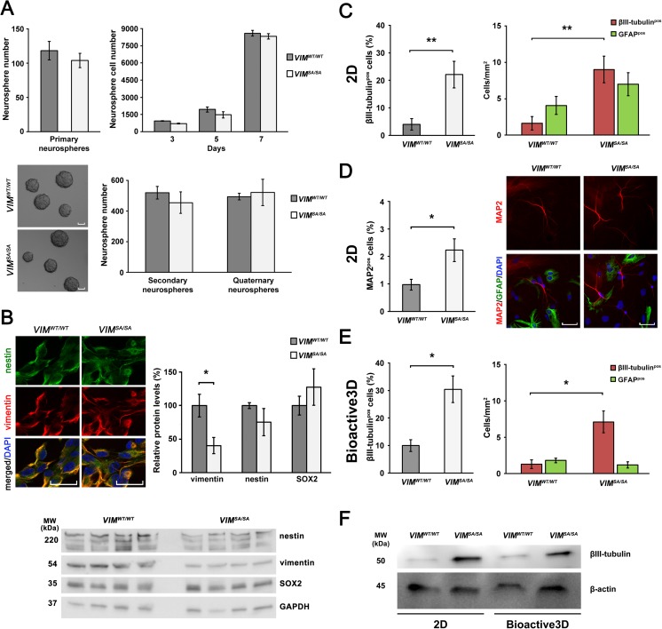
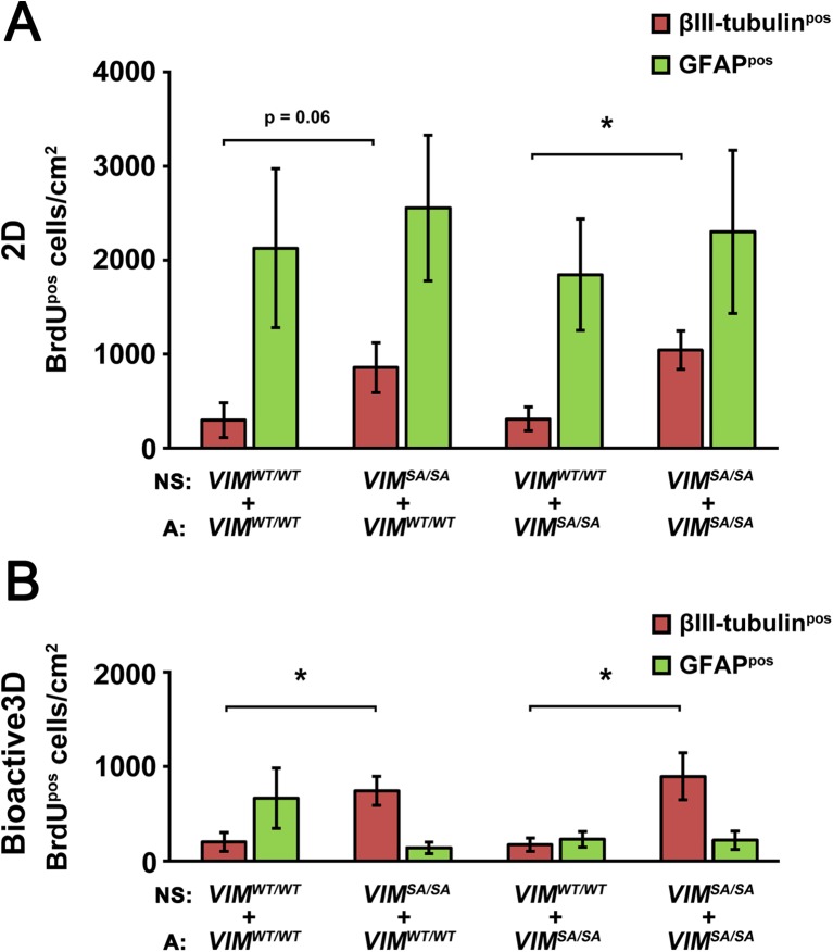
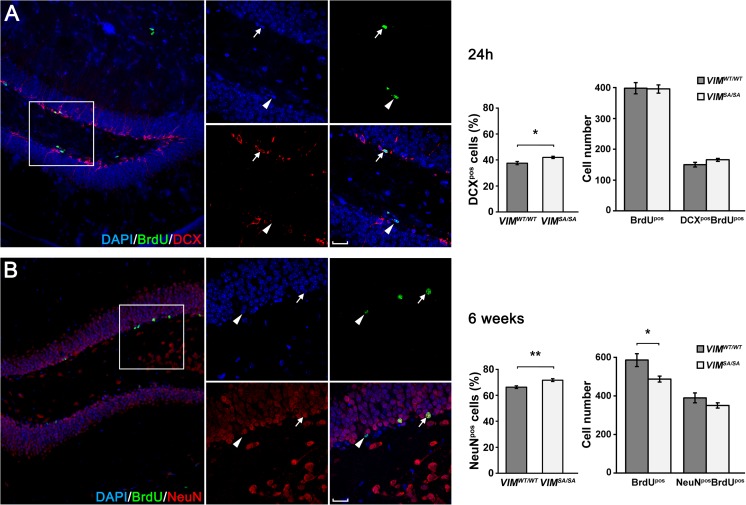
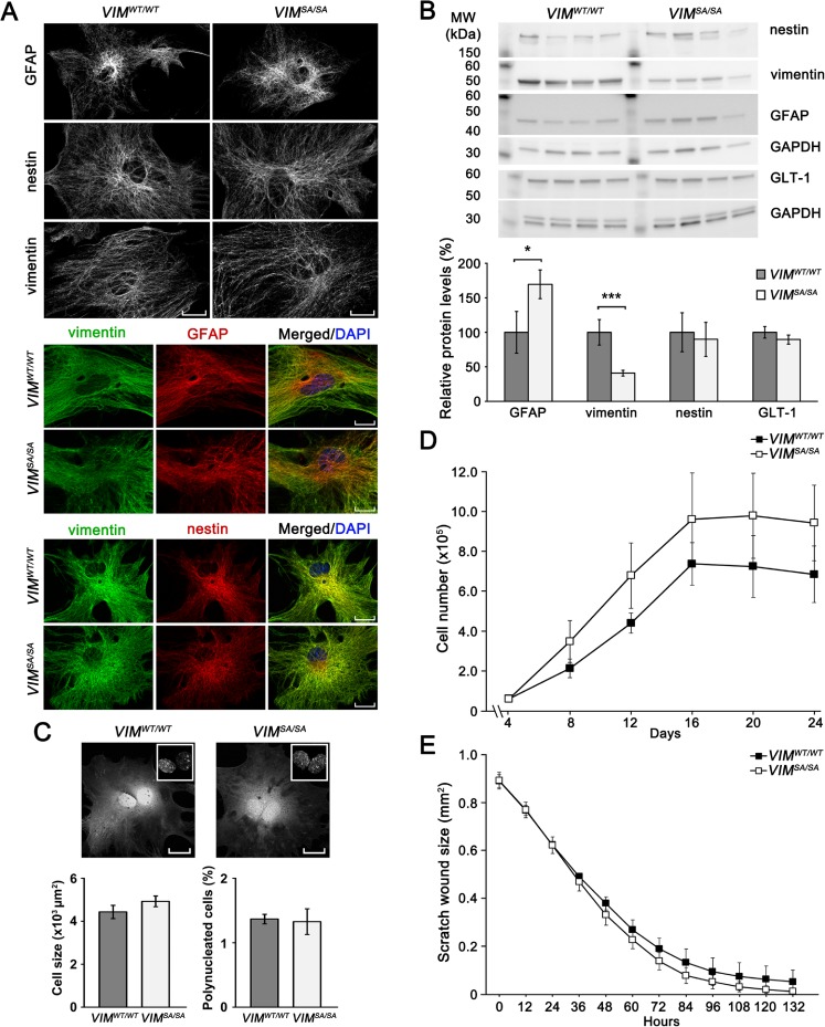
Vimentin is an intermediate filament (also known as nanofilament) protein expressed in several cell types of the central nervous system, including astrocytes and neural stem/progenitor cells. Mutation of the vimentin serine sites that are phosphorylated during mitosis (VIM SA/SA ) leads to cytokinetic failures in fibroblasts and lens epithelial cells, resulting in chromosomal instability and increased expression of cell senescence markers. In this study, we investigated morphology, proliferative capacity, and motility of VIM SA/SA astrocytes, and their effect on the differentiation of neural stem/progenitor cells. VIM SA/SA astrocytes expressed less vimentin and more GFAP but showed a well-developed intermediate filament network, exhibited normal cell morphology, proliferation, and motility in an in vitro wound closing assay. Interestingly, we found a two- to fourfold increased neuronal differentiation of VIM SA/SA neurosphere cells, both in a standard 2D and in Bioactive3D cell culture systems, and determined that this effect was neurosphere cell autonomous and not dependent on cocultured astrocytes. Using BrdU in vivo labeling to assess neural stem/progenitor cell proliferation and differentiation in the hippocampus of adult mice, one of the two major adult neurogenic regions, we found a modest increase (by 8%) in the fraction of newly born and surviving neurons. Thus, mutation of the serine sites phosphorylated in vimentin during mitosis alters intermediate filament protein expression but has no effect on astrocyte morphology or proliferation, and leads to increased neuronal differentiation of neural progenitor cells.
Keywords: Astrocytes; Bioactive3D culture system; GFAP; Intermediate filaments; Nanofilaments; Neural stem/progenitor cells; Vimentin.
Conflict of interest statement
The authors declare that they have no conflict of interest.
Figures
Similar articles
-
Vimentin Phosphorylation Is Required for Normal Cell Division of Immature Astrocytes.Cells. 2019 Sep 1;8(9):1016. doi: 10.3390/cells8091016. Cells. 2019. PMID: 31480524 Free PMC article.
-
Increased neurogenesis and astrogenesis from neural progenitor cells grafted in the hippocampus of GFAP-/- Vim-/- mice.Stem Cells. 2007 Oct;25(10):2619-27. doi: 10.1634/stemcells.2007-0122. Epub 2007 Jul 12. Stem Cells. 2007. PMID: 17628017
-
Astrocytes negatively regulate neurogenesis through the Jagged1-mediated Notch pathway.Stem Cells. 2012 Oct;30(10):2320-9. doi: 10.1002/stem.1196. Stem Cells. 2012. PMID: 22887872
-
[Glial cells function as neural stem cells and progenitor cells].Sheng Li Xue Bao. 2017 Apr 25;69(2):207-217. Sheng Li Xue Bao. 2017. PMID: 28435980 Review. Chinese.
-
Intermediate filaments in the nervous system: implications in cancer.Cancer Metastasis Rev. 1996 Dec;15(4):483-97. doi: 10.1007/BF00054014. Cancer Metastasis Rev. 1996. PMID: 9034605 Review.
Cited by
-
An in vitro 3-dimensional Collagen-based Corneal Construct with Innervation Using Human Corneal Cell Lines.Ophthalmol Sci. 2024 May 6;4(6):100544. doi: 10.1016/j.xops.2024.100544. eCollection 2024 Nov-Dec. Ophthalmol Sci. 2024. PMID: 39139547 Free PMC article.
-
Cleft palate and minor metabolic disturbances in a mouse global Arl15 gene knockout.FASEB J. 2023 Nov;37(11):e23211. doi: 10.1096/fj.202201918R. FASEB J. 2023. PMID: 37773757 Free PMC article.
-
α-Enolase and γ-Enolase Expression in Enriched S- and N-Type SH-SY5Y Cells: Regulatory Role of Cathepsin X.Mol Neurobiol. 2025 Aug;62(8):10006-10019. doi: 10.1007/s12035-025-04898-2. Epub 2025 Apr 3. Mol Neurobiol. 2025. PMID: 40180688 Free PMC article.
-
Vimentin Diversity in Health and Disease.Cells. 2018 Sep 21;7(10):147. doi: 10.3390/cells7100147. Cells. 2018. PMID: 30248895 Free PMC article. Review.
-
Ash1l loss-of-function results in structural birth defects and altered cortical development.Brain. 2025 Jan 7;148(1):55-68. doi: 10.1093/brain/awae218. Brain. 2025. PMID: 38943682 Free PMC article.
References
MeSH terms
Substances
Grants and funding
LinkOut - more resources
Full Text Sources
Other Literature Sources
Miscellaneous
