. 2018 Feb;24(1):4-13.
doi: 10.1177/1591019917733125. Epub 2017 Sep 28.

The Medina Embolic Device: Karolinska experience

Affiliations

The Medina Embolic Device: Karolinska experience

P Bhogal et al. Interv Neuroradiol. 2018 Feb.

Abstract

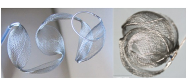
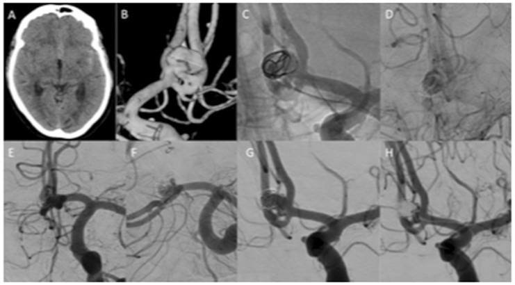
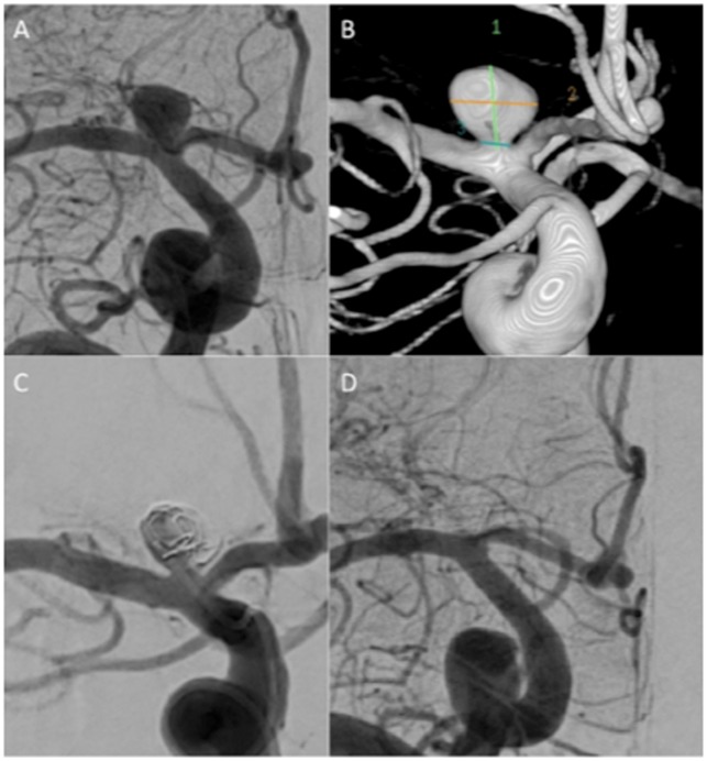
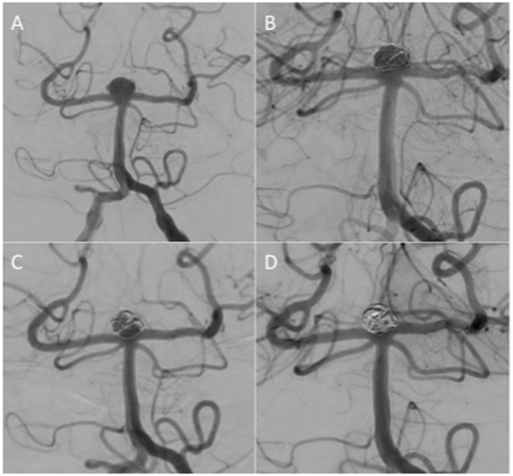
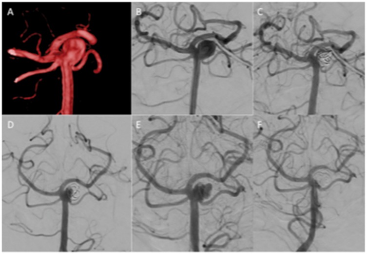
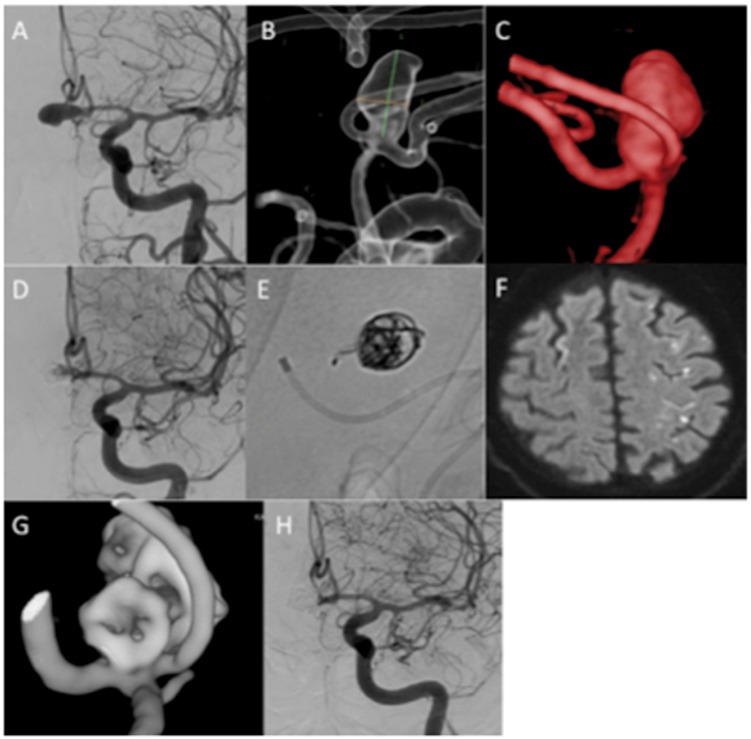
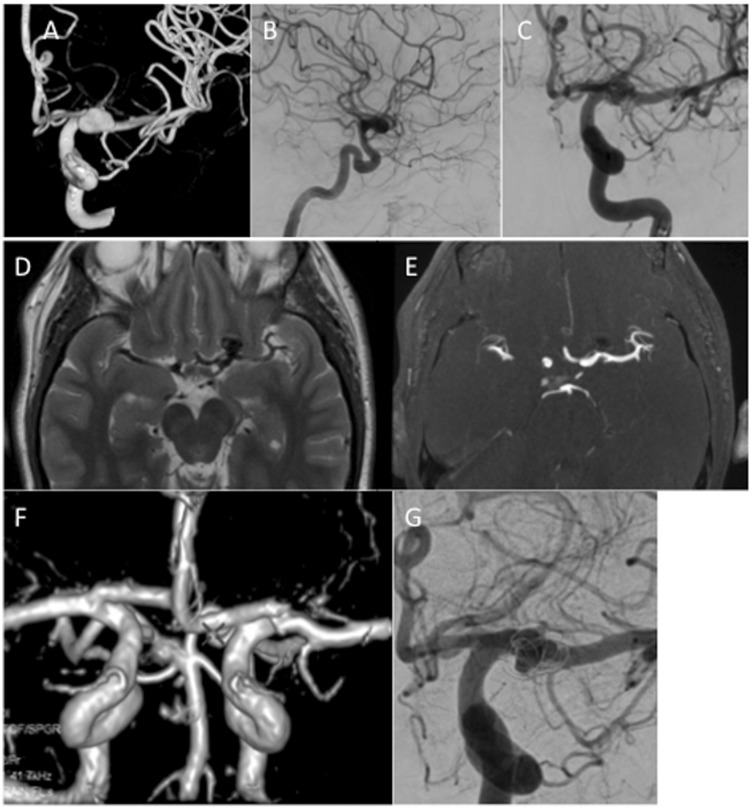
Background The aim of this study was to report our single centre experience with the Medina Embolic Device (MED). Methods We performed a retrospective analysis of prospectively collected data to identify all patients treated with the MED. A total of 14 aneurysms (non-consecutive), in 13 patients, were treated including one ruptured and one partially thrombosed aneurysm. Fundus diameter was ≥5 mm in all cases. We evaluated the angiographic appearances, the clinical status, complications, and the need for adjunctive devices or repeat treatments. Results Aneurysm location was cavernous internal carotid artery (ICA; n = 1), supraclinoid ICA ( n = 1), terminal ICA ( n = 2), anterior communicating artery (AComA; n = 4), A2-3 ( n = 1), M1-2 junction ( n = 1), posterior communicating artery (PComA; n = 1), superior cerebellar artery (SCA; n = 1), and basilar tip ( n = 2). The average aneurysm fundus size was 8.6 mm (range 7-10 mm) and average neck size 3.75 mm (range 1.9-6.9 mm). Immediate angiographic results were modified Raymond-Roy occlusion classification (mRRC) I n = 2, mRRC II n = 1, mRRC IIIa n = 2, mRRC IIIb n = 2, the remaining 7 aneurysms showed complete opacification. At follow-up angiography (mean 5 months) mRRC I n = 5, mRRC II n = 5, mRRC IIIa n = 3, and persistent filling was seen in 1 aneurysm. Overall, four patients had repeat treatment and one is pending further treatment. Of the aneurysms treated with more than one MED, 75% showed complete occlusion at 6-month follow up whereas only one aneurysm treated with a single device showed complete occlusion. Overall, three patients had temporary complications and there were no deaths. Conclusions The MED is an intra-saccular flow-diverting device with satisfactory angiographic results and an acceptable safety profile. Use of a single MED cannot be recommended and further longer term studies are needed prior to widespread clinical use.

Keywords: Aneurysm; Medina Embolic Device; flow diverter; intra-saccular.


Figures

Figure 1.
Figure 1.
The Medina Embolic Device is deployed in a similar fashion to standard endovascular coils. It has a shape set core wire and flow diverter ‘leaflets’ (a). When unconstrained it adopts a spherical shape (b).
Figure 2.
Figure 2.
A patient with a ruptured AComA aneurysm (a and b) was treated with a single MED (c). The MED had to be repositioned several times before stagnation of contrast was achieved (d). At control angiography there was still contrast media filling of the aneurysm (e) although this appeared incomplete (f and g). Retreatment was performed at 3 months with conventional coiling within the MED frame. There was still a neck remnant (h). AComA: anterior communicating artery; MED: Medina Embolic Device.
Figure 3.
Figure 3.
An unruptured terminal carotid aneurysm (a and b) was treated with 2 MEDs at the end of the procedure there was still filling in the aneurysm (c, mRRC IIIb) however, at follow up at 6 months the aneurysm was completely excluded from the circulation (d). MED: Medina Embolic Device; mRRC: modified Raymond–Roy occlusion classification.
Figure 4.
Figure 4.
An unruptured basilar tip aneurysm (patient 12) (a) was treated with a single MED framer with persistent contrast media circulation in the aneurysm at the end of the procedure (b). Follow up and 6 months showed persistent filling of the aneurysm (c) and therefore repeat treatment at 9 months was performed with balloon assistance in a Y-configuration. Delayed follow-up angiography is awaited. MED: Medina Embolic Device.
Figure 5.
Figure 5.
An unruptured aneurysm of the superior cerebellar artery (a and b) was treated initially with a single MED with mRRC IIIb occlusion at the end of the procedure (c). Follow-up angiography at 6 months showed a slight change in the configuration of the MED and a residual neck remnant (d). Delayed angiography demonstrated a large recurrence (e). The patient was retreated with conventional coils (f). MED: Medina Embolic Device; mRRC: modified Raymond–Roy occlusion classification.
Figure 6.
Figure 6.
A patient with an unruptured AComA aneurysm (a–c) was treated with 2 MEDs. There was good stasis of contrast in the aneurysm at the end of the procedure (d) however some persistent filling was seen. Upon removal of the microcatheter a single MED loop was displaced and could be seen in the parent artery (e). The patient was started on dual anti-platelet agents. He developed symptoms consistent with an ACA territory infarction and MRI demonstrated multiple small embolic infarctions within the ACA territory (f). He recovered completely and follow-up angiography showed complete occlusion of the aneurysm (g and h). ACA: anterior cerebral artery; AComA: anterior communicating artery; MED: Medina Embolic Device; MRI: magnetic resonance imaging.
Figure 7.
Figure 7.
A patient with an unruptured left ICA bifurcation aneurysm (a–c) was treated with a single MED. At initial follow up performed at 6 months axial T2-weighted MRI (d) showed a signal void in the aneurysm suggestive of continued filling however, on TOF MRA (e) there was no evidence flow which was confirmed on 3D reconstruction of the MRA data (f). On angiography, there was persistent filling of the aneurysm (mRRC IIIa). The discrepancy between the MRI and the angiographic data was thought to be due to a ‘Faraday cage’ like effect of the MED. ICA: internal carotid artery; MED: Medina Embolic Device; MRA: Magnetic Resonance Angiography; MRI: magnetic resonance imaging; mRRC: modified Raymond–Roy occlusion classification; TOF: Time of Flight.

References

    1. Henkes H, Weber W. The past, present and future of endovascular aneurysm treatment. Clin Neuroradiol 2015; 25(Suppl. 2): 317–324. - PubMed
    1. Turk AS, Maia O, Ferreira CC, et al. Periprocedural safety of aneurysm embolization with the Medina Coil System: the early human experience. J Neurointerventional Surg 2016; 8: 168–172. - PMC - PubMed
    1. Aguilar Perez M, Bhogal P, Martinez Moreno R, et al. The Medina Embolic Device: early clinical experience from a single center. J Neurointerventional Surg 2017; 9: 77–87. - PMC - PubMed
    1. Mascitelli JR, Moyle H, Oermann EK, et al. An update to the Raymond–Roy Occlusion Classification of intracranial aneurysms treated with coil embolization. J Neurointerventional Surg 2015; 7: 496–502. - PubMed
    1. Aguilar Perez M, Bhogal P, Martinez Moreno R, et al. The Medina Embolic Device: early clinical experience from a single center. J Neurointerventional Surg 2017; 9: 77–87. - PMC - PubMed

LinkOut - more resources