. 2017 Nov 14;91(23):e01234-17.
doi: 10.1128/JVI.01234-17. Print 2017 Dec 1.

Novel Role for Interleukin-17 in Enhancing Type 1 Helper T Cell Immunity in the Female Genital Tract following Mucosal Herpes Simplex Virus 2 Vaccination

Affiliations

Novel Role for Interleukin-17 in Enhancing Type 1 Helper T Cell Immunity in the Female Genital Tract following Mucosal Herpes Simplex Virus 2 Vaccination

Puja Bagri et al. J Virol. .

Abstract

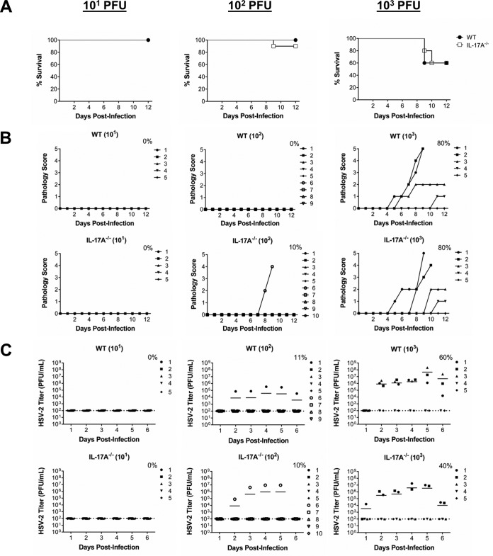
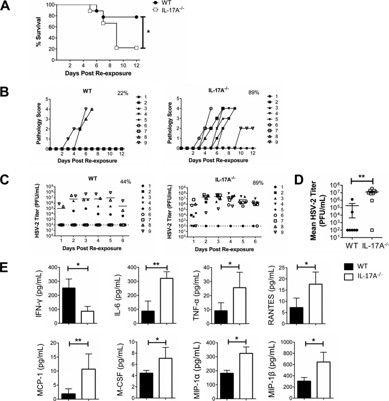
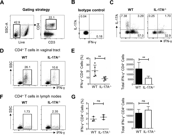
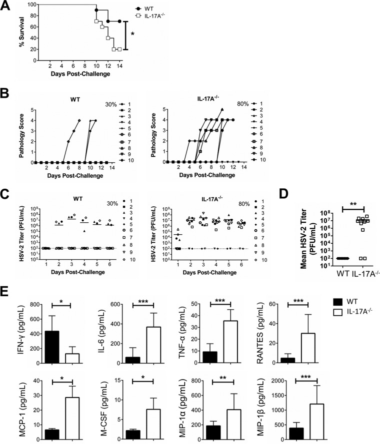
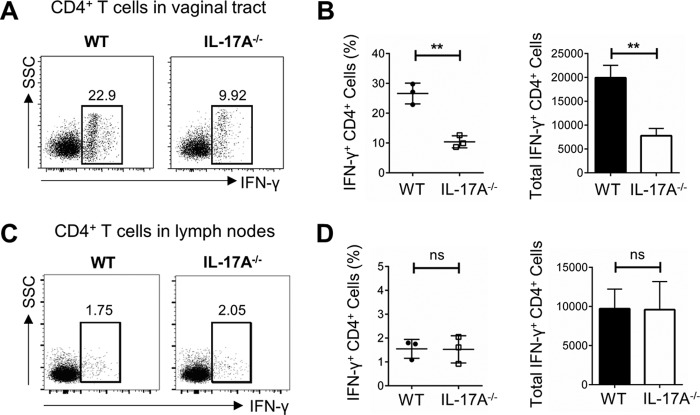
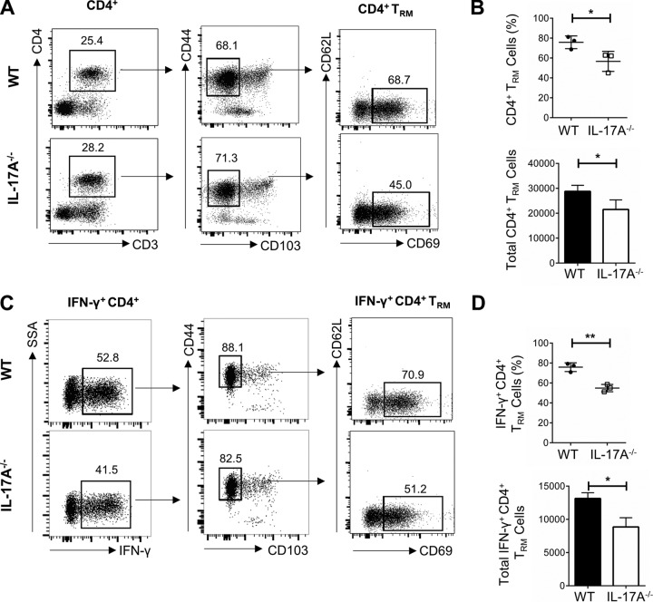
It is well established that interferon gamma (IFN-γ) production by CD4+ T cells is critical for antiviral immunity against herpes simplex virus 2 (HSV-2) genital infection. However, the role of interleukin-17A (IL-17A) production by CD4+ T cells in HSV-2 antiviral immunity is yet to be elucidated. Here we demonstrate that IL-17A plays an important role in enhancing antiviral T helper type 1 (Th1) responses in the female genital tract (FGT) and is essential for effective protection conferred by HSV-2 vaccination. While IL-17A did not play a critical role during primary genital HSV-2 infection, seen by lack of differences in susceptibility between IL-17A-deficient (IL-17A-/-) and wild-type (WT) C57BL/6 mice, it was critical for mediating antiviral responses after challenge/reexposure. Compared to WT mice, IL-17A-/- mice (i) infected intravaginally and reexposed or (ii) vaccinated intranasally and challenged intravaginally demonstrated poor outcomes. Following intravaginal HSV-2 reexposure or challenge, vaccinated IL-17A-/- mice had significantly higher mortality, greater disease severity, higher viral shedding, and higher levels of proinflammatory cytokines and chemokines in vaginal secretions. Furthermore, IL-17A-/- mice had impaired Th1 cell responses after challenge/reexposure, with significantly lower proportions of vaginal IFN-γ+ CD4+ T cells. The impaired Th1 cell responses in IL-17A-/- mice coincided with smaller populations of IFN-γ+ CD4+ tissue resident memory T (TRM) cells in the genital tract postimmunization. Taken together, these findings describe a novel role for IL-17A in regulating antiviral IFN-γ+ Th1 cell immunity in the vaginal tract. This strategy could be exploited to enhance antiviral immunity following HSV-2 vaccination.IMPORTANCE T helper type 1 (Th1) immunity, specifically interferon gamma (IFN-γ) production by CD4+ T cells, is critical for protection against genital herpesvirus (HSV-2) infection, and enhancing this response can potentially help improve disease outcomes. Our study demonstrated that interleukin-17A (IL-17A) plays an essential role in enhancing antiviral Th1 responses in the female genital tract (FGT). We found that in the absence of IL-17A, preexposed and vaccinated mice showed poor disease outcomes and were unable to overcome HSV-2 reexposure/challenge. IL-17A-deficient mice (IL-17A-/-) had smaller populations of IFN-γ+ CD4+ tissue resident memory T (TRM) cells in the genital tract postimmunization than did wild-type (WT) mice, which coincided with attenuated Th1 responses postchallenge. This has important implications for developing effective vaccines against HSV-2, as we propose that strategies inducing IL-17A in the genital tract may promote more effective Th1 cell immunity and better overall protection.

Keywords: CD4 T cell immunity; IL-17; genital tract immunity; herpes simplex virus; mucosal immunity; sexually transmitted diseases.


Figures

FIG 1
FIG 1
IL-17A−/− mice demonstrate no significant difference in susceptibility to primary intravaginal HSV-2 infection. OVX WT (C57BL/6) and IL-17A−/− mice (n = 5 to 10/group) were infected intravaginally with sublethal doses of WT HSV-2 (101, 102, or 103 PFU/mouse). Survival was monitored (A) and pathology scores were recorded on a scale of 0 to 5 (B) for 12 days postinfection. Data points superimposed on the x axes of panel B indicate mice without genital pathology, and the percentages represent maximum numbers of mice that demonstrated pathology. (C) Vaginal washes were collected daily for 6 days postinfection and HSV-2 shedding was assessed using a Vero cell-based assay. The bars in panel C indicate mean PFU per milliliter of shed virus. The dotted lines in panel C indicate the lower detection limit of the assay, and data points on this line indicate undetectable viral shedding. The percentages in panel C represent maximum numbers of mice that shed virus on any given day. Each symbol represents a single animal. The results are representative of those from two independent experiments.
FIG 2
FIG 2
Preexposed IL-17A−/− mice are more susceptible to intravaginal HSV-2 reexposure. OVX WT (C57BL/6) and IL-17A−/− mice (n = 9/group) were intravaginally exposed to WT HSV-2 (102 PFU/mouse), and 6 weeks later, they were reexposed intravaginally with a higher dose of WT HSV-2 (5 × 10³ PFU/mouse). Survival was monitored (A) and pathology scores were recorded on a scale of 0 to 5 (B) for 12 days after reexposure. Significance in difference in survival (A) was calculated using the log rank (Mantel-Cox) test (*, P < 0.05). Data points superimposed on the x axes of panel B indicate mice without genital pathology, and the percentages represent maximum numbers of mice that demonstrated pathology. Vaginal washes were collected daily for 6 days after reexposure; HSV-2 viral shedding was calculated using a Vero cell-based assay (C and D), and cytokine and chemokine (IFN-γ, IL-6, TNF-α, RANTES, MCP-1, M-CSF, MIP-1α, and MIP-1β) concentrations were measured by multianalyte assays (E). The bars in panel C indicate mean PFU per milliliter of shed virus. The dotted lines in panel C indicate the lower detection limit of the assay, and data points on this line indicate undetectable viral shedding. The percentages in panel C represent maximum numbers of mice that shed virus on any given day. Data shown in panel D represent the viral loads (means ± SEMs) over 6 days. Each symbol represents a single animal. Data shown in panel E represent the means ± SEMs from two independent experiments, done in duplicate (n = 4 to 7/group). Data were analyzed using the unpaired, nonparametric, two-tailed Mann-Whitney test with 95% confidence interval, with the ROUT method used to identify outliers and the Bonferroni correction used to correct for multiple measures.*, P < 0.05; **, P < 0.01.
FIG 3
FIG 3
Preexposed IL-17A−/− mice have lower proportions of IFN-γ+ Th1 cells in the vaginal tract following intravaginal HSV-2 reexposure. OVX WT (C57BL/6) and IL-17A−/− mice (n = 4 to 6/group) were intravaginally exposed to WT HSV-2 (102 PFU/mouse) and 6 weeks later reexposed intravaginally with a higher dose of WT HSV-2 (5 × 10³ PFU/mouse). Vaginal tissue and lymph nodes were isolated at day 3 following reexposure, pooled, processed, stained with a panel of antibodies, and examined by flow cytometry. (A) CD4+ T cells were gated among total live CD3+ T cells in the vaginal tissue. (B) Isotype controls for intracellular staining of IL-17A and IFN-γ. (C) Vaginal cells were stimulated in vitro with a cell stimulation cocktail (CSC) containing Golgi inhibitors and PMA plus ionomycin for 16 h to detect intracellular staining of IL-17A and IFN-γ. (D) Intracellular staining for IFN-γ was used to examine the in vivo response to HSV-2 (without in vitro stimulation) and the differentiation of CD4+ T cells into Th1 cells in the vaginal tract. (E) The differences in percentages and total cell numbers of IFN-γ-producing CD4+ T cells after HSV-2 reexposure in the vaginal tract were compared across five independent experiments (n = 4 to 6/group). (F and G) Intracellular staining for IFN-γ (F) and the differences in percentages and total cell numbers of IFN-γ-producing CD4+ T cells after HSV-2 reexposure in the lymph nodes (G) were compared across three independent experiments (n = 5 or 6/group). Data shown in panels E and G represent means ± SEMs. Significant difference in IFN-γ expression was calculated using the unpaired, two-tailed t test with 95% confidence interval. **, P < 0.01. ns, no significance.
FIG 4
FIG 4
Intranasally immunized IL-17A−/− mice are more susceptible to intravaginal HSV-2 challenge. OVX WT (C57BL/6) and IL-17A−/− mice (n = 10/group) were immunized intranasally with TK HSV-2 (102 PFU/mouse) and 6 weeks later challenged intravaginally with WT HSV-2 (5 × 10³ PFU/mouse). Survival was monitored (A) and pathology scores were recorded on a scale of 0 to 5 (B) for 14 days postchallenge. Significance in difference in survival (A) was calculated using the log rank (Mantel-Cox) test (*, P < 0.05). Data points superimposed on the x axes of panel B indicate mice without genital pathology, and the percentages represent maximum numbers of mice that demonstrated pathology. Vaginal washes were collected daily for 6 days postchallenge; HSV-2 shedding was calculated using a Vero cell-based assay (C and D), and cytokine and chemokine (IFN-γ, IL-6, TNF-α, RANTES, MCP-1, M-CSF, MIP-1α, and MIP-1β) concentrations were measured by multianalyte assays (E). The bars in panel C indicate mean PFU per milliliter of shed virus. The dotted lines in panel C indicate the lower detection limit of the assay, and data points on this line indicate undetectable viral shedding. The percentages in panel C represent maximum numbers of mice that shed virus on any given day. Data shown in panel D represent the viral loads (means ± SEMs) over 6 days. Each symbol represents a single animal. Data shown in panel E represents means ± SEMs from two independent experiments, done in duplicate (n = 4 to 7/group). Data were analyzed using the unpaired, nonparametric, two-tailed Mann-Whitney test with 95% confidence interval, with the ROUT method used to identify outliers and the Bonferroni correction used to correct for multiple measures. *, P < 0.05; **, P < 0.01; ***, P <0.001.
FIG 5
FIG 5
Intranasally immunized IL-17A−/− mice have lower proportions of IFN-γ+ Th1 cells in the vaginal tract following intravaginal HSV-2 challenge. OVX WT (C57BL/6) and IL-17A−/− mice (n = 4 to 6/group) were immunized intranasally with TK HSV-2 (102 PFU/mouse) and 6 weeks later challenged intravaginally with WT HSV-2 (5 × 10³ PFU/mouse). Vaginal tissues and lymph nodes were isolated at day 3 postchallenge, pooled, processed, stained with a panel of antibodies, and examined by flow cytometry. Intracellular staining for IFN-γ was used to examine the in vivo response to HSV-2. (A and B) The differentiation of CD4+ T cells into Th1 cells in the vaginal tract (A) and the differences in percentages and total numbers of IFN-γ-producing CD4+ T cells post-HSV-2 challenge in the vaginal tract (B) were compared across three independent experiments (n = 4 to 6/group). (C and D) The differentiation of CD4+ T cells into Th1 cells (C) and the differences in percentages and total numbers of IFN-γ-producing CD4+ T cells after HSV-2 challenge in the lymph nodes (D) were compared across three independent experiments (n = 4 to 6/group). Data shown in panels B and D represent means ± SEMs. Significant difference in IFN-γ expression was calculated using the unpaired, two-tailed t test with 95% confidence interval. **, P < 0.01.
FIG 6
FIG 6
Phenotypic and functional characteristics of CD4+ TRM cells localized in the vaginal tracts of WT and IL-17A−/− mice following HSV-2 vaccination. OVX WT (C57BL/6) and IL-17A−/− mice (n = 5 to 7/group) were vaccinated intravaginally with TK HSV-2 (104 PFU/mouse), and 3 weeks later, vaginal tissues were collected, pooled, and processed. Vaginal cells were stimulated in vitro with a cell stimulation cocktail (CSC) containing Golgi inhibitors and PMA plus ionomycin for 16 h, stained with a panel of antibodies, and examined by flow cytometry. (A) CD4+ T cells were gated among total live CD3+ T cells in the vaginal tissue, and CD4+ TRM cells were detected using surface markers CD44, CD103, CD69, and CD62L. CD4+ TRM cells were defined as CD4+ CD44+ CD103 CD69+ CD62L. (B) The differences in percentages and total numbers of CD4+ TRM cells postimmunization in the vaginal tract were compared across three independent experiments (n = 5 to 7/group). (C) IFN-γ+ CD4+ TRM cells were further gated based on detection of surface markers associated with TRM cells. (D) The differences in percentages and total numbers of IFN-γ+ CD4+ TRM cells postimmunization in the vaginal tract were compared across three independent experiments (n = 5 to 7/group). Data shown in panels B and D represent means ± SEMs. Significant difference in IFN-γ expression was calculated using the unpaired, two-tailed t test with 95% confidence interval. *, P < 0.05; **, P < 0.01.

References

    1. Looker KJ, Magaret AS, Turner KM, Vickerman P, Gottlieb SL, Newman LM. 2015. Global estimates of prevalent and incident herpes simplex virus type 2 infections in 2012. PLoS One 10:e114989. doi:10.1371/journal.pone.0114989. - DOI - PMC - PubMed
    1. Roth K, Ferreira VH, Kaushic C. 2013. HSV-2 vaccine: current state and insights into development of a vaccine that targets genital mucosal protection. Microb Pathog 58:45–54. doi:10.1016/j.micpath.2012.11.001. - DOI - PubMed
    1. Petro C, Gonzalez PA, Cheshenko N, Jandl T, Khajoueinerjad N, Benard A, Sengupta M, Herold BC, Jacobs WR. 2015. Herpes simplex type 2 virus deleted in glycoprotein D protects against vaginal, skin and neural disease. eLife 4:e06054. - PMC - PubMed
    1. Milligan GN, Bernstein DI, Bourne N. 1998. T lymphocytes are required for protection of the vaginal mucosae and sensory ganglia of immune mice against reinfection with herpes simplex virus type 2. J Immunol 160:6093–6100. - PubMed
    1. Harandi AM, Svennerholm B, Holmgren J, Eriksson K. 2001. Differential roles of B cells and IFN-γ-secreting CD4+ T cells in innate and adaptive immune control of genital herpes simplex virus type 2 infection in mice. J Gen Virol 82:845–853. doi:10.1099/0022-1317-82-4-845. - DOI - PubMed

Publication types

MeSH terms

Grants and funding

LinkOut - more resources