. 2018 Jan:77:250-257.
doi: 10.1016/j.jmbbm.2017.09.018. Epub 2017 Sep 14.

Adaptive responses of murine osteoblasts subjected to coupled mechanical stimuli

Affiliations

Adaptive responses of murine osteoblasts subjected to coupled mechanical stimuli

Jean C Serrano et al. J Mech Behav Biomed Mater. 2018 Jan.

Abstract

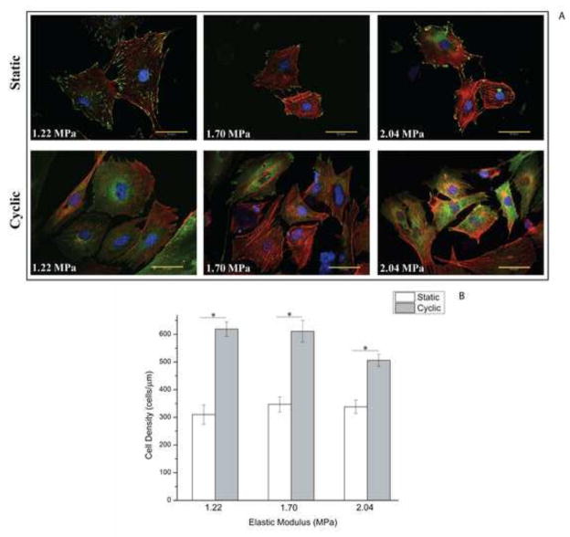
Restitution of the natural organization and orientation of cells is imperative for the construction of functional tissue scaffolds. While numerous studies have exploited mechanical methods to engineer tissues with the desired cellular architecture, fundamental knowledge is still lacking in understanding the manner in which morphological features can be modulated through coupled mechanical cues. To address this knowledge gap, the adhesion and alignment response of murine osteoblast cells under the synergistic effects of matrix rigidity and cyclic mechanical loading was investigated. This was accomplished by applying cyclic mechanical strain (1% at 0.05Hz) to MC3T3-E1 cells seeded on PDMS substrates of different elastic moduli (1.22, 1.70 and 2.04MPa). Results demonstrate that the overall cell density and expression of inactive vinculin increased on substrates subjected to cyclic stimulus in comparison to substrates under static loading. Conversely, in terms of the adhesion response, osteoblasts exhibited an increased growth of focal adhesion complexes under static substrates. Interestingly, results also elucidate that substrates of a stiffer matrix exposed to cyclic stimulus, had a significantly higher percentage of osteoblasts aligned parallel to the direction of the applied strain, as well as a higher degree of internal order with respect to the strain axis, in comparison to both cells seeded on substrates of lower stiffness under cyclic loading or under static conditions. These findings suggest the role of cyclic mechanical strain coupled with matrix rigidity in eliciting mechanosensitive adaptations in cell functions that allow for the reconstitution of the spatial and orientational assembly of cells in vivo for tissue engineering.

Keywords: Cell orientation; Cyclic load; MC3T3-E1 cells; Mechanical stimulus; Vinculin cloud.


Conflict of interest statement

7. Conflict of Interest and Ethics Statement

None of the authors have a conflict in interest in carrying out or based on the results in this study. The study has been carried out under the strict guidelines of research ethics and integrity.

Figures

Figure 1
Figure 1. Schematic diagram of the cellular orientation analysis
An ellipse was fitted around the cell’s periphery thus providing two axes (major and minor) to utilize for subsequent analysis. Cellular orientation was analyzed by measuring the angle between the cell’s major axis and the strain axis. The elongation analysis was performed from the measured major and minor axes of the fitted ellipse.
Figure 2
Figure 2. Substrate Elastic Modulus dependent on PDMS curing ratio
Mean values of the measured elastic moduli of PDMS substrates as a function of base-to-crosslinker ratio. (*= p-value <0.05)
Figure 3
Figure 3. Cell density influenced by the loading condition of the substrates
(A) Representative images of MC3T3-E1 cell density under different substrate stiffness and loading condition. F-actin was stained with TRITC-phalloidin appearing as red, vinculin protein appears as green and blue as nuclei staining with DAPI. (Upper Row: static conditions // Lower Row: 1% cyclic strain at 0.05 Hz). Scale bar represents 50.0μm. (B) Mean values for cell density as a function of substrate stiffness and loading condition. (*= p-value <0.05)
Figure 4
Figure 4. Focal adhesions and vinculin expression influenced by mechanical loading condition
(A) Mean values for the total focal adhesion (active vinculin) area as a function of substrate stiffness and mechanical loading condition. (*= p-value <0.05) (B) Mean values for the total (active + inactive) vinculin area as a function of substrate stiffness and mechanical loading condition. (*= p-value <0.05) (C) Representative images of vinculin expression (green) in MC3T3-E1 cells cultured under different mechanical parameters. Each row shows the fluorescent images of the vinculin expression for MC3T3-E1 cells cultured on PDMS substrates of different elastic modulus. (Upper Row: static conditions // Lower Row: 1% cyclic strain at 0.05 Hz)(Cora et al., 2016). Note strong expression of diffuse vinculin “cloud” in the cells subjected to cyclic stimulus. Scale bar represents 50.0μm.
Figure 5
Figure 5. Increase in the cell population and internal order preferentially aligned with the strain direction as a response to substrate stiffness and mechanical loading condition
Cell Major Axis orientation (with respect to the applied strain direction) as a function of substrate stiffness. (A, B) Cells exposed to static loading conditions and cells exposed to cyclic tensional loading, respectively. A dotted line is drawn at 45° indicating the upper limit for preferential orientation. Preferential orientation with respect to the strain axis was defined as cell’s oriented between 0°–45° with respect to the strain axis. Cells with an orientation beyond 45° are considered to be preferentially oriented with the perpendicular axis (perpendicular to the induced strain direction). Percentages shown to the right of each box corresponds to the percentage of cells exhibiting an orientation above 45° thus preferentially oriented with the strain direction. (A) For static loading, each box corresponds to at least 24 cells. (B) For cyclic loading, each box corresponds to at least 48 cells. (*= p-value <0.05) The black dot (•) represents the mean value in each box and the reduced center represents the median value. Each box ends represent the second and third interquartile, while each whisker end represents the values within 1.5 of the interquartile. (C) Mean values for the cell’s order parameter as a function of substrate elastic modulus and loading condition. The implemented order parameter < S=[(3/2)cos2(θ)]-(1/2)> yields a value of one when the cells are perfectly aligned with the strain direction, a value of zero corresponds to a random orientation and a value of negative one represents a perpendicular orientation with respect to the strain axis. (*= p-value <0.05) (D) Mean cell elongation as function of substrate stiffness and mechanical loading condition. A value of 0% corresponds to a cell with a perfectly spherical area while a value of 100% describes a cell as a perfectly elongated line.

References

    1. Albrecht DR, Underhill GH, Wassermann TB, Sah RL, Bhatia SN. Probing the role of multicellular organization in three-dimensional microenvironments. Nature Methods. 2006;3:369–375. - PubMed
    1. Bershadsky AD, Ballestrem C, Carramusa L, Zilberman Y, Gilquin B, Khochbin S, Alexandrova AY, Verkhovsky AB, Shemesh T, Kozlov MM. Assembly and mechanosensory function of focal adhesions: experiments and models. European Journal of Cell Biology. 2006;85:165–173. - PubMed
    1. Boada-Lopez J, DeJesus-Maldonado I, Jerez J, Romañach R, Diffoot-Carlo N, Sundaram P. Collagen abundance in mechanically stimulated osteoblast cultures using near infrared microscopy. Journal of Biomechanics. 2013;46:2442–2450. - PubMed
    1. Bowles RD, Williams RM, Zipfel WR, Bonassar LJ. Self-Assembly of Aligned Tissue-Engineered Annulus Fibrosus and Intervertebral Disc Composite Via Collagen Gel Contraction. Tissue Engineering Part A. 2010;16:1339–1348. - PMC - PubMed
    1. Brunette DM, Chehroudi B. The Effects of the Surface Topography of Micromachined Titanium Substrata on Cell Behavior in Vitro and in Vivo. Journal of Biomechanical Engineering. 1999;121(1):49–57. - PubMed

Publication types

LinkOut - more resources